Download presentation
Presentation is loading. Please wait.

1
بسم الله الرحمن الرحيم

2
INTRODUCTION TO HISTOLOGY

3
HISTOLOGY (MICROSCOPIC ANATOMY) Definition.
 Definition.")
4
Light Microscope (L/M) 1- Illumination. 2- Magnification. 3- Resolution. N.B. Resolving power ( It is the least distance between 2 particles at which they will appear separated). R.P. for L/M is 250 nm
. R.P. for L/M is 250 nm.")
5
Light microscope

6
STAINING FOR L/M Staining with Hematoxylin and Eosin (H&E) or (Hx&E): Basophilic structures. Acidophilic structures.

7
(B) Electron microscopy: 1-Transmission E/M: Resolving power 0.2 nm. * Electron-dense structure **Electron-lucent structure 2-Scanning E/M: Resolving power 10 nm.

8
Transmission Electron Microscope

9
Scanning Electron microscope

10
THE CELL

11
THE CELL NUCLEUS (INTERPHASE NUCLEUS)
")
12
Shape of nuclei

13
Dark Nucleus (Deeply-stained nucleus)
")
15
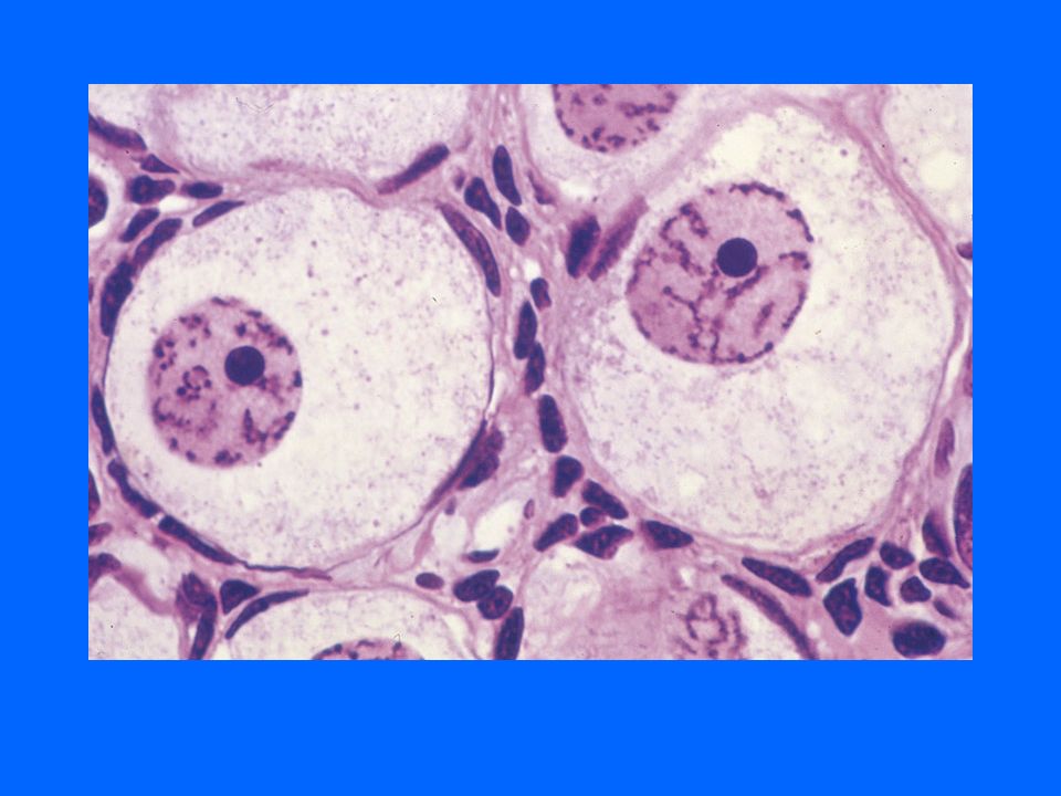
Vesicular (open face) Nucleus
 Nucleus")
17
L/M: Appearance (Type): - Light nucleus (vesicular) (open face) - Dark nucleus (deeply-stained) Number: 1, 2, or more. Position: Central, eccentric, peripheral, basal. CELL NUCLEUS (Interphase Nucleus)
.")
18
Cell Nucleus L/M (cont.): Size: Small, medium, large ( Nucleus/cell ratio) Shape: e.g. Rounded, oval, rod-shaped.

19
Nucleus (E/M diagram) RER------- Hc
 RER Hc")
20
Nucleus (Electron Micrograph) PRACTICAL
 PRACTICAL")
21
Nuclear pores PRACTICAL

22
Cell Nucleus (Interphase nucleus) E/M: (1) Nuclear envelope Inner nuclear membrane. Outer " ". Nuclear pores. Nuclear pore complex. Perinuclear cisterna. Nuclear lamina. N.B. Rough Endoplasmic reticula (Relation with,).
..")
23
(2) Chromatin: ( Classification ): According to Metabolic activity: a- Euchromatin(Extended chromatin) b- Heterochromatin( Condensed chrom. ) According to Position: a- Peripheral chromatin. b- Nucleolus-associated chromatin. c- Chromatin islands.
 According to Position: a- Peripheral chromatin. b- Nucleolus-associated chromatin. c- Chromatin islands..")
24
Nucleolus (E/M)
")
25
(3) Nucleolus: L/M: 1-5 basophilic bodies E/M: 1- Pale-staining fibrillar center. 2- Pars fibrosa: containing rRNA (nRNA) being transcribed.
 being transcribed..")
26
Nucleolus (cont.): 3- Pars granulosa: maturing ribosomal subunits are assembled. 4- Nucleolar matrix. N.B. Nucleolus is a non-membranous structure. Function of nucleolus: rRNA synthesis.

27
NUCLEOPLASM 1- Nuclear matrix. 2- Ribonucleoprotein. 3- Interchromatin granules. 4- Perichromatin granules.

28
CYTOPLASM (1)Organelles. (2)Inclusions. (3)Cytosol.
Organelles. (2)Inclusions. (3)Cytosol.")
29
Cytoplasmic organelles 1- Cell Membrane. 2- Ribosomes. 3- Endoplasmic Reticulum. 4- Golgi Apparatus. 5- Endosomes 6- Lysosomes. 7- Peroxisomes. 8- Mitochondria. 9- Cytoskeleton. 10-Centrioles. 11-Cilia & flagella. 12-Filaments (thin f., intermediate f., thick f.).
..")
30
Cytoskeleton 1- Thin Filaments (actin filaments). 2- Intermediate Filaments. 3- Microtubules.
. 2- Intermediate Filaments. 3- Microtubules.")
31
Specializations of cell membranes (1)Microvilli. (2)Cilia. (3)Intercellular junctions.
Microvilli. (2)Cilia. (3)Intercellular junctions.")
32
Cytoplasmic Inclusions 1- Glycogen. 2- Lipids. 3- Pigments: e.g. lipofuscin pigments, melanin. 4- Crystals. 5- (Secretory granules).
..")
33
CELL MEMBRANE (PLASMALEMMA)
")
34
Cell Membrane (Plasmalemma)
")
35
CELL MEMBRANE & GLYCOCALYX

37
GLYCOCALYX (CELL COAT)
")
38
MICROVILLI

39
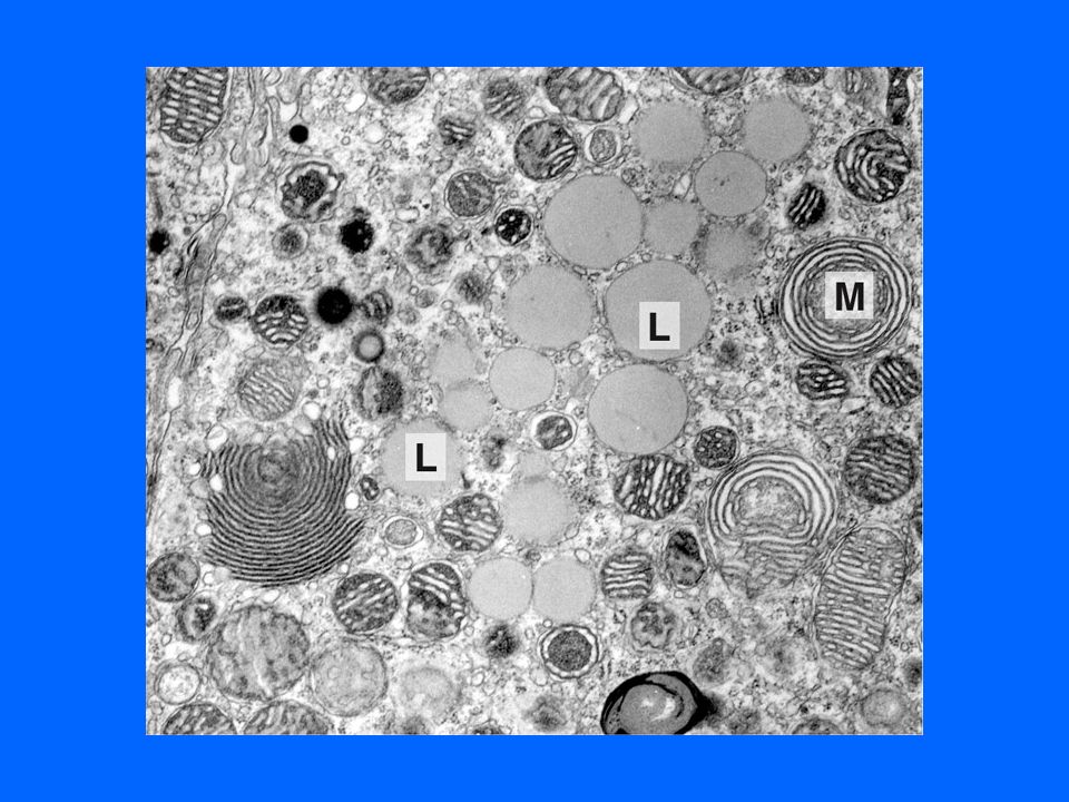
MITOCHONDRIA

40
PRACTICAL

41
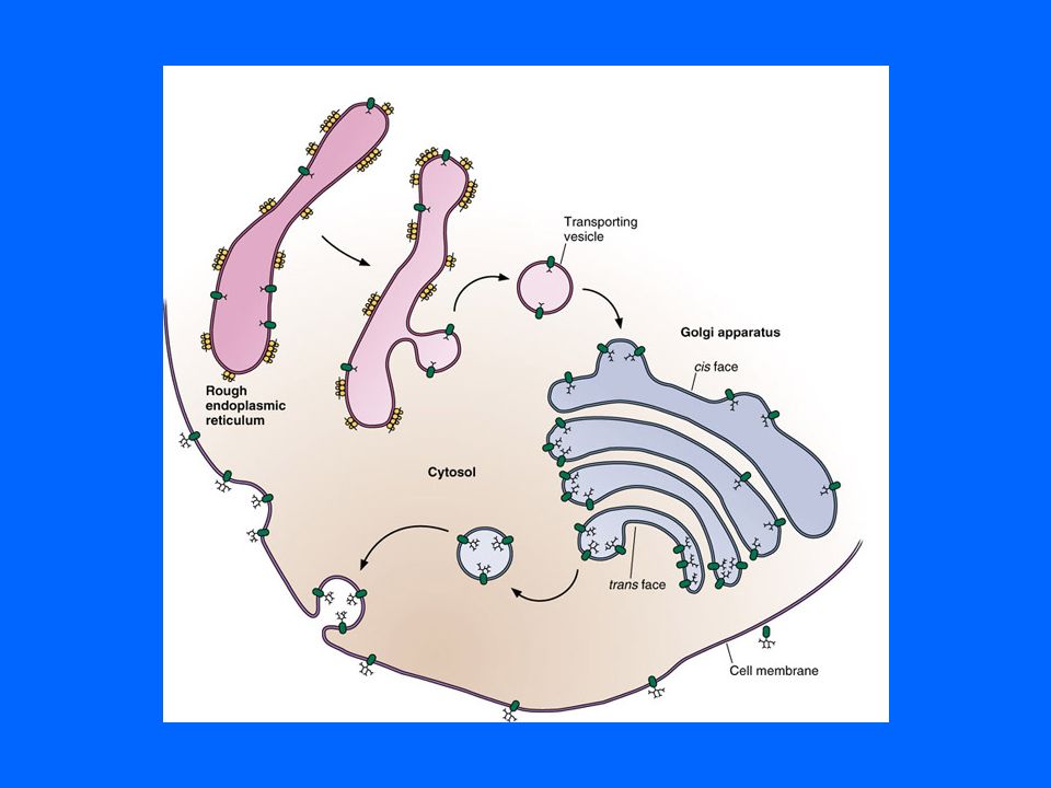
ENDOPLASMIC RETICULUM

42
ROUGH ENDOPLASMIC RETICULUM

43
RER & SECRETORY GRANULES

44
GOLGI APPARATUS

47
LYSOSOMES

49
CILIA

51
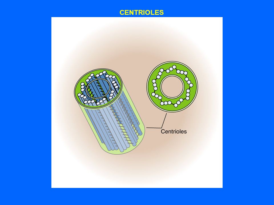
CILIA & CENTRIOLES

52
CENTRIOLES

54
MICROTUBULES & ACTIN FILAMENTS

55
DISTRIBUTION OF MICROTUBULES IN INTERPHASE CELLS 1- Cytoplasmic microtubules. 2- Cilia. 3- Flagella. 4- Centrioles.

57
Glycogen granules in hepatocyte

58
INTERCELLULAR JUNCTIONS

59
(1)Occludent junction. (2)Adherent junction. (3)Gap junction (Nexus).
Occludent junction. (2)Adherent junction. (3)Gap junction (Nexus).")
60
OCCLUDENT JUNCTION (TIGHGT JUNCTION)
")
61
DESMOSOMES (Macula adherent junctions)
")
62
GAP JUNCTION (NEXUS)
")
63
JUNCTIONAL COMPLEX

Similar presentations


, visible light passes through a specimen and then through glass lenses, which magnify the image The quality of an.>")
 Robert Hooke -- 1665: examined thinly sliced cork and coined term “cell”>")


 nuclear.>")
 (b) Structure of the plasma membrane Outside of cell Inside of cell 0.1 µm Hydrophilic region Hydrophobic region.>")


