Download presentation
Presentation is loading. Please wait.

1
The Physiology of Vision part 2

2
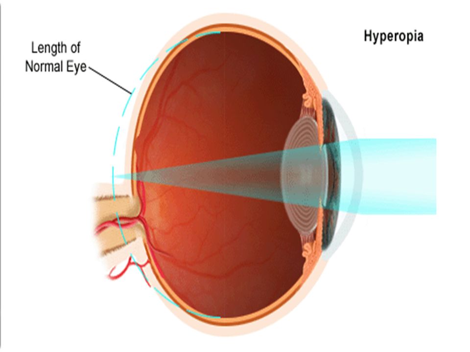
Defects of image forming 1- Hyperopia ( farsightedness) : -Is a defect in which the eye-ball is shorter than normal. -Parallel rays are focused behind the retina, so the image is formed behind the retina. -Sustained accommodation partially compensates for the defect, but it may lead to strabismus because of muscle fatigue. -It is corrected using convex lenses.

4
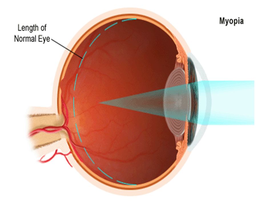
2- Myopia (nearsightedness) : - The anteroposterior axis of the eyeball is too long. -It is mainly a genetic disorder ( look at my family !!! ) -The image is formed in front of the retina. -Corrected using concave lenses.
 -The image is formed in front of the retina. -Corrected using concave lenses..")
6
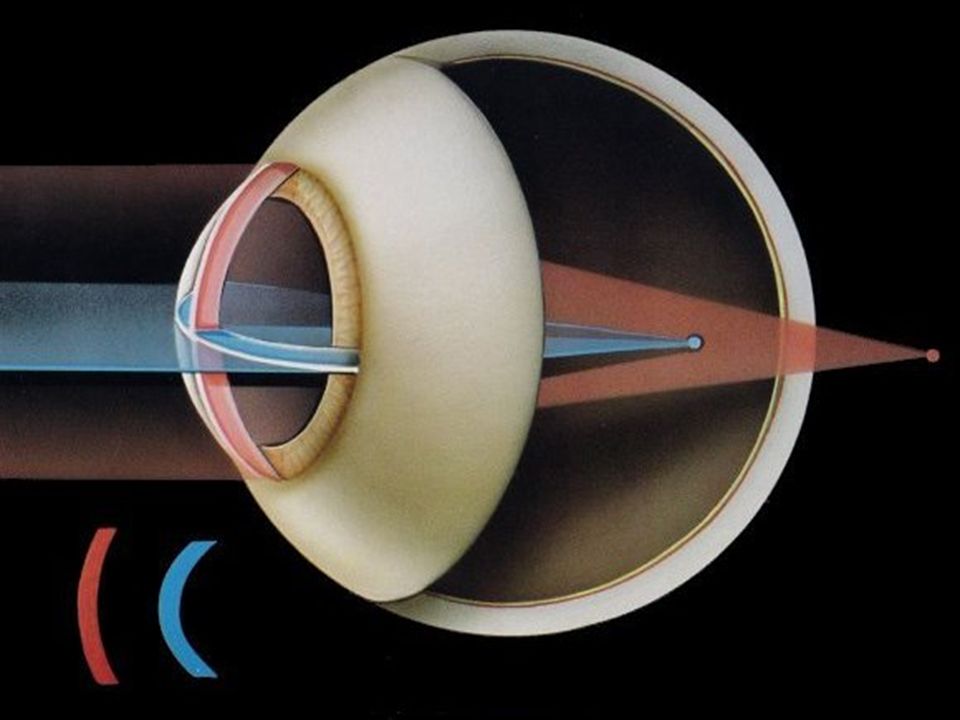
3- Astigmatism: -Curvature of the cornea is not uniform. -Some light rays are refracted to other spots making this part of the image blurry. -Corrected using cylindrical lenses.

9
Photoreceptor mechanisms Light acts on photosensitive compounds in the rods & cones of the retina, triggering action potentials. This is mainly due to the chemical changes that occur in these photosensitive compounds. Receptor potentials of photoreceptors ( & most other neural elements) are local & graded. Only ganglion cells produce all-or-none potentials
 are local & graded. Only ganglion cells produce all-or-none potentials.")
10
Photoreceptor mechanisms Rods, cones & horizontal cells are hyperpolarizing. Bipolar cells maybe either hyper- or hypo- polarizing. Amacrine cells produce depolarizing potentials that act as generator potentials for propagated spikes in ganglion cells. Cones have a sharp onset & offset of action potentials Rods have a sharp onset & a slow offset.

11
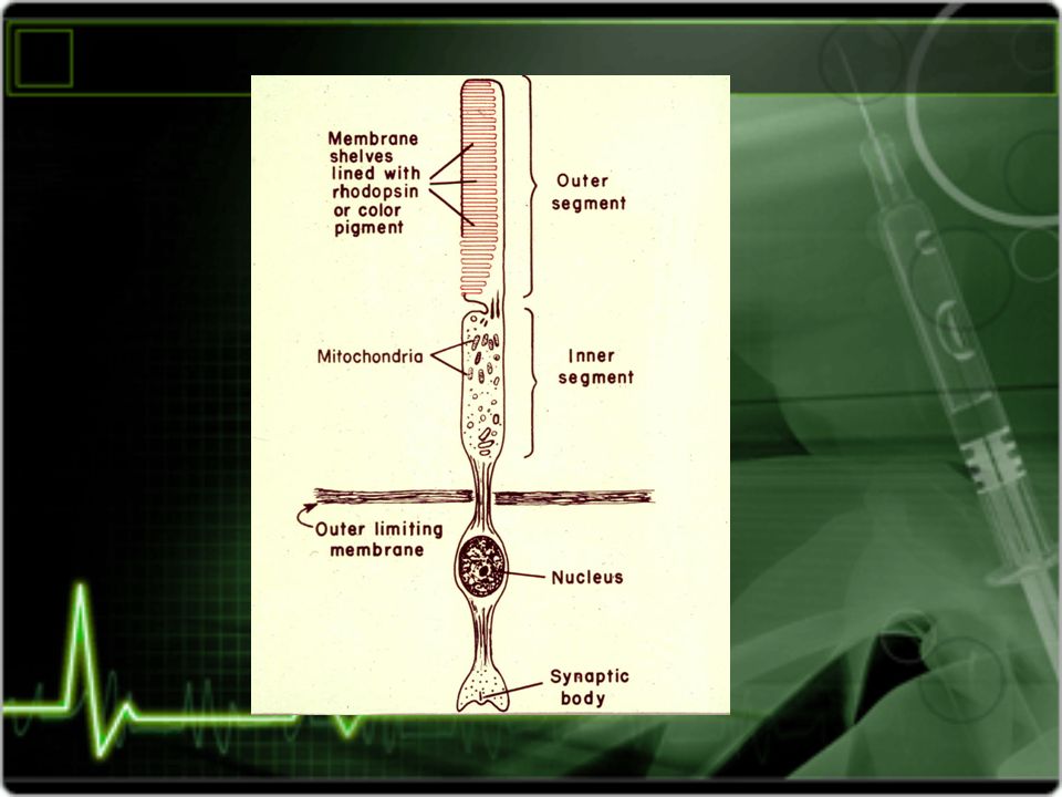
Ionic events 1- In the dark : Na + channels are open. Na + flows from the inner segment to the outer segment & the synaptic end of the receptor. Na + - K + pump maintains the equilibrium of this state. Neurotransmitter release is steady.

13
2- when light strikes the outer segment : -Chemical reactions occur near the sodium channels & close them, leading to hyperpolarization of the membrane. -Hyperpolarization decreases neurotransmitter release. -The decrease in neurotransmitter release triggers a signal in the bipolar cells. -Bipolar cells generate action potentials in ganglion cells.

15
Photosensitive compounds Are found in rods & cones. Mainly opsin ( a protein ) & retinine1 ( a form of vitamin A ). Rhodopsin : -Made up of retinine + scotopsin. -Also called visual purple. -Found in the membranes of rods. -Has a peak sensitivity to light at a wavelength of 505 nm.
 & retinine1 ( a form of vitamin A ). Rhodopsin : -Made up of retinine + scotopsin. -Also called visual purple. -Found in the membranes of rods. -Has a peak sensitivity to light at a wavelength of 505 nm..")
16
In the dark, rhodopsin’s retinine is in the 11- cis form. When light strikes it, it is transformed to an all-trans form. Metarhodopsin II is formed & leads to closure of Na+ channels by decreasing cGMP levels in the cell. This causes hyperpolarization, leading to decreased release of neurotransmitters & triggering an action potential.

17
After turning into the all-trans conformation, retinine is separated from scotopsine. Some of it is converted back to 11-cis & reassociates with scotopsin ( recycling ). Some retinine is synthesized de novo from vitamin A.
. Some retinine is synthesized de novo from vitamin A..")
18
Light Change in photopigment Metarodhopsin II Activation of transducin ( g-protein) Activation of phosphdiestrases Decreased cGMP Closure of NA channels Hyperpolarization, decreased release of NTs Action potential.
 Activation of phosphdiestrases Decreased cGMP Closure of NA channels Hyperpolarization, decreased release of NTs Action potential.")
19
What happens. When retinine is converted to its all-trans form, it dissociates from scotopsin. Scotopsin then activates transducin ( g- protein). Transducin’s alpha-subunit activates cyclic GMP phosphodiestrase. The phosphodiestrase converts cGMP to 5’- GMP. This causes closure of the sodium channel, because cGMP is what keeps them open.
. Transducin’s alpha-subunit activates cyclic GMP phosphodiestrase. The phosphodiestrase converts cGMP to 5’- GMP. This causes closure of the sodium channel, because cGMP is what keeps them open..")
20
Image formation Is a 3-stage process : 1- the image is formed on the retina’s photoreceptors. 2- it is changed to a second image in the bipolar cells. 3- then it is changed into a third image in the ganglion cells. - The third image is altered by the horizontal amacrine cells, then it reaches the occipital visual cortex.

21
Color vision Red, green & blue are the primary colors. Other colors are produced by mixing them. Young – helmoholtz theory : -Postulates that humans possess 3 types of cones, each containing a different photopigment. -Each photopigment is maximally sensitive to one of the three primary colors. -The sensation of any given color is determined by the relative frequency of the impulses from each of the 3 cone systems.

22
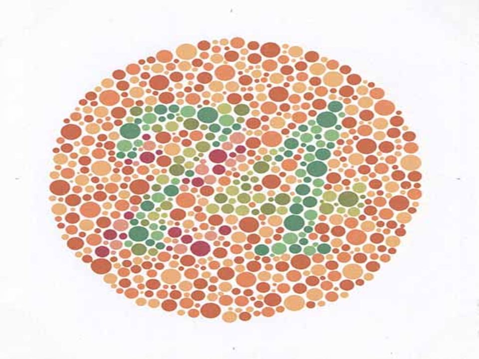
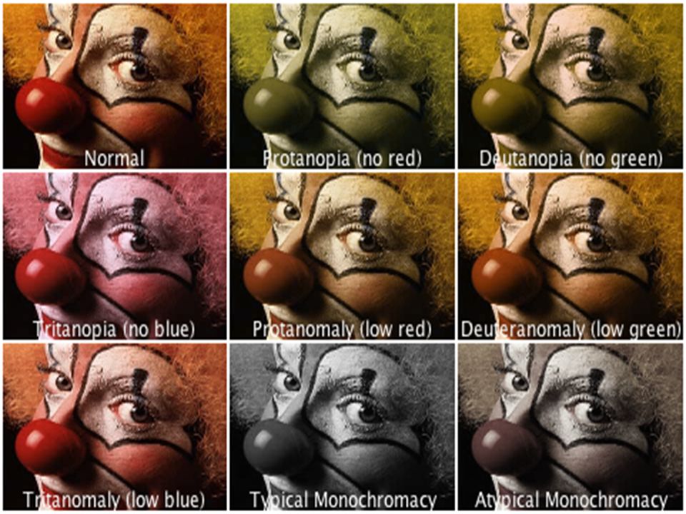
Color blindness Ishihara charts are the most common method of diagnosing color blindness. Terminology : 1) -anomaly : means weakness. 2) -anopia : means blindness. 3) Prot- : is red. 4) Deuter- : is green. 5) Trit- : is blue.
 -anomaly : means weakness. 2) -anopia : means blindness. 3) Prot- : is red. 4) Deuter- : is green. 5) Trit- : is blue..")
24
Normal people are trichromats, they can see the three primary colors clearly. Dichromats have only two cone systems, they may have protanopia, deuteranopia, or tritanopia. Monochromats have only one system ( extremely rare ). Color blindness is mainly inherited, but can be caused by a lesion in V8 ( the part of the visual cortex that is responsible for color vision). V8 lesions cause achromatopsia ( loss of color vision). In Caucasians, 8% of the males & 0.4% of the females inherit color blindness.
. Color blindness is mainly inherited, but can be caused by a lesion in V8 ( the part of the visual cortex that is responsible for color vision). V8 lesions cause achromatopsia ( loss of color vision). In Caucasians, 8% of the males & 0.4% of the females inherit color blindness..")
26
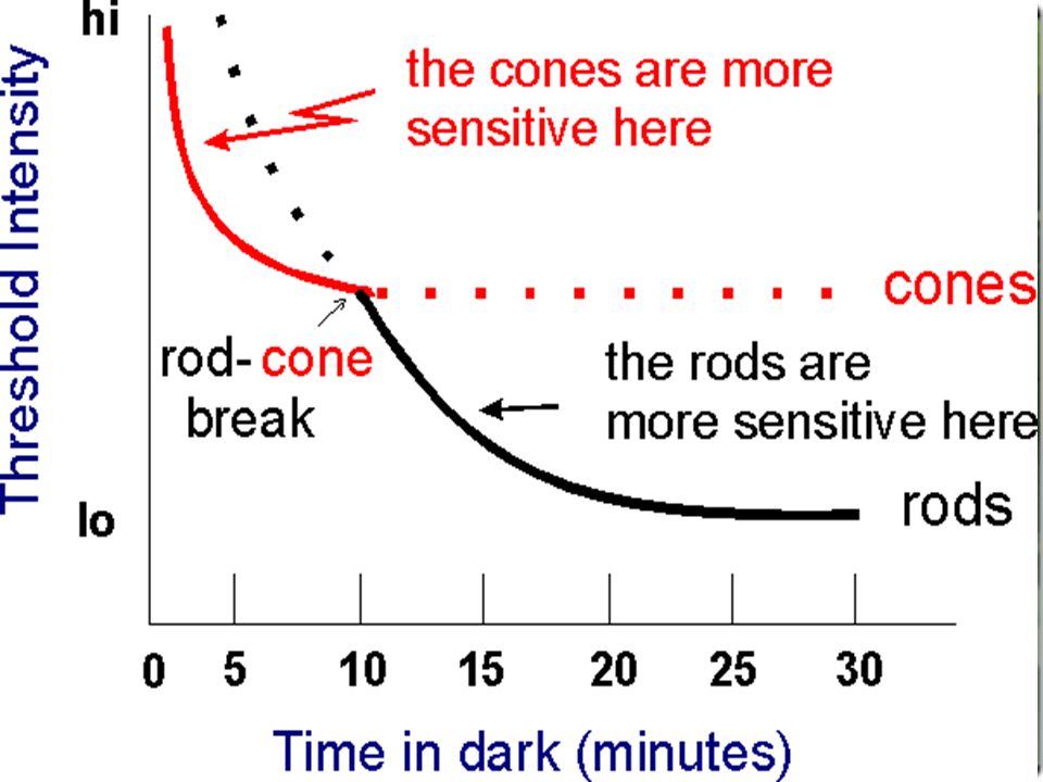
Dark adaptation. When an individual leaves a bright lighted space to a dim lighted one, his retinas become more sensitive to light. This phenomena is known as dark adaptation. In contrast, leaving a dark area to a bright one causes light adaptation, which only requires 5 minutes. Dark adaptation reaches its maximum in 20 minutes. It has two components : 1- adaptation of the cones : rapid ( 5 mins.) but small in magnitude. 2- adaptation of the rods : slower ( 15-20), with great magnitude.
 but small in magnitude. 2- adaptation of the rods : slower ( 15-20), with great magnitude..")
Similar presentations


 Middle layer (nutritive) iris cilliary body choroid Inner layer (retina-photosensetive)>")
 of retinal ganglion cells (RGC) Neural.>")


>")


