Download presentation
Presentation is loading. Please wait.

1
Sexual Reproduction in the Human

2
Learning Objectives(1/3)
Outline the general structure of the reproductive system (Male & Female) State the functions of the main parts of the reproductive system Outline the role of meiosis to produce sperm & ova (egg) cells Define the term secondary sexual characteristics Outline the role of oestrogen, progesterone & testosterone Outline the nature of birth control to include natural, mechanical, chemical and surgical methods State the location of fertilisation
 State the functions of the main parts of the reproductive system. Outline the role of meiosis to produce sperm & ova (egg) cells. Define the term secondary sexual characteristics. Outline the role of oestrogen, progesterone & testosterone. Outline the nature of birth control to include natural, mechanical, chemical and surgical methods. State the location of fertilisation.")
3
Learning Objectives(2/3)
Outline the events & outline the role of oestrogen and progesterone of the menstrual cycle Explain copulation Outline infertility State one cause of male infertility State the availability of corrective measures for male infertility State one cause female infertility State the availability of corrective measures for female infertility

4
Learning Objectives(3/3)
Explain implantation, placenta formation & function Outline the birth process Explain In-vitro fertilisation & implantation Outline milk production & breastfeeding including biological benefits

5
Sexual Reproduction in Humans
In mammals male reproductive organs produce haploid motile sperm (n) The female non-motile egg is also haploid (n) These fuse at fertilisation to produce a diploid zygote (2n)
 The female non-motile egg is also haploid (n) These fuse at fertilisation to produce a diploid zygote (2n)")
6
Structure of the male reproductive system

7
Bladder Seminal Vesicle Prostate gland Cowper’s Gland Penis Sperm duct Urethra Epididymis Testis Scrotum

8
Testes A gonad is an organ that produces sex cell in animals.
Male gonads are called testes Testes develop inside the body at first, but a few weeks before birth descend into the scrotum. This means they are kept at slightly lower than body temperature (35°) which is the ideal temperature for sperm production.
 which is the ideal temperature for sperm production.")
9
Testes Seminiferous tubules inside the testes are lined with sperm producing cells. Cells between the tubules produce the hormone testosterone.

10
Internal structure of testes
Seminiferous tubule Sperm producing cells (2n) Sperm Sertoli cell – nourishes sperm Blood capillary Interstitial cells – produce testosterone
 Sperm. Sertoli cell – nourishes sperm. Blood capillary. Interstitial cells – produce testosterone.")
11
Epididymis All the seminiferous tubules join to form the epididymis. Sperm mature and are stored here. The epididymis leads to the sperm duct (vas deferens) The sperm duct brings sperm to the urethra. The urethra is responsible for carrying sperm and urine out of the body
 The sperm duct brings sperm to the urethra. The urethra is responsible for carrying sperm and urine out of the body.")
12
Glands in the male reproductive system
Seminal Vesicle Prostate Gland Cowper’s Gland These glands produce seminal fluid which nourishes the sperm and provides a medium in which to swim. Seminal fluid + Sperm = Semen

13
Functions of the main parts of the Male reproductive system
Testis Epididymis Sperm duct Seminal Vesicle Prostate gland Bladder Urethra Scrotum Cowper’s Gland Penis Produces 1. Sperm 2 Testosterone

14
Functions of the main parts of the Male reproductive system
Testis Epididymis Sperm duct Seminal Vesicle Prostate gland Bladder Urethra Scrotum Cowper’s Gland Penis Stores sperm

15
Functions of the main parts of the Male reproductive system
Testis Epididymis Sperm duct Seminal Vesicle Prostate gland Bladder Urethra Scrotum Cowper’s Gland Penis Carries sperm from the epididymis to the urethra

16
Functions of the main parts of the Male reproductive system
Testis Epididymis Sperm duct Seminal Vesicle Prostate gland Bladder Urethra Scrotum Cowper’s Gland Penis Produces Seminal Fluid - For sperm to swim in - Nourishment for sperm

17
Tube through which the sperm travel through the penis
Functions of the main parts of the Male reproductive system Testis Epididymis Sperm duct Seminal Vesicle Prostate gland Bladder Urethra Scrotum Cowper’s Gland Penis Tube through which the sperm travel through the penis

18
Keeps testes at a lower temperature
Functions of the main parts of the Male reproductive system Testis Epididymis Sperm duct Seminal Vesicle Prostate gland Bladder Urethra Scrotum Cowper’s Gland Penis Keeps testes at a lower temperature

19
Functions of the main parts of the Male reproductive system
Testis Epididymis Sperm duct Seminal Vesicle Prostate gland Bladder Urethra Scrotum Cowper’s Gland Penis Places sperm in the females body

20
Seminal vesicles, Cowper’s gland and Prostate gland
Summary of functions of main parts of male reproductive system Part Function Testis Produces sperm and testosterone Epididymis Matures and stores sperm Sperm duct Carries sperm from the epididymis to the urethra Seminal vesicles, Cowper’s gland and Prostate gland Produces seminal fluid which feeds the sperm and allows them to swim. Sperm and seminal fluid are collectively called semen. Urethra Allows the passage of either urine or sperm. Penis Places sperm inside the body of a female Scrotum Keeps testes at a lower temperature (35°). This is the optimum temperature for Meiosis to occur.
. This is the optimum temperature for Meiosis to occur.")
21
Sperm

22
Sperm Structure Acrosome (contains digestive enzymes) Head
Nucleus (contains 23 chromosomes) Collar (contains mitochondria) Middle Flagellum (allows sperm to swim) Tail
 Collar (contains mitochondria) Middle. Flagellum (allows sperm to swim) Tail.")
23
Role of meiosis in sperm and egg production
Sperm and egg producing cells are diploid i.e. they contain 46 chromosomes. They divide by meiosis to form sperm and egg cells. Each sperm and egg cell, therefore, has a haploid number of chromosomes i.e. they have 23 each

24
Role of meiosis in sperm and egg production
As both a sperm nucleus and an egg nucleus are haploid they combine in fertilisation to form a diploid zygote i.e. the new zygote has 46 chromosomes. 23 chromosomes + 23 chromosomes = 46 chromosomes The zygote now grows by mitosis division ensuring that each new cell has a diploid number of chromosomes.

25
Role of meiosis in sperm and egg production

26
Testosterone: male hormone responsible for the development of the primary and secondary male sexual characteristics The primary sexual characteristics are the presence of the male and female reproductive parts Secondary sexual characteristics refer to features that distinguish males from females e.g. presence of facial hair, deep voice

27
Male Secondary Sexual characteristics
The growth of pubic, facial and body hair The enlargement of larynx and ‘breaking’ of the voice Increased muscular development and bone development A growth spurt at puberty An increased secretion of sebum in the skin

28
Structure of the female reproductive system

29
The Ovary Produce the eggs and female hormones. All the eggs in an ovary are present at birth. After puberty 20 eggs mature each month. Only one will be released from the ovary – the rest will die.

30
The Fallopian tube (oviduct)
The Fallopian tubes are muscular and approx 12cm long. Funnels at the tip of each tube catch the egg after it is released from the ovary. The egg is moved along the tube by cilia and muscular peristalsis. The egg is either fertilised or dies in the fallopian tube.

31
The Uterus (womb) Muscular structure approximately the size of your fist. Outer wall made of involuntary muscle. Inner lining is called the endometrium This lining thickens each month with cells and blood vessels to nourish the embryo. The cervix separates the uterus from the vagina.

32
The vagina Elastic muscular tube 10cm long. Allows entry of sperm. Is the birth canal for the exit of a baby. Lined with cells that produce mucous. This serves to protect against the entry of pathogens.

33
Fallopian tube (Oviduct)
Structure of the Female reproductive system Fallopian tube (Oviduct) Funnel Ovary Ovarian Ligament Uterus Lining of uterus (endometrium) Cervix Wall of uterus Vagina Vulva
 Funnel. Ovary. Ovarian Ligament. Uterus. Lining of uterus (endometrium) Cervix. Wall of uterus. Vagina. Vulva.")
34
Functions of the main parts of the female reproductive system
Fallopian tube (Oviduct) Funnel Ovary Ovarian Ligament Uterus Lining of uterus (endometrium) Cervix Wall of uterus Vagina Produces : Egg Oestrogen Progesterone Vulva
 Funnel. Ovary. Ovarian Ligament. Uterus. Lining of uterus (endometrium) Cervix. Wall of uterus. Vagina. Produces : Egg. Oestrogen. Progesterone. Vulva.")
35
Functions of the main parts of the female reproductive system
Fallopian tube (Oviduct) Funnel Ovary Ovarian Ligament Uterus Lining of uterus (endometrium) Cervix Wall of uterus Vagina Catches the egg after release from ovary Transports egg from ovary to womb Site of fertilisation Vulva
 Funnel. Ovary. Ovarian Ligament. Uterus. Lining of uterus (endometrium) Cervix. Wall of uterus. Vagina. Catches the egg after release from ovary. Transports egg from ovary to womb. Site of fertilisation. Vulva.")
36
Functions of the main parts of the female reproductive system
Fallopian tube (Oviduct) Funnel Ovary Ovarian Ligament Uterus Lining of uterus (endometrium) Cervix Wall of uterus Vagina Implantation Hold foetus Forms placenta Vulva
 Funnel. Ovary. Ovarian Ligament. Uterus. Lining of uterus (endometrium) Cervix. Wall of uterus. Vagina. Implantation. Hold foetus. Forms placenta. Vulva.")
37
Functions of the main parts of the female reproductive system
Fallopian tube (Oviduct) Funnel Ovary Ovarian Ligament Uterus Lining of uterus (endometrium) Cervix Wall of uterus Vagina Vulva Allows entry of sperm into female system Birth canal to allow exit of baby
 Funnel. Ovary. Ovarian Ligament. Uterus. Lining of uterus (endometrium) Cervix. Wall of uterus. Vagina. Vulva. Allows entry of sperm into female system. Birth canal to allow exit of baby.")
38
Summary of functions of main parts of female reproductive system
Ovary To produce the egg (ova). To produce the hormones oestrogen and progesterone Fallopian tube (oviduct) Catches the egg from the ovary and transports it to uterus. Site of fertilisation. Uterus Site of implantation. Holds the developing embryo. Has a lining (endometrium) enriched with blood vessels to nourish the embryo. Forms the placenta. Vagina Allows entry of sperm and exit of baby at birth.
. To produce the hormones oestrogen and progesterone. Fallopian tube. (oviduct) Catches the egg from the ovary and transports it to uterus. Site of fertilisation. Uterus. Site of implantation. Holds the developing embryo. Has a lining (endometrium) enriched with blood vessels to nourish the embryo. Forms the placenta. Vagina. Allows entry of sperm and exit of baby at birth.")
39
Female Hormones Oestrogen and progesterone are the female hormones
A combination of oestrogen and progesterone at puberty causes the development of the secondary female characteristics: Maturing and enlargement of the breasts. Widening of the pelvis to allow for birth. The growth of pubic and underarm hair. A growth spurt.

40
The Ovary Produce the eggs and the female hormones oestrogen and progesterone. The ovaries of a female foetus contains all the potential eggs at birth. These eggs have not yet divided by meiosis and as a result are diploid After puberty a number of eggs are produced by meiosis each month. Usually only one egg continues to grow … the rest die

41
The Ovary Once meiosis is complete the egg is surrounded within a structure called the Graafian follicle. This structure produces the female hormone oestrogen When mature the follicle forms a swelling on the outside of the ovary. It bursts at ovulation to release the egg After ovulation the follicle fills with yellow cells and becomes the Corpus luteum (yellow body). This secretes the hormone progesterone
. This secretes the hormone progesterone.")
42
Summary of events in the menstrual cycle
The menstrual cycle is a 28 day sequence of events that produces an egg and prepares the body for pregnancy. This cycle begins at puberty and continues until the menopause (the end of the woman’s reproductive life). Summary of events in the menstrual cycle Days 1 – 5 Old lining of the uterus (endometrium) breaks down and is shed from the body. The loss of this blood and tissue is called menstruation (period). A new egg is produced in the ovary by meiosis. This new egg is surrounded by the Graafian follicle.
. Summary of events in the menstrual cycle. Days 1 – 5. Old lining of the uterus (endometrium) breaks down and is shed from the body. The loss of this blood and tissue is called menstruation (period). A new egg is produced in the ovary by meiosis. This new egg is surrounded by the Graafian follicle.")
43
- Oestrogen also prevents the development of any more eggs.
The Menstrual Cycle Days Hormone oestrogen is produced by the developing Graafian follicle. This has two functions: - It causes the lining of the uterus (endometrium) to build up again in preparation for implantation. - Oestrogen also prevents the development of any more eggs. Day 14 Ovulation. This occurs when the Graafian follicle bursts to release the egg into the fallopian tube.
 to build up again in preparation for implantation. - Oestrogen also prevents the development of any more eggs. Day 14. Ovulation. This occurs when the Graafian follicle bursts to release the egg into the fallopian tube.")
44
- It causes the endometrium to thicken even further.
Days The Graafian follicle now develops into the Corpus Luteum (yellow body). This has two functions: - It causes the endometrium to thicken even further. - It also prevents new eggs from forming. The released egg will die by day 16 if it is not fertilised. Thus days 12 – 16 of the menstrual cycle are referred to as the Fertile Period. (Even though the egg is not released until day 14, sperm, which can survive for a period of time in the female body, may already be present. Thus the fertile period begins on day 12).
. This has two functions: - It causes the endometrium to thicken even further. - It also prevents new eggs from forming. The released egg will die by day 16 if it is not fertilised. Thus days 12 – 16 of the menstrual cycle are referred to as the Fertile Period. (Even though the egg is not released until day 14, sperm, which can survive for a period of time in the female body, may already be present. Thus the fertile period begins on day 12).")
45
If fertilisation does not take place the Corpus Luteum starts to degenerate around day 22.
This results in a reduction in progesterone levels. As a result the lining of the uterus breaks down again on day 28. The menstrual cycle begins again with day 1.

46
If fertilisation has not occurred the cycle begins again with the breakdown of the endometrium.
The Menstrual Cycle 1 2 3 4 5 6 7 8 9 10 11 12 13 14 15 16 17 18 19 20 21 22 23 24 25 26 27 28 DAY 1-5 : Blood from the womb lining is shed from the body After day 5 the lining of the uterus repairs and builds up again Fertile period Ovulation occurs on Day 14 Implantation may happen

47
Inside the Ovary Developing Graafian follicle – secretes oestrogen
Potential egg Egg is released from ovary (ovulation) Graafian follicle now changes to the Corpus luteum which secretes progesterone
 Graafian follicle now changes to the Corpus luteum which secretes progesterone.")
48
Graafian follicle Corpus Luteum Hormones Endometrium 5 14 Days 28
Oestrogen________ Progesterone _____ Endometrium 5 14 Days 28

49
Learning Check Name the main parts of the male reproductive system?
Give a function for each part named? Name the main parts of the female reproductive system and give a function for each part named. Outline what is happening on each of the following days of the menstrual cycle: 1,5,12,14,26? Outline the role played by oestrogen and progesterone in the cycle?

50
Copulation – Sexual intercourse
Sexual arousal The penis becomes erect The vagina becomes lubricated Copulation The penis is inserted into and moved inside the vagina Orgasm Sperm is released from the penis (Ejaculation) Contraction of vagina and uterus
 Contraction of vagina and uterus.")
51
Insemination Insemination is the release of sperm into the female
Contractions of uterus and fallopian tubes move the sperm to the fallopian tubes within 5 minutes If an egg is present it releases chemicals to attract the sperm this is called chemotaxis

52
Fertilisation Fertilisation is the fusion of the egg and sperm nuclei to form a diploid zygote.

53
Fertilisation usually occurs in the fallopian tube.

54
Fertilisation A number of sperm may reach the egg at the same time.
The acrosome releases enzymes to digest the egg membrane A number of sperm may reach the egg at the same time. The sperm loses its tail and the head enters the egg. The sperm and egg nuclei fuse to form a zygote A chemical reaction at the membrane prevents other sperm cells entering.

55
Implantation Implantation is the embedding of the fertilised egg into the lining of the uterus This occurs days after fertilisation. By this time the zygote has grown into an embryo. During implantation a membrane called the amnion develops around the embryo. This secretes amniotic fluid which will surround the developing embryo and act as a shock absorber. After implantation the placenta forms.

56
Zygote Implanted embryo

57
Placenta Formation After implantation the embryo forms an outer membrane called a chorion This chorion develops projections (chorionic villi) which, together with the blood vessels of the mother in the endometrium, form the placenta The placenta allows nutrients, wastes, gases, antibodies and hormones to be exchanged between the blood of the mother and the embryo
 which, together with the blood vessels of the mother in the endometrium, form the placenta. The placenta allows nutrients, wastes, gases, antibodies and hormones to be exchanged between the blood of the mother and the embryo.")
58
Placenta Formation The blood of the mother and embryo do not mix
This is important as: - The blood groups of mother and baby might not be compatible. - The blood pressure of the mothers system may cause damage to the embryo

59
Placenta Formation The placenta also makes the hormone progesterone
The umbilical cord connects the placenta with the embryo. It contains blood vessels which circulate blood between the embryo and the placenta

60
The Placenta Chorion Embryo Placenta Mother’s blood Amnion
Nutrients, Oxygen, antibodies Wastes, Carbon Dioxide, Water Amnion Amniotic fluid Umbilical cord Embryo’s blood Embryo

62
Development of Embyro 1.The diploid zygote divides by mitosis to form a Morula (a ball of undifferentiated cells) after 3-5 days 2. Morula continues to divide by mitosis to form a blastocyst (fluid filled sac containing an inner cell mass that gives rise to an embryo) which moves into the uterus and implants around day 9 3. The outer layer of the blastocyst is called the trophoblast. The inner cell mass of the blastocyst organises itself into three distinct germ layers: ectoderm, mesoderm and endoderm Ectoderm: Gives rise to skin and nervous system Mesoderm: Gives rise to musuloskeletal system, kidneys, lungs, heart etc. Endoderm: Gives rise to liver, pancreas and inner linings of breathing, digestive and excretory system.
 which moves into the uterus and implants around day The outer layer of the blastocyst is called the trophoblast. The inner cell mass of the blastocyst organises itself into three distinct germ layers: ectoderm, mesoderm and endoderm. Ectoderm: Gives rise to skin and nervous system. Mesoderm: Gives rise to musuloskeletal system, kidneys, lungs, heart etc. Endoderm: Gives rise to liver, pancreas and inner linings of breathing, digestive and excretory system.")
63
Development of Foetus 1. A protective sac, the amnion, begins to form around the embryo. It contains amniotic fluid which protects and acts as a shock absorber around the embryo. 2. By the end of the eighth week of pregnancy all the major organs have formed and the embryo is now called a foetus. 3. The skeleton is made of cartilage. From the 3rd month onward bone begins to form. 4. Arms and legs begin to move and sex organs are visible. The foetus urinates into the amniotic fluid. 5. The placenta forms from the trophoblast layer of the embryo and tissue from the uterus. It is fully formed at 3 months into pregnancy.

64
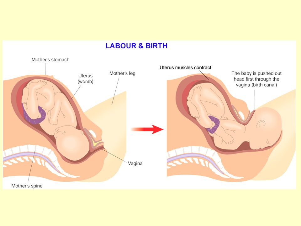
Birth The hormones oestrogen and progesterone are produced throughout pregnancy firstly by the corpus luteum (3 months) and then by the placenta. The placenta acts as an endocrine gland. Immediately before birth the placenta stops making progesterone. The walls of the uterus begin to contract as a result. The pituitary gland releases the hormone called oxytocin. This causes further contractions of the uterus Labour has now begun
 and then by the placenta. The placenta acts as an endocrine gland. Immediately before birth the placenta stops making progesterone. The walls of the uterus begin to contract as a result. The pituitary gland releases the hormone called oxytocin. This causes further contractions of the uterus. Labour has now begun.")
65
There are three main stages:
Stage 1 - (about 12 hours) Labour The contraction of the uterus pushes the foetus towards the cervix. This causes the cervix to open (dilate). During this stage the contractions cause the amnion to break releasing the amniotic fluid through the vagina. (The ‘waters break’).
 Labour. The contraction of the uterus pushes the foetus towards the cervix. This causes the cervix to open (dilate). During this stage the contractions cause the amnion to break releasing the amniotic fluid through the vagina. (The ‘waters break’).")
66
Stage 2 - (20 minutes to 1 hour) Parturition
The foetus passes through the cervix and the birth canal head first.

67
Stage 2 - (20 minutes to 1 hour) After birth
The foetus passes through the cervix and the birth canal head first.

68
Stage 2 - (20 minutes to 1 hour)
The foetus passes through the cervix and the birth canal head first.

69
Stage 2 - (20 minutes to 1 hour)
The foetus passes through the cervix and the birth canal head first.

70
Stage 2 - (20 minutes to 1 hour)
The foetus passes through the cervix and the birth canal head first.

71
The foetus passes through the cervix and the birth canal head first.
Stage 2 - (20 minutes to 1 hour) The foetus passes through the cervix and the birth canal head first. The umbilical cord is tied and cut. This leaves a scar which will eventually become the navel (belly button).
 The foetus passes through the cervix and the birth canal head first. The umbilical cord is tied and cut. This leaves a scar which will eventually become the navel (belly button).")
72
Stage 3 - (10 to 15 minutes) The baby is now born. The uterus now contracts again and expels the afterbirth (the umbilical cord and placenta.

73
Stage 3 - (10 to 15 minutes) The baby is now born. The uterus now contracts again and expels the afterbirth (the umbilical cord and placenta.

74
Stage 3 - (10 to 15 minutes) The baby is now born. The uterus now contracts again and expels the afterbirth (the umbilical cord and placenta.

75
Stage 3 - (10 to 15 minutes) The baby is now born. The uterus now contracts again and expels the afterbirth (the umbilical cord and placenta.

77
Umbilical cord is cut

78
Breastfeeding Lactation The secretion of milk from the mammary glands
The first days after birth colostrum produced Milk production triggered by release of prolactin by pituitary

79
Breastfeeding Breastfeeding has the following advantages for the baby:
Colostrum and breast milk provides the baby with essential antibodies protecting it against infection Ideal balance of nutrients for baby Has little fat making it is easier to digest than milk

80
Birth control Birth control refers to the methods employed to limit the number of children that are born Removing the possibility of conception is called contraception. This is achieved by preventing the egg and sperm from meeting There are a number of methods:

81
Mechanical contraception - male
The use of condoms Surgical contraception Sperm ducts are cut and tied

82
Mechanical contraception - female
The use of diaphragms

83
Chemical contraception
Use of ‘the pill’. The pill contains oestrogen and progesterone which prevents ovulation and hence conception. Use of spermicide

84
Surgical contraception
The fallopian tubes and sperm ducts can be cut and tied

85
Natural contraception
Not having sexual intercourse during the fertile period of the menstrual cycle Natural methods of contraception try to identify the time of ovulation based on: - monitoring the body temperature. This rises slightly after ovulation - mucous secreted in the cervix (which changes its texture after ovulation)
")
86
Infertility is the inability of a couple to achieve conception.

87
Male infertility disorders
Low sperm count – Refers to a low number of sperm per ml of seminal fluid. Low sperm mobility - If movement of the sperm is slow, not in a straight line or both, the sperm may have difficulty passing through the cervical mucous or penetrating the shell of the egg. Endocrine gland failure – A failure of the testes to produce sperm

88
Low sperm count Causes:
The persistent use of drugs such as alcohol, cigarettes and anabolic steroids. Abnormalities in sperm production or obstruction of the tubes through which sperm travels. Stress

89
Low sperm count Treatment A change in diet.
A change in lifestyle e.g. stopping alcohol consumption, smoking. A reduction in stress levels.

90
Female infertility disorders
Blockage of the Fallopian Tube Scarring of the fallopian tube can block the passage of the egg to the uterus Endocrine gland failure A failure of the ovaries to produce an egg

91
Blockage of the fallopian tubes
Causes: Fragments of the uterus lining may spread to the fallopian tube Inflammation as a result of infection Treatment In-vitro fertilisation (I.V.F.)
")
92
In-vitro fertilisation (I.V.F.)
")
93
IVF is a method of treating infertility It involves removing eggs from an ovary and fertilising them outside the body

94
During the natural menstrual cycle an egg is produced by the ovary every month

95
During IVF fertility drugs are given to the female to stimulate the ovaries to produce more than one egg

96
These eggs are then taken from the females body and into the laboratory

97
In the meantime a sperm sample is taken from the male

98
The eggs and sperm are mixed together in the hope that fertilisation will occur

100
The sample is placed in the most ideal conditions for fertilisation to occur

101
The main aim of the procedure is to obtain a zygote
The main aim of the procedure is to obtain a zygote. If successful the zygotes development will be monitored closely

102
If successful the zygote develops into a morula, blastocyst and eventually becomes an embryo

103
The developing embryo can now be placed back into the females body for implantation to take place

104
Babies born as a result of IVF are often incorrectly called ‘test tube’ babies.
While fertilisation takes place in the laboratory (‘in vitro’ – in glass) the fertilised egg is re-inserted into the mother’s body and develops naturally in the uterus
 the fertilised egg is re-inserted into the mother’s body and develops naturally in the uterus.")
105
If analysis of a couples eggs and sperm suggests that IVF treatment is unsuitable, other methods of assisted fertility treatment are available

106
Depth of treatment(1/2) General structure of the reproductive system – male and female Functions of the main parts Role of meiosis in the production of sperm cells and egg (ova) Definition of “secondary sexual characteristics” Role of oestrogen, progesterone and testosterone The menstrual cycle: the events and outlined role of oestrogen and progesterone
 Definition of secondary sexual characteristics Role of oestrogen, progesterone and testosterone. The menstrual cycle: the events and outlined role of oestrogen and progesterone.")
107
Depth of treatment(1/2) Copulation Location of fertilisation
Implantation, placenta formation and function Birth – outline of process Milk production and breastfeeding

108
Contemporary Issues and Technology
Birth control – natural, mechanical, chemical and surgical methods of contraception Infertility One cause of male infertility from the following disorders: low sperm count, low sperm mobility, endocrine gland failure Availability of corrective measures

109
Contemporary Issues and Technology
One cause of female infertility from the following disorders: blockage of the Fallopian tube, endocrine gland failure Availability of corrective measures In-vitro fertilisation and implantation Biological benefits of breastfeeding

Similar presentations


 and.>")


 – Oestrogen – LH (Luteinising.>")

