Download presentation
Presentation is loading. Please wait.

1
07 Lab: What are the characteristics of cells? Interactive Lab Manual

2
Begin on a new page in your lab notebook: Experimental Question: What are the characteristics of cells? Partner: Objectives: 1.Compare structure of prokaryotic and eukaryotic cells. 2.Identify key features of eukaryotic cells such as protists, plants and animals. 3.Identify unknown cells.

4
You can choose 4 samples to view: 1 Prokaryote 1 Protist 1 Plant 1 Animal 1.View the slide under the microscope at 400X. Click here for instructions on focusing your sample.here 2.Draw, label and identify the cells. Click here for instructions to draw and label your sample.here 3.Describe each sample in words. Click here to see important tips to describe your sample.here

5
Need to draw into lab notebook 4 of these: Slide: Mag: Description:

6
You will be graded on your sketches and descriptions. The purpose of your drawing is to communicate information about the characteristics of each type of cell. Make sure your work is : Colorful Labeled Accurate Neat Detailed In Pencil

7
1. View the slide under the microscope. A.Start on scanning power (lowest magnification = 40X) B.Adjust the stage using the large knob to focus on the edge of the coverslip. C.Move slide to find the sample. D.Turn the revolving nosepiece to 100X objective and adjust focus using the large knob. When your sample is focused at 100 X: E.Move the revolving nosepiece to 400X and adjust focus using the small knob.
 B.Adjust the stage using the large knob to focus on the edge of the coverslip. C.Move slide to find the sample. D.Turn the revolving nosepiece to 100X objective and adjust focus using the large knob. When your sample is focused at 100 X: E.Move the revolving nosepiece to 400X and adjust focus using the small knob..")
8
2. Draw, label and identify the cells. Label your drawing with the following information: Draw at 400x. Draw several cells, but you do not have to draw the entire field. Label important cell structures Identify the Domain: Prokaryote or Eukaryote Identify the Kingdom: Prokaryote: bacteria or cyanobacteria Eukaryote: protist, plant or animal Identify the organism (on the slide label) Use color! This will help you identify structures within the cells.
 Use color. This will help you identify structures within the cells..")
9
3. Describe each sample Include 2 descriptors that identify characteristics of the sample Descriptions may include: Unicellular or Multicellular Size of the cells Shape of the cells Nucleus present Cell wall present Organelles visible in the cell Arrangement of cells

10
Answer these Pre-lab questions in your lab notebook. 1.Draw a Venn diagram comparing prokaryotic and eukaryotic cells. 2.What magnification will you use to make a sketch of your slide? 3.What are some characteristics of the cell you should look for?

11
Prokaryote – Bacteria - Bacillus Look for individual rod-shaped cells and groups of cells that are linked together. Draw at 400X. You do not have to draw every cell you see! Describe 2 characteristics you see for these cells Click here for helphere Label: Cell wall – The cell wall is the only part of these cells you can see. Use color! Note: Bacillus means “rod-shaped” bacteria

12
Prokaryote – Nostoc / Cyanobacteria Nostoc are photosynthetic bacteria that can be seen on the piece of leaf on your slide. Look for tiny strands of red/pink cells on the leaf. These cells are very small – you need to view them at 400X. If you cannot find the “beads on a string” structure for colonies of these cells, ask your instructor for help. Draw at 400X. Label individual cell and a colony. Use color! Include 2 descriptors

13
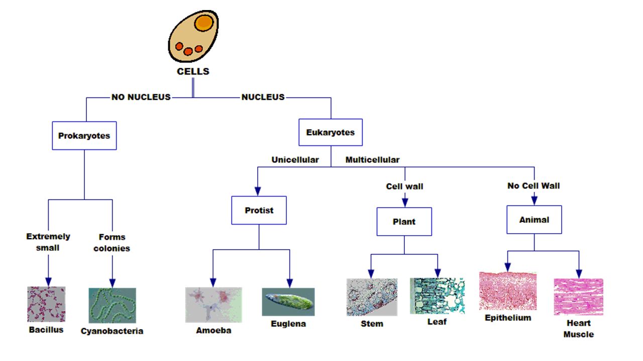
Eukaryotes – 4 Kingdoms Unicellular Eukaryotes: Protists – Single celled organisms. Found in ponds and other aquatic environments. Variety of shapes with specialized organelles. Multicellular Eukaryotes: Plants – Thick cell wall, central vacuole, and nucleus. Leaf cells may have chloroplasts Animals – Flexible cell membrane and nucleus that stains darkly.

14
Eukaryote – Protist - Amoeba Draw two different amoeba at 400X. Label the darkly stained nucleus, cell membrane and cytoplasm. Write 2 descriptors. Amoeba are one-celled eukaryotes able to change their shape. They move by extending their cytoplasm in pseudopods. Amoeba live in fresh water ponds, and eat by surrounding their food with pseudopods to create a food vacuole. Digestive enzymes then break down the food. Play the video to see how amoeba move.

15
Eukaryote – Protist - Euglena Draw at 400X Label the eyespot, cell membrane, nucleus, flagellum and chloroplast. Write 2 descriptors. Euglena are one-celled eukaryotes that have characteristics of both plants and animals. Euglena move using a whip-like flagellum (see video below). They have a red eye-spot that allows them to detect light. And Euglena have chloroplasts - so they can photosynthesize – like plants!
. They have a red eye-spot that allows them to detect light. And Euglena have chloroplasts - so they can photosynthesize – like plants!.")
16
Eukaryote – Protist - Paramecium Draw at 400X Label the nucleus, cell membrane and cilia. Write 2 descriptors. Paramecium are one-celled eukaryotes. Paramecium live in ponds, lakes and streams. They have tiny cilia around the other portion of their bodies. Paramecium eat algae, bacteria and dead plant matter.

17
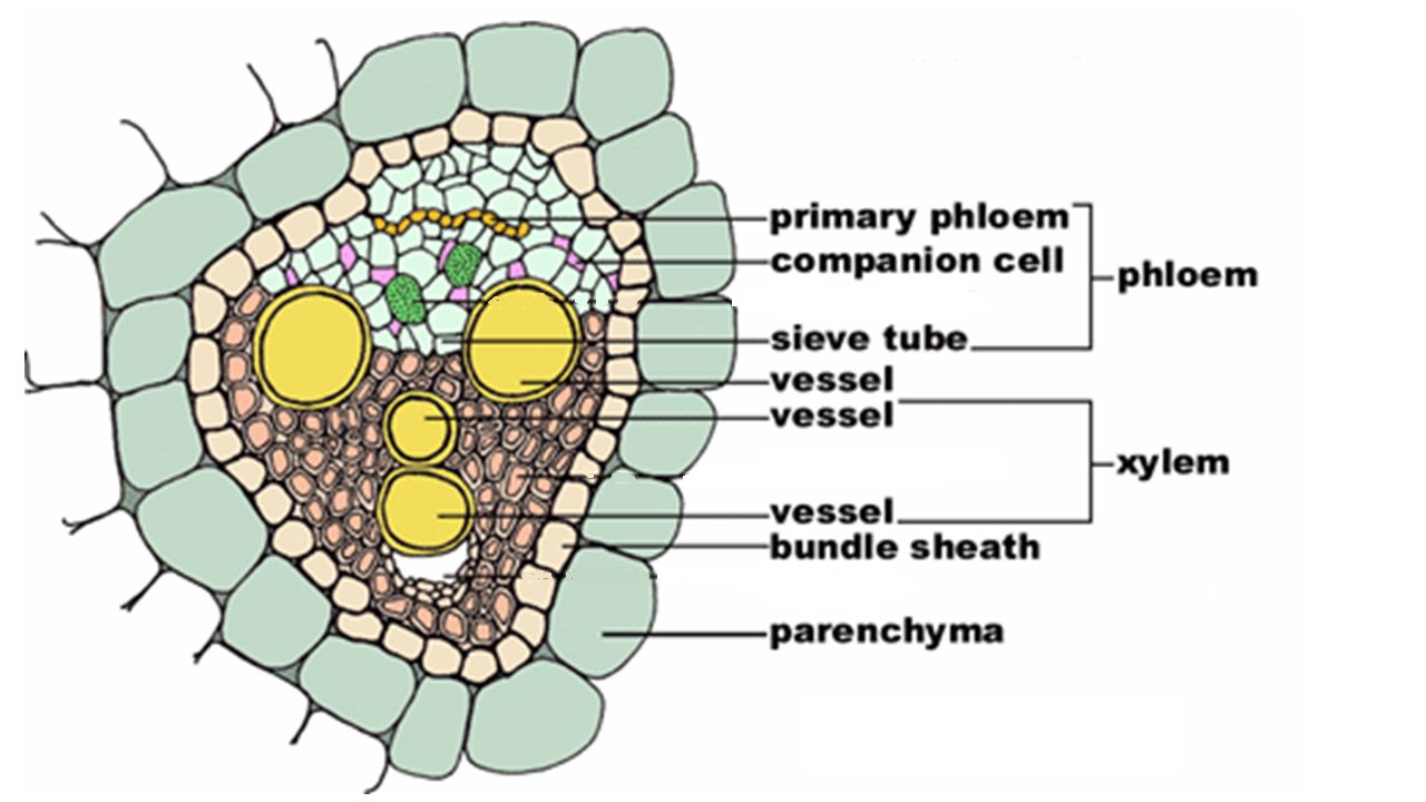
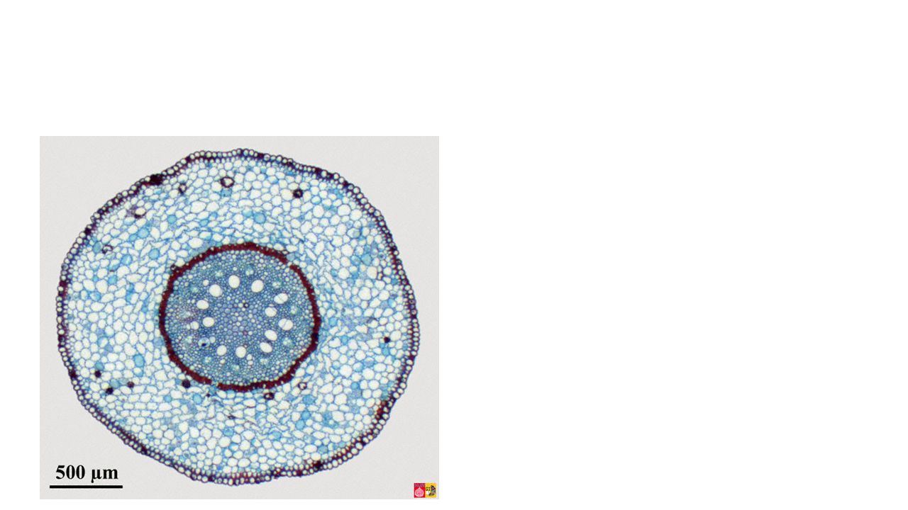
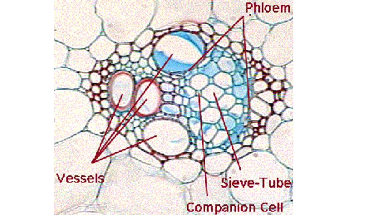
Eukaryote – Plant – Corn or Sweet Potato Stem This is a cross section of a plant stem. The large round structures in the image are empty tubes used by the plant to move water from the roots to the leaves or move food made in the leaves to the rest of the plant. Living cells are actually the smaller structures such as parenchyma, companion cells and primary phloem shown in the diagram to the left and enlarged on the next slide. Look for a small nucleus in the primary phloem and companion cells near the sieve tubes. View the plant slides and draw at 400X. Label the cell walls that surround the plant cells and a nucleus. Include 2 descriptions.

21
Eukaryote – Plant – Leaf (Privet or Corn leaf) This is a cross section of a leaf. The palisade cells are the best cells to examine; these are the cells that carry out photosynthesis. These cells have rectangular cells with thick cell walls. Look for a nucleus and a central vacuole that pushes the nucleus to the side. Chloroplasts appear around the edge of the cell. They do not look green because of the stain used to color the sample. The air spaces in the leaf provide the cells with a way to exchange CO 2 and O 2. Cell View and draw at 400X. Label the cell wall, nucleus, the central vacuole, and chloroplasts. Include 2 descriptions.

22
Eukaryote – Animal – Stratified squamous epithelium (peithelium on the slide label is a spelling mistake!) These are flat cells that line your mouth and throat. The top image shows individual cells that have been scraped from the inside of someone’s cheek. Look for a darkly stained nucleus. The lower image is a slice of tissue showing how the cells form sheets of tissue many layers deep. It is difficult to see the borders between individual cells. Label the nucleus and cell membrane. Write 2 descriptors for these cells.

23
Eukaryote – Animal – Cardiac Muscle Cells This is the muscle that makes up your heart. The nucleus is very obviously darker than the rest of the cell. The dark line across the cell is a special connection between cells that allows them to beat in rhythm. The cells are spread slightly in this image to show detail; in the heart, the cells are tightly packed together. Draw at 400X. Label: nucleus and connection between cells. Cardiac Muscle cell

Similar presentations


 tissue found throughout the plant. Xylem tissue consists of 1. Xylem vessels.>")


