Download presentation
Presentation is loading. Please wait.

1
Chapter 10 Atomic Emission Spectrometry

2
10A EMISSION SPECTROSCOPY BASED ON PLASMA SOURCES

3
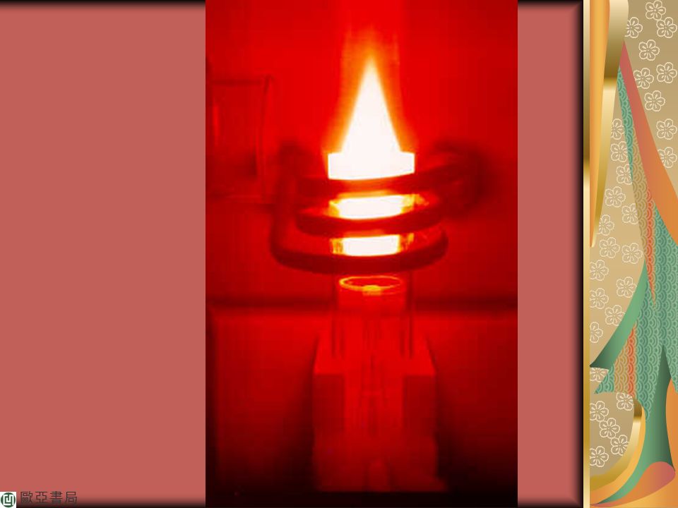
Inductively coupled plasma (ICP)
Direct current plasma (DCP) Microwave induced plasma (MIP)
 Microwave induced plasma (MIP)")
4
10A-1 The Inductivity Coupled Plasma Source

5
FIGURE 10-1 A typical ICP source. Position A shows radial viewing of
the torch, and position B shows axial viewing.

9
Sample introduction

10
FIGURE 10-2 The Meinhard nebulizer. The nebulizing gas flows
through an opening that surrounds the capillary concentrically. This causes a reduced pressure at the tip and aspiration of the sample. The high-velocity gas at the tip breaks up the solution into a mist.

11
Plasma Appearance and Spectra
Analyte Atomization and Ionization

12
FIGURE 10-3 Device for electrothermal vaporization.

13
FIGURE 10-4 Temperatures in a typical ICP course.

14
10A-2 The Direct Current Plasma Source

15
FIGURE 10-5 A three-electrode DC plasma jet.

16
10A-3 Plasma Source Spectrometers

17
Instruments for emission spectroscopy are of three
basic types: sequential, simultaneous multichannel, and Fourier transform.

18
TABLE 10-1 Desirable Properties of an Emission Spectrometer

19
Sequential Instruments
Slew-Scan Spectrometers Scanning Echelle Spectrometers

20
FIGURE 10-6 Optical diagram of a sequential ICP optical emission spectrometer. All
moving parts are under computer control, and their modes of motion are indicated by the three-dimensional arrow. Moving parts include the grating, a mirror for transducer selection, a refractor plate for optimizing signal throughput, and a viewing mirror to optimize the plasma viewing position. The spectrometer contains a mercury lamp for automatic wavelength calibration. Notice the axial viewing geometry.

22
FIGURE 10-7 Schematic of an echelle spectrograph system.

23
Multichannel Spectrometers
Polychromators.

24
FIGURE 10-8 Direct- reading ICP emission spectrometer. The
polychromator is of the Paschen-Runge design. It features a concave grating and produces a spectrum around a Rowland circle. Separate exit slits isolate each spectral line, and a separate photomulitiplier tube converts the optical information from each channel into an electrical signal. Notice the radial viewing geometry. PMT= photomultiplier tube.

25
ICP-AES

26
A Charge-Injection Device Instrument

27
FIGURE 10-9 Optical diagram of an echelle spectrometer
with a charge-injection detector.

28
FIGURE 10-10

29
FIGURE 10-10(a) Schematic representing the surface of a CID. The short
horizontal lines represent the read windows. A magnified image of one of the read windows is also shown. The nine central elements form the examination window, where a line is positioned.

30
FIGURE 10-10(b) Intensity profile for an iron line. All of the radiation from the line falls on the 3 × 3 examination window.

31
A Charge-Coupled Device Instrument
A Combination Instrument

32
FIGURE 10-11 An echelle spectrometer with segmented
array of CCDs.

33
FIGURE 10-12 Schematic of an array segment showing
phototransducers, storage and output registers, and readout circuitry.

34
Fourier Transform Spectrometers
Fourier, Joseph

35
10A-4 Applications of Plasma Sources

36
Sample Preparation Elements Determined

37
Line Selection Calibration Curves

38
FIGURE 10-13 Periodic table characterizing the detection power and number of
useful emission lines of ICP by using a pneumatic nebulizer. The color and degree of shading indicate the range of detection limits for the useful lines. The area of shading indicates the number of useful lines.

39
FIGURE 10-14 Typical calibration curves in ICP emission
spectrometry.

40
FIGURE 10-15 Internal standard calibration curves with an ICP
source. Here, an yttrium line at nm served as an internal standard. Notice the lack of interelement interference.

41
Interferences Detection Limits

43
10B EMISSION SPECTROSCOPY BASED ON ARC AND SPARK SOURCES

44
These spectra permitted the qualitative and quantitative determination of metallic elements in
a variety of sample types, including metals and alloys, soils, minerals, and rocks,

45
TABLE 10-2 Effect of Standardization Frequency on Precision of ICP Data

46
10B-1 Sample Types and Sample Handling

47
Metals Nonmetallic Solids

48
TABLE 10-3 Comparison of Detection Limits for Several Atomic spectral Methods

49
FIGURE 10-16 Some typical graphite electrode shapes.
Narrow necks are to reduce thermal conductivity.

50
10B-2 Instruments for Arc and
Spark Source Spectroscopy

51
Spectrographs

52
FIGRUE 10-17 The Eagle mounting for a grating spectrograph.

53
Multichannel Photoelectric Spectrometers
Multichannel Photomultiplier Instruments. Array-Based Multichannel Instruments.

54
10B-3 Arc Source Emission Spectroscopy

55
Characteristics of Arc Sources
Cyanogen Spectral Bands. Rates or Emission.

56
Applications of Arc Sources

59
10B-4 Spark Sources and Spark Spectra

60
Applications of Spark Source Spectroscopy

61
10C MISCELLANEOUS SOURCES FOR OPTICAL EMISSION SPECTROSCOPY

62
10C-1 Flame Emission Sources

63
10C-2 Glow-Discharge Sources

64
10C-3 Laser Microprobe Sources

Similar presentations


 is created by an electrical discharge between two electrodes. A plasma support gas is necessary,>")

 Lab: Should finish Set 2 Period 4 lab today Thursday is make-up.>")

: It should have high efficiency in generating ions of the element of.>")


>")

(‰)(%) (‰)(cm) Gr. 3-5A5-24.701.62%0.17%-24.70-5 Gr. 3-1515-24.141.02%0.12%-24.14-15 Gr. 3-2525-24.050.71%0.09%-24.05-25.>")


>")

 Atomic X-ray Spectrometry (Ch 12) Atomic Mass.>")