Download presentation
Presentation is loading. Please wait.

1
Stimulus Presentation and Image Acquisition Valentina Petre

2
How do we Design an Experiment Event related Block design

3
* Very good statistical power * Good for “all or none” phenomena (example visual, or acoustic stim) * Good for an initial experimental probe – pilot study (exceptions) * The optimal block length 14 to 20 s * Do not depend much on the assumption of a certain hemodynamic function * Subject might anticipate the stimulus
 * Good for an initial experimental probe – pilot study (exceptions) * The optimal block length 14 to 20 s * Do not depend much on the assumption of a certain hemodynamic function * Subject might anticipate the stimulus")
4
Event related * Multiple trials presentation in one run * Optimum time 16s * Close trials 4s –still can be separated * You must assume linearity * Depend on the model of the hemodynamic function * Reduced in sensitivity – vary the inter trial interval

5
Stim on Slice i Frame aq Event Related stimulus presentation desynchronized from scanner acquisition

6
Protocols available 128 x 12864 x 64 Mosaic Resolution Signal to Noise Time betw. Acq. Max # of slices for 128 frames Time to acquire one slice Delay time Excitation order high 2.3 x 2.3 mm low long (~10 min) 16 slices with 3.5s TR 120-130ms 140-150ms low 5 x 5 mm high short 27 slices with 3 s TR 100ms 140ms Protocol Prop InterleavedAscending
 16 slices with 3.5s TR ms ms low 5 x 5 mm high short 27 slices with 3 s TR 100ms 140ms Protocol Prop InterleavedAscending.")
7
Small voxel volume, low signal Large voxel, high signal Air tissue interface Air Tissue The field inhomogeneity is large, thus it will cause large phase dispersion across the voxel. The signal is reduced.

8
How to avoid intravoxel dephasing Reduce the slice thickness. The longest dimension of your voxel should not be parallel with the direction of field variation Orient the slices oblique For spin echo sequences, the effect is smaller

9
BoBo

10
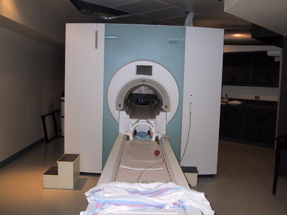
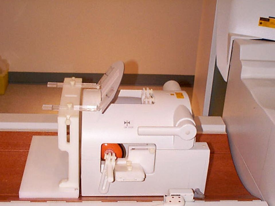
Equipment available 1.5 T Vision Siemens scanner head coil surface coil Head holder device ear protection nose piece bite bar Mirror and mirror stand Mouse & joy stick Trigger box Projector cables RGB VGA screen Physiological monitoring

14
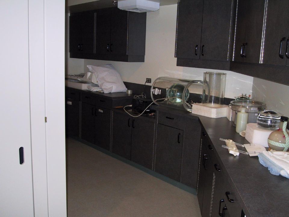
Accessories

17
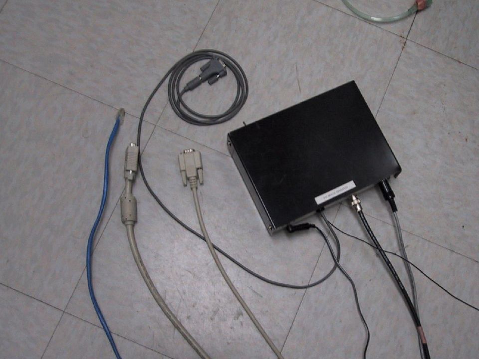
MR compatible mouse

18
Outside the scanner room Inside the scanner room Penetration panel

19
Screens

21
Types of stimulation used at the MNI Visual Acoustic Pain Somatosensory EEG

22
Programs used for stimulus presentation GLstim available on the SGI (R. Hoge ) Media Control Function on PC (Windows) Pierre Ahad SuperLab http://www.superlab.com Make your own.
 Media Control Function on PC (Windows) Pierre Ahad SuperLab Make your own..")
23
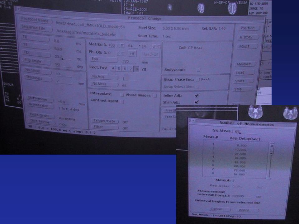
Parameters for the protocol to be used

25
Artifacts Patient wearing a metal studded beltBraces http://www1.stpaulshosp.bc.ca/stpaulsstuff/MRartifacts.html

27
RF Artifacts

28
Safety issues Implants Pacemaker Metallic part left after surgery, or metallic fragments in their eyes Belts Tools (screw drivers, scissors, pens…) Credit cards, bus pass. Certain type of makeup, tattoos Avoid loops

29
Safety issues

30
Safety issues contd..

Similar presentations


 By Mike Toulis November 12, 2002.>")


>")


 exhibit a wide range of response.>")

