Download presentation
Presentation is loading. Please wait.

1
The University of Manchester Faculty of Life Sciences Andreas Prokop BIOL20332/20972 GENETICS / Dev. Biol. RSM MODULE 2 Embryo staining, embedding, documentation & filing

2
- fixation - detergent - 1° ab - wash - 2° ab Today's procedures

3
- fixation - detergent - 1° ab - wash - 2° ab - wash 15‘ - ABC 1hr - wash 15‘ - DAB/H 2 0 2 Today's procedures

4
rinse with PBT in centrifuge tube (2 x for 3-5 min. each) in parallel prepare your dissecting scope (light from top, white side of bottom plate) remove PBT and add H 2 0 2 /DAB solution (handed out) - You must wear gloves! close centrifuge tube and shake, then quickly open and pour its content into a glass well; take up some liquid from the glass well to wash out embryos that got stuck in the tube observe staining progress under the dissecting microscope - stop on time to avoid excessive background staining - ask course assistants remove the H 2 0 2 /DAB solution with a pulled-out Pasteur pipette into the original 15ml Falcon tube and replace with PBT one more time rinse with PBT (dispose off in original 15ml Flacon tube - DAB-contaminated liquid, washing solutions and tips will be collected in yellow bags for incineration) by gently rotating the well gather embryos at bottom of well – transfer back to centrifuge tube with Eppendorf pipette (blue tip) remove PBT from centrifuge tube and add 700µl of 90% glycerol Today's procedures
 in parallel prepare your dissecting scope (light from top, white side of bottom plate) remove PBT and add H /DAB solution (handed out) - You must wear gloves. close centrifuge tube and shake, then quickly open and pour its content into a glass well; take up some liquid from the glass well to wash out embryos that got stuck in the tube observe staining progress under the dissecting microscope - stop on time to avoid excessive background staining - ask course assistants remove the H /DAB solution with a pulled-out Pasteur pipette into the original 15ml Falcon tube and replace with PBT one more time rinse with PBT (dispose off in original 15ml Flacon tube - DAB-contaminated liquid, washing solutions and tips will be collected in yellow bags for incineration) by gently rotating the well gather embryos at bottom of well – transfer back to centrifuge tube with Eppendorf pipette (blue tip) remove PBT from centrifuge tube and add 700µl of 90% glycerol Today s procedures.")
5
in a microfuge at 3000 rpm for 2 min, spin embryos down for several minutes; they will settle on one side of the tube cut off the very tip of a yellow pipette tip with a razor blade, set the pipette to 100 μl, and pick up as many embryos as possible into this volume; to achieve this, place the pipette tip in the area of the tube wall with the highest embryo density, then slowly release the button of your pipette moving the tip gently forward. repeat this process for each slide, and spin down embryos again, if they dispersed away from the tube wall. Today's procedures

6
Embedding of larval preparations - transfer embryos in a small drop of glycerol (70 µl) - put on cover slip, then let more oil flow in from the fringe, if required - seal all around the cover slip with nail varnish
 - put on cover slip, then let more oil flow in from the fringe, if required - seal all around the cover slip with nail varnish")
7
Filing of slides

8
The formation of neuronal cicuits

9
Structure of the Drosophila NS

10
Beware of embryo orientation

11
germ cell migration http://flymove.uni-muenster.de/Organogenesis/Gonads/OrgGonMov/pole_cells_lat120.mov Beware of developmental stages

12
http://flybase.org/.bin/fbimage?FBbt:00005093 Beware of developmental stages gut structured, CNS shorter than embryo length

13
Optimising microscope settings

14
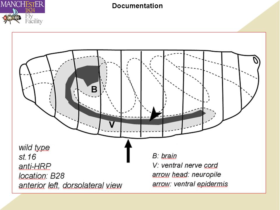
Documentation

16
Templates are provided

17
mutant gene BP102 (mouse) anti-A (mouse) anti-C (mouse) anti-βGal (rabbit) anti-Fas2 (mouse) A (x12) groups 1-3 groups 4-5, 22 B (x12) groups 7-9 groups 10-12 C (x14) groups 13-15, 6 groups 16-18 C-lacZ (x7) groups 19-21, 31 wt (x16) groups 23-26 groups 27-29,32 aliquots (x20)(x8) (x7)(x18) Fly stocks and antibodies by group
 anti-A (mouse) anti-C (mouse) anti-βGal (rabbit) anti-Fas2 (mouse) A (x12) groups 1-3 groups 4-5, 22 B (x12) groups 7-9 groups C (x14) groups 13-15, 6 groups C-lacZ (x7) groups 19-21, 31 wt (x16) groups groups 27-29,32 aliquots (x20)(x8) (x7)(x18) Fly stocks and antibodies by group")
Similar presentations


 Developed from Slijk.>")


