Download presentation
Presentation is loading. Please wait.

1
Molecular Mechanisms of Pediatric Kidney Disease Nader Gordjani MD PhD Professor of Pediatrics Universities of Freiburg and Frankfurt

2
Molecular origins of renal diseases H2OH2O Hypophasphatemic rickets Bartter-Syndrome Hypokalemic alkalosis: Gitelman-S. Diabetes insipidus renalis

3
kidney Glomerular structure Protein loss

4
Lesions sites of the glomerulus Filtration barrier T H -Lypmphocytes Capillaries Mesangial cells s. Biopsie Deposits of antibodies and complement Podocyte Autoantibodies Nephrotic Syndrome Nephritic Syndrome kidney

5
Proteinuria > 40 mg/m 2 * h > 1000 mg/m 2 * 24 h Plasma Albumin < 25 g/l Nephrotic Syndrome in Children

6
80% Steroid-sensitive Stable function 20% Steroid-resistant deterioration Nephrotic Syndrome in Children

7
Somlo S Nat Genet 24: 333 (2000) The glomerulus in health & NS
 The glomerulus in health & NS")
8
Somlo S Nat Genet 24: 333 (2000) Morphology of the Filtration Barriere normal Nephroc syndrome
 Morphology of the Filtration Barriere normal Nephroc syndrome")
9
Geheimnisse des „Podozyten“ Molecular structures of the Podocyte

10
What is the role of the podocyte in nephrotic syndrome ? Chemokines cytokines complement autoantibodies Inflammatory agents Inflammatory agents

11
Das kongenitale NS vom finnischen Typ Nephrin Congenital nephrotic syndrome Finnish Type

12
Familiäres Steroid-resistentes NS Podocin Familial Steroid resistant Nephrotic Syndrome

13
Die familiäre Glomerulosklerose a-act 4 Familial Glomerulosclerosis

14
What is the role of the podocyte in nephrotic syndrome and as target of immunmodulation? Chemokines cytokines complement autoantibodies Inflammatory agents Inflammatory agents Immunosuppression

15
Ca 2+ - Signalling in the cell Stimulation of: Signalling pathways Gene expression Channel opening/closing …

16
Calcium signalling in the Podocyte Fluorescence microscopy with Fura-2 AM Single-Photon Tube Video-Imaging Laser-Scanning RT-PCR Isolation of rat glomeruli by sieving method Statistical analysis: student´s t-test

17
Mouse Podocytes in Culture 33 o C 37 o C

18
CCR 3 CCR 4 CCR 1 CCR 2 CCR 5 GAPDH 334 605 409 563 410 334 500 bp CC-receptors of cultured mouse podocytes

23
--

25
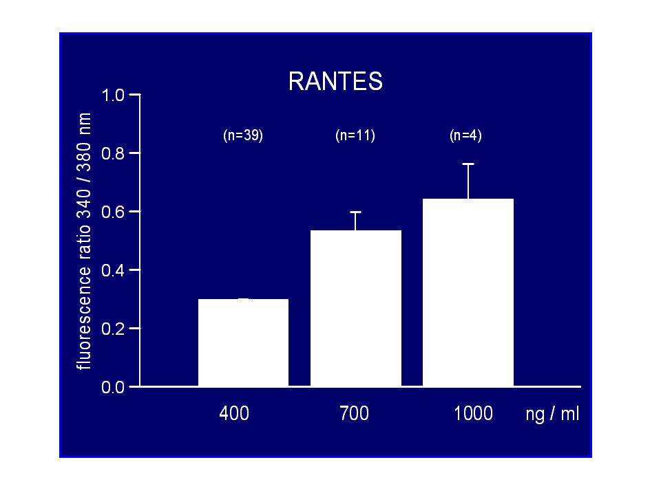
(n=5) (n=8) CyA ATPA II CyA
 (n=8) CyA ATPA II CyA")
27
Freshly isolated rat glomerulum 25 µm decapsulated 25 µm decapsulated

28
Immunofluorescence staining for podosynapsin in freshly isolated rat glomerulum 25 µm

29
Immunofluorescence staining for WT-1 in freshly isolated rat glomerulum

30
Freshly isolated rat glomerulum

31
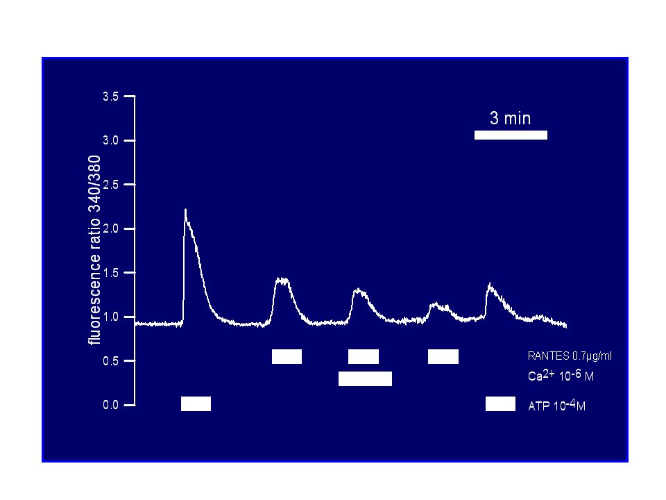
Conclusions Chemokines induce an increase of [Ca 2+ ] i in mouse podocytes by release from cytosolic Ca 2+ -stores and thus stimulate Ca 2+ -mediated signal transduction. Therefore podocytes are target cells of these proinflammatory factors. ATP also causes a characteristic rise of [Ca 2+ ] i, which is mediated by Ca 2+ -influx from the extracellular space in addition to store release. Cyclosporine A did not influence the chemokine- or ATP-associated Ca 2+ -effects. These findings could partly be confirmed in podocytes from freshly isolated intact rat glomeruli. Chemokines induce an increase of [Ca 2+ ] i in mouse podocytes by release from cytosolic Ca 2+ -stores and thus stimulate Ca 2+ -mediated signal transduction. Therefore podocytes are target cells of these proinflammatory factors. ATP also causes a characteristic rise of [Ca 2+ ] i, which is mediated by Ca 2+ -influx from the extracellular space in addition to store release. Cyclosporine A did not influence the chemokine- or ATP-associated Ca 2+ -effects. These findings could partly be confirmed in podocytes from freshly isolated intact rat glomeruli.
![Conclusions Chemokines induce an increase of [Ca 2+ ] i in mouse podocytes by release from cytosolic Ca 2+ -stores and thus stimulate Ca 2+ -mediated signal transduction.](http://images.slideplayer.com/23/6821870/slides/slide_31.jpg "Therefore podocytes are target cells of these proinflammatory factors. ATP also causes a characteristic rise of [Ca 2+ ] i, which is mediated by Ca 2+ -influx from the extracellular space in addition to store release. Cyclosporine A did not influence the chemokine- or ATP-associated Ca 2+ -effects. These findings could partly be confirmed in podocytes from freshly isolated intact rat glomeruli. Chemokines induce an increase of [Ca 2+ ] i in mouse podocytes by release from cytosolic Ca 2+ -stores and thus stimulate Ca 2+ -mediated signal transduction. Therefore podocytes are target cells of these proinflammatory factors. ATP also causes a characteristic rise of [Ca 2+ ] i, which is mediated by Ca 2+ -influx from the extracellular space in addition to store release. Cyclosporine A did not influence the chemokine- or ATP-associated Ca 2+ -effects. These findings could partly be confirmed in podocytes from freshly isolated intact rat glomeruli..")
32
Physiology Andreas Benesic Ruth Freudinger Michael Gekle Gerald Schwerdt Pediatrics Nader Gordjani Antje Kirchhoff Brigitte Wollny University of Würzburg University of Freiburg Physiologie Rainer Greger Hermann Pavenstädt Jens Leipziger Roland Nitschke Viktoria Munzinger

33
Rainer Greger 1946 - 2004 Institute of Physiology/Freiburg Rainer Greger 1946 - 2004 Institute of Physiology/Freiburg

34
What causes Cyst formation in kidneys?

35
… and damage in other organs? M. Waters et al. Pediatr Nephrol (2011) 6:1039–1056
 6:1039–1056")
36
… and other organs? M. Waters et al. Pediatr Nephrol (2011) 6:1039–1056
 6:1039–1056")
37
Hildebrandt F et al. JASN 2009;20:23-35 Juvenile/infantile Nephronophthisis

38
Hildebrandt F et al. JASN 2009;20:23-35 Ciliar defects: a unifying theory of cystic kidney disease?

39
Lin et al. PNAS 2002

40
Subcellular localization of nephrocystins Hildebrandt F et al. JASN 2009;20:23-35

42
Chapin et al. JCB 2010 Ciliar defects: a unifying theory of cystic kidney disease?

43
Hildebrandt F et al. JASN 2009;20:23-35 Correct mitotic spindle orientation False mitotic spindle orientation

44
Hildebrandt F et al. JASN 2009;20:23-35

45
The hedgehog signaling pathway may be involved in renal cystogenesis. Hildebrandt F et al. JASN 2009;20:23-35

Similar presentations


 RENAL DISEASE: OVERVIEW AND ACUTE RENAL FAILURE Pathophysiology of Disease: Chapter 16 (388-394) Jack.>")


 Caused by renal diseases that increase the permeability across the glomerular filtration.>")


 Urinary space surrounds glomerulus.>")
 Most prevalent, potentially lethal, monogenic disorder Prevalence.>")

