Download presentation
Presentation is loading. Please wait.

1
Eukaryotic Cell Division
Purpose Increase cell number for growth, maintenance, repair, or reproduction Cell Types Somatic Germ Cell Division Cycle Interphase Nuclear Division (Mitosis or Meiosis) Cytokinesis
 Cytokinesis.")
2
Fig. 5.1a

3
TERMINOLOGY Genetic Material Chromatin
Structural Types Chromatin Uncoiled genetic material not visible with a light microscope. Chromosome Coiled genetic material visible with a light microscope.

4
Fig. 5A

5
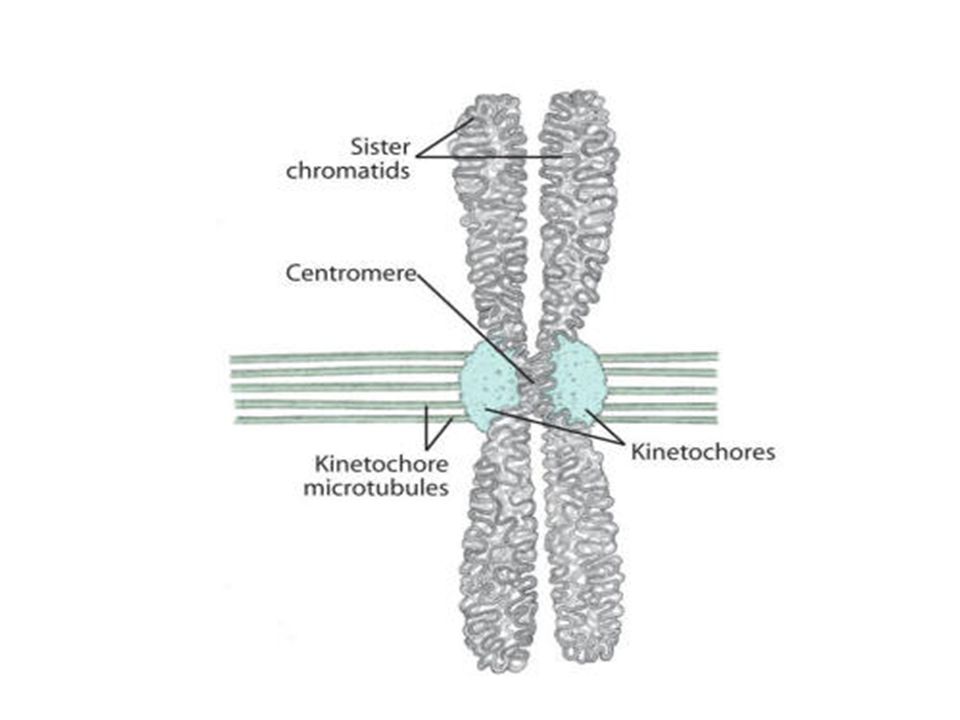
Terminology Genetic Material Structural Apparatus Sister Chromatids
One of two identical copies of genetic material attached at the centromere. Centromere Constricted portion of a chromosome that holds sister chromatids together. Kinetochore Protein structure at the centromere to which spindle fibers attach.

7
Terminology Centrioles
Cylindrical structure within a centrosome producing spindle fibers.

8
Terminology Mitotic Spindles (Spindle Fibers) Microtubules that bring about chromosomal movement.
 Microtubules that bring about chromosomal movement.")
9
SOMATIC CELL DIVISION Somatic Cells Definition All diploid cells that are not involved in gamete formation. Examples Skin, Heart, Liver, Intestinal, Bone, Muscle Type of Cell Division Mitotic (Mitosis) Steps Interphase Mitosis Cytokinesis
 Steps Interphase Mitosis Cytokinesis")
10
SOMATIC CELL DIVISION Mitotic Cell Division Outcome
Each daughter cell is a clone of the parent. Each cell has homologous pairs (diploid, N) of chromosomes. Human Karyotype 46 chromosomes . 22 paired autosomes sex chromosomes ( XX or XY)
 of chromosomes. Human Karyotype. 46 chromosomes paired autosomes . 2 sex chromosomes ( XX or XY)")
11
KARYOTYPE

12
Homologous Pairs, Diploid, 2N
Autosomes #1-22 Sex Chromosomes

13
22.9 Humanoid Daughter’s Old Boyfriend

14
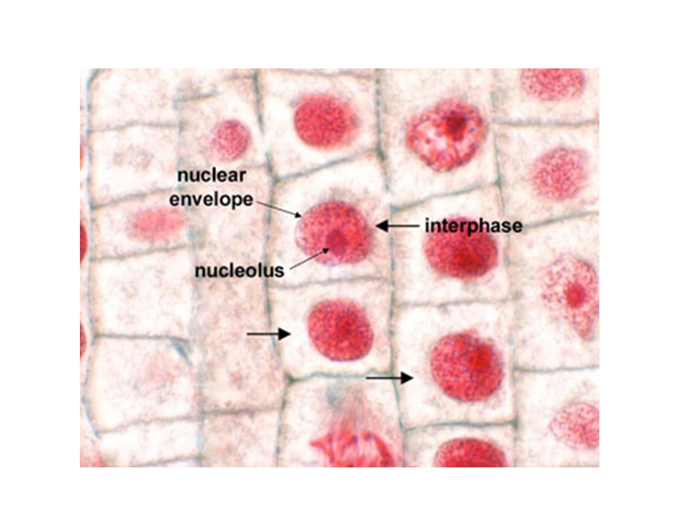
MITOTIC CELL DIVISION Interphase Purpose
Preparation for cell division. Steps G1 (Gap 1) Phase S Phase (Synthesis) G2 (Gap 2) Phase Genetic Material Chromatin
 Phase. S Phase (Synthesis) G2 (Gap 2) Phase. Genetic Material. Chromatin.")
15
INTERPHASE G1 (Gap 1) Phase Undividing Cell 1) Cell growth
2) Cell metabolism Dividing Cell 1) Organelles begin to replicate 2) Preparation for S phase a) DNA is checked for damage b) Environment is checked for adequacy c) Cell size is checked
 Cell metabolism. Dividing Cell. 1) Organelles begin to replicate. 2) Preparation for S phase. a) DNA is checked for damage b) Environment is checked for adequacy. c) Cell size is checked.")
16
Fig. 5.1b

17
G1 PHASE

19
INTERPHASE S (Synthesis) Phase DNA replication
Formation of Sister Chromatids

20
DNA REPLICATION Sister Chromatids

21
INTERPHASE G2 (Gap 2) Phase Final preparation for mitosis.
Check for DNA damage Check to see that S phase is complete Check the environment for adequacy Finish Organelle Replication

22
Fig. 5.1b

23
MITOTIC CELL DIVISION Mitosis Purpose Nuclear division
Separation of sister chromatids Steps Prophase Metaphase Anaphase Telophase

24
MITOSIS Prophase 1) Chromatin coils becoming chromosome.
2) Nuclear envelope disintegrates. 3) Centrioles produce mitotic spindles and move toward the spindle poles 4) Kinetochore fibers attach to kinetochores 5) Polar fibers run from pole to pole 6) Asters attach centrioles to plasma membrane
 Nuclear envelope disintegrates. 3) Centrioles produce mitotic spindles and. move toward the spindle poles. 4) Kinetochore fibers attach to kinetochores. 5) Polar fibers run from pole to pole. 6) Asters attach centrioles to plasma. membrane.")
27
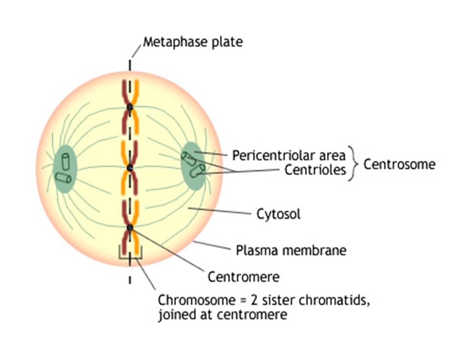
MITOSIS Metaphase Centriole movement aligns chromosomes at
the spindle equator. M Checkpoint for spindle fiber attachment.

29
MITOSIS Anaphase 1) Kinetochores pull on Kinetochore fibers
2) Sister chromatids separate. a) Chromatids are now referred to as “Daughter Chromosomes”. 3) Daughter chromosomes move toward opposite spindle poles.
 Sister chromatids separate. a) Chromatids are now referred to as Daughter Chromosomes . 3) Daughter chromosomes move toward opposite spindle poles.")
30
Daughter Chromosome

32
MITOSIS Telophase 1) Chromosomes arrive at spindle poles.
2) Nuclear membrane regenerates. 3) Spindle fibers disintegrate. 4) Chromosomes become chromatin.
 Nuclear membrane regenerates. 3) Spindle fibers disintegrate. 4) Chromosomes become chromatin.")
34
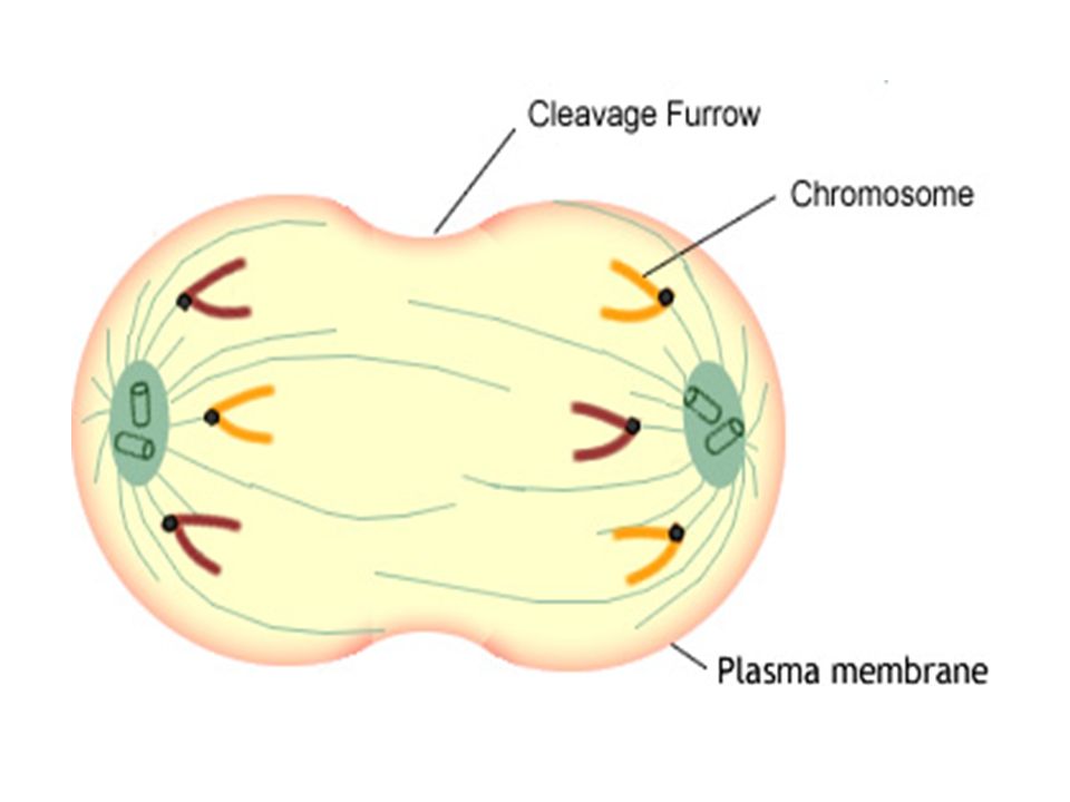
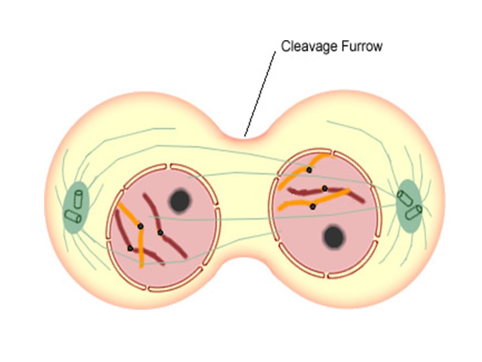
CYTOKINESIS Types Purpose Cleavage Furrow
Formation of two daughter cells. Types Animal Cell Cleavage Furrow Formation of a contractile ring at the cell equator which will "pinch“ the cell in two. Plant Cell Cell Plate Construction of a new cell wall inside of the cell creating two cells.

35
ANIMAL CYTOKINESIS

36
ANIMAL CYTOKINESIS

37
DAUGHTER CELLS

38
PLANT CYTOKINESIS

39
PLANT CYTOKINESIS

Similar presentations


 Chromosomes – Rod-shaped structures composed.>")
