Download presentation
Presentation is loading. Please wait.

1
Bleeding Disorders Meera Shreedhara 8/25/08

2
What is it? A bleeding disorder is an acquired or inherited tendency to bleed excessively

5
Mechanisms of bleeding
Vascular Integrity Platelets Clotting factors Fibrinolysis Derangement of any of these factors can cause abnormal bleeding

6
Key to diagnosis History

7
Bleeding history Epistaxis Gingival hemorrhage Mucosal Bleeding
Heavy Menses Child birth Easy bruisability Bleeding following tooth extractions Hematomas Bleeding following surgery Hemarthrosis

8
Medication History Aspirin Warfarin NSAIDS B- Lactam antibiotics
Clopidogrel and other antiplatelet agents Herbal medications.

9
Nutritional history Vit K deficiency Vit C deficiency
Broad spectrum antibiotics

10
Clinical Characterisitc
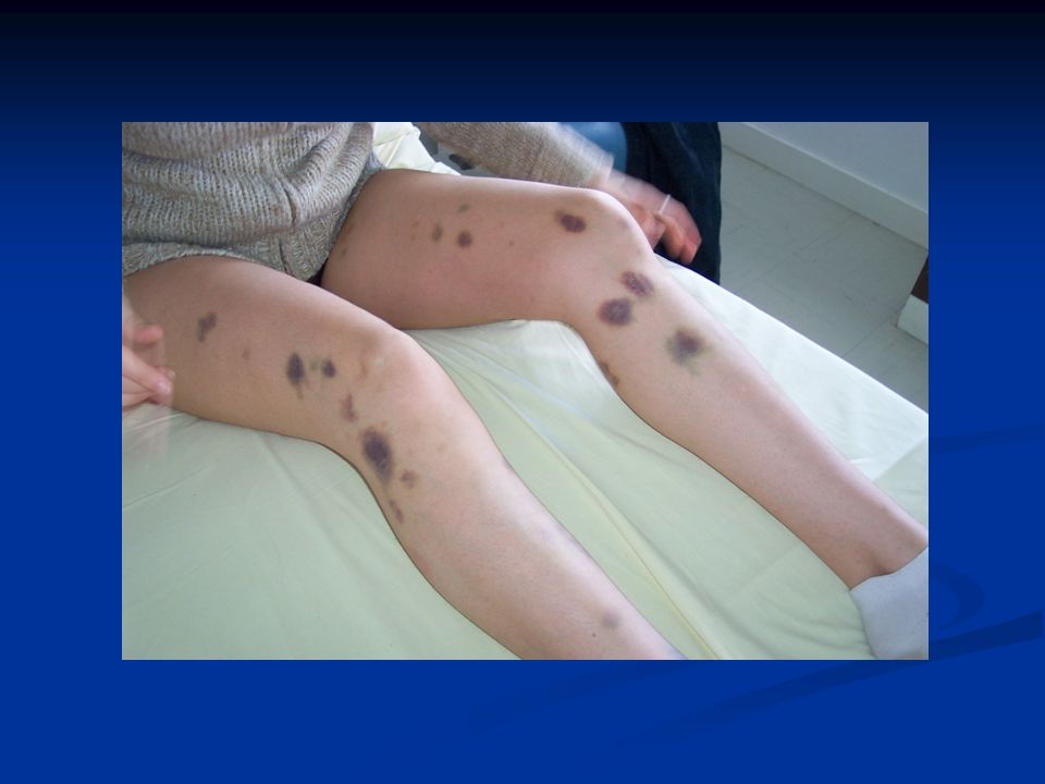
Platelet defect Clotting factor deficiency Site of bleeding Skin, mucous membranes (gingivae, nares, GI and genitourinary tracts) Deep in soft tissues (joints, muscles) Bleeding after minor cuts Yes Not usually Petechiae Present Absent Ecchymoses Small, superficial Large, palpable Hemarthroses, muscle hematomas Rare Common Bleeding after surgery Immediate, mild Delayed, severe
 Deep in soft tissues (joints, muscles) Bleeding after minor cuts. Yes. Not usually. Petechiae. Present. Absent. Ecchymoses. Small, superficial. Large, palpable. Hemarthroses, muscle hematomas. Rare. Common. Bleeding after surgery. Immediate, mild. Delayed, severe.")
15
History Should the pt undergo a limited or extensive workup?
Is this acquired or hereditary? Is this likely a disorder of clotting factors,platelets, fibrinolysis or vWF? Do medications or intercurrent illnesses play a role? What is the immediate cause for which a workup is being done?

16
Hereditary Deficiency of coagulation factors Platelet disorders
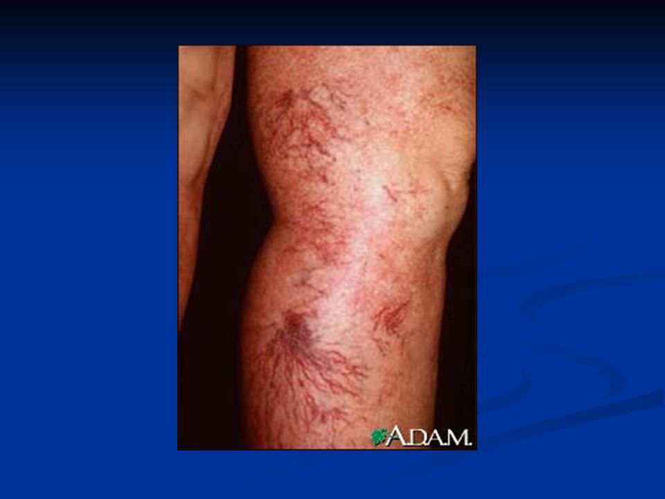
Hemophilia Fibrinogen deficiency Von Willebrand disease Platelet disorders Glanzmann thrombasthenia Bernard-Soulier syndrome Platelet granule disorders Fibrinolytic disorders Alpha 2 antiplasmin deficiency PAI 1 deficiency Structural disorders Hemorrhagic Telangiectasias Ehler Danlos syndrome

17
Acquired Thrombocytopenis Liver disease Renal failure Vit K deficiency
Acquired antibodies to coagulation factors DIC Drugs Vascular

18
Lab testing Platelet count
Bleeding time-Measure of the interaction of platelets with the blood vessel wall. Thrombocytopenia (platelet count usually below 50,000/microL), Qualitative platelet abnormalities (eg, uremia), von Willebrand disease (VWD), Vascular purpura, Severe fibrinogen deficiency
, Qualitative platelet abnormalities (eg, uremia), von Willebrand disease (VWD), Vascular purpura, Severe fibrinogen deficiency.")
19
Platelet function assay
Expose platelets within citrated whole blood to high shear (5,000 to 6,000/sec) within a capillary tube and monitor the drop in flow rate as the platelets form a hemostatic plug within the center of a membrane coated with collagen and either ADP or epinephrine Abnormal closure times are an indication of platelet dysfunction, they are not specific for any disorder The test is coagulation factor independent PFA-100™ is more sensitive (>70 percent) than the bleeding time (20 to 30 percent) in detecting all subtypes of von Willebrand's disease (vWD) Exception is type 2N vWD, in which the hemostatic defect resides in the Factor VIII binding site on vWF
 within a capillary tube and monitor the drop in flow rate as the platelets form a hemostatic plug within the center of a membrane coated with collagen and either ADP or epinephrine. Abnormal closure times are an indication of platelet dysfunction, they are not specific for any disorder. The test is coagulation factor independent. PFA-100™ is more sensitive (>70 percent) than the bleeding time (20 to 30 percent) in detecting all subtypes of von Willebrand s disease (vWD) Exception is type 2N vWD, in which the hemostatic defect resides in the Factor VIII binding site on vWF.")
20
Platelet function assay
Collagen/epinephrine closure time (CEPI-CT)- Abnormal in Aspirin intake Collagen/adenosine diphosphate (CADT-CT)-Normal in aspirin intake
- Abnormal in Aspirin intake. Collagen/adenosine diphosphate (CADT-CT)-Normal in aspirin intake.")
21
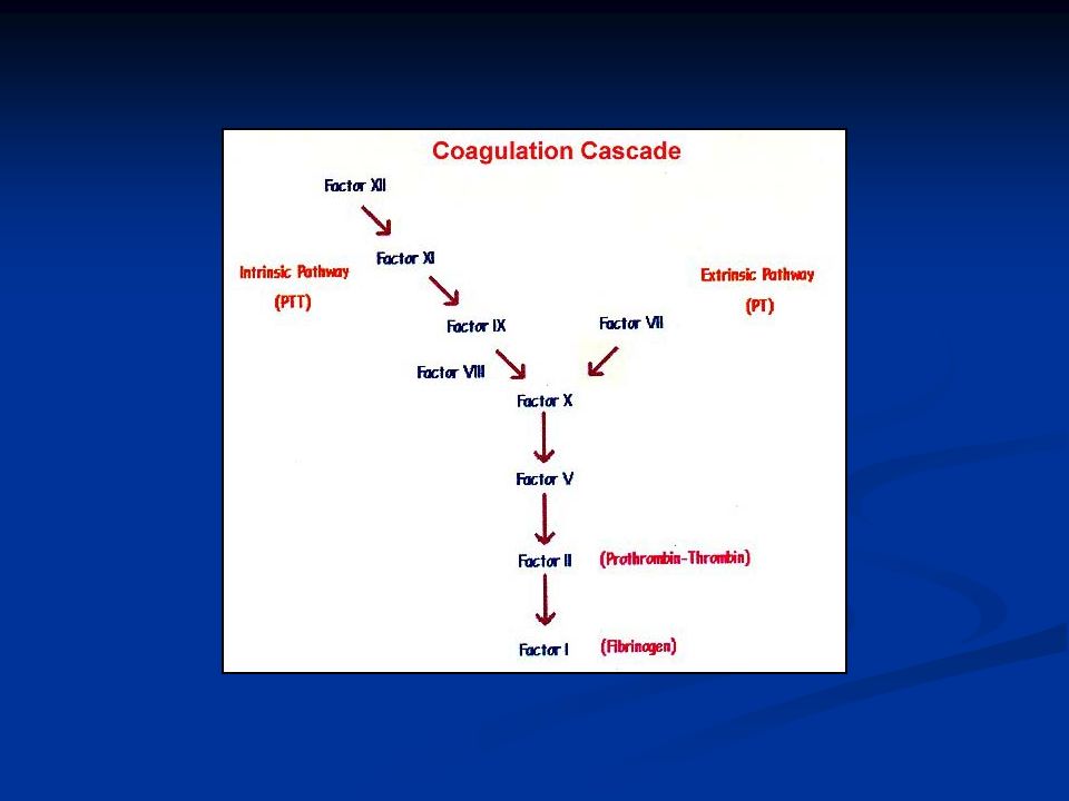
Prothrombin time Measure of the extrinsic pathway and common pathway
Bypasses the intrinsic pathway and uses thromboplastins to substitute for platelets Within the combined pathway, factors VII, X, and prothrombin are vitamin-K dependent and are altered by warfarin

23
Prolonged PT Vitamin K deficiency
Liver disease, which decreases the synthesis of both vitamin K-dependent and -independent clotting factors. Deficiency or inhibition of factors VII, X, II (prothrombin), V, or fibrinogen The infrequent antiphospholipid antibodies (lupus anticoagulant phenomenon) with antiprothrombin activity Heparin does NOT prolong the PT
, V, or fibrinogen. The infrequent antiphospholipid antibodies (lupus anticoagulant phenomenon) with antiprothrombin activity. Heparin does NOT prolong the PT.")
24
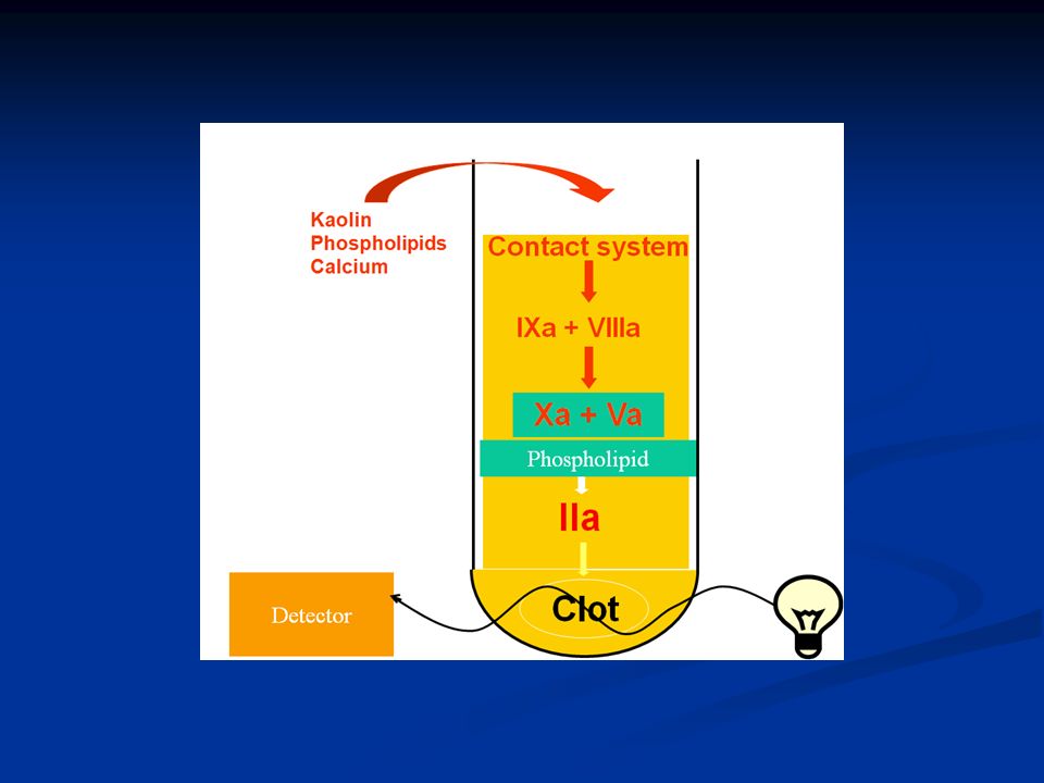
aPTT Measures the intrinsic and common pathways of coagulation
Uses partial thromboplastins; they are incapable of activating the extrinsic pathway Prolonged in deficiency of, or an inhibitor to, any of the clotting factors except for factor VII Prolonged in the presence of Lupus Anticoagulant. Used to monitor heparin activity

27
Thrombin time Measure conversion of fibrinogen to fibrin monomers and the formation of initial clot by thrombin Hypofibrinogenemia, Dysfibrinogens Increased fibrin split products Heparin increases TT but not RT

29
Factor deficiencies/ inhibitors
A prolonged aPTT can be due to a deficiency (or absence) of a coagulation factor or the presence of a coagulation factor inhibitor Mixing studies help differentiate this Lupus anticoagulants can result in a prolonged aPTT that is not correctable by the addition of normal plasma Overcome by adding excess platelet phospholipid (particularly a hexagonal phase phospholipid) or by assessing the diluted Russell's viper venom time
 of a coagulation factor or the presence of a coagulation factor inhibitor. Mixing studies help differentiate this. Lupus anticoagulants can result in a prolonged aPTT that is not correctable by the addition of normal plasma. Overcome by adding excess platelet phospholipid (particularly a hexagonal phase phospholipid) or by assessing the diluted Russell s viper venom time.")
30
Fibrinolysis Fibrin and fibrinogen degradation products (FDP) are protein fragments resulting from the action of plasmin on fibrin or fibrinogen
 are protein fragments resulting from the action of plasmin on fibrin or fibrinogen.")
31
Fibrinolysis FDP assays do not differentiate between fibrin degradation products and fibrinogen degradation products Fibrin D-dimers are degradation products of cross-linked fibrin D-dimers specifically reflect fibrinolysis of cross-linked fibrin (ie, the fibrin clot) – so are more reliable indicators of thrombosis
 – so are more reliable indicators of thrombosis.")
32
Fibrinolysis Assays for plasminogen,
Tissue plasminogen activator (t-PA), Alpha-2 antiplasmin, Plasminogen activator inhibitor-1 (PAI-1), Thrombin-activatable fibrinolysis inhibitor (TAFI).
, Alpha-2 antiplasmin, Plasminogen activator inhibitor-1 (PAI-1), Thrombin-activatable fibrinolysis inhibitor (TAFI).")
33
Normal PT and PTT Thrombocytopenia vWD Factor 13 deficiency
Platelet dysfunction Vascular purpuras Psychogenic purpura

34
Normal PT and Prolonged aPTT
Hemophilia A Hemophilia B Factor XI deficiency Factor VIII inhibitor Malignancy, Clonal lymphoproliferative disorders, Pregnancy, Rheumatologic disorders

35
Prolonged PT and normal aPTT
Factor VII deficiency Warfarin therapy Early liver disease Early DIC

36
Prolonged PT and PTT Vit K deficiency Liver disease Warfarin treatment
Acquired inhibitor to factor V Factor X deficiency- seen in Amyloidosis DIC

37
Acute Promyelocytic Leukemia
DIC is often seen at presentation or during treatment Medical Emergency as Cerebral hemorrhage can occur in upto 4% of untreated pts Promyelocytes seen on smear Reciprocal translocation between the long arms of chromosomes 15 and 17, with the creation of a fusion gene, PML/RAR-alpha Immediate initiation of ATRA induces de deifferentiation

38
Hemophilia Hemophilia A and B are X-linked recessive diseases
Severe disease <1 % factor activity, Moderate disease- 1 to 5 % Mild disease >5 % The most common sites are into joints and muscles and from the gastrointestinal tract

39
Treatment The two components to therapy are treatment of active bleeding and inhibitor ablation via immune tolerance induction Cryoprecipitate has high levels of factor VIII Porcine Factor VIII Recombinant human Factor VIII The choice of factor VIII product usually is based upon safety, purity, and cost.

40
Dosing One international unit (IU) of clotting factor is that amount present in 1 mL of pooled normal plasma Dose of F VIII (IU) = Weight (kg) x (Desired % increase) x 0.5 Depends on the clinical indication and the presence of inhibitors
 = Weight (kg) x (Desired % increase) x 0.5. Depends on the clinical indication and the presence of inhibitors.")
41
von Willebrand’s disease
Most common of the inherited bleeding disorders In 1926, Erik von Willebrand described the first patient with the disease Von Willebrand factor (VWF) binds to both platelets and endothelial components, forming an adhesive bridge between platelets and vascular subendothelial structures and between adjacent platelets at sites of endothelial injury
 binds to both platelets and endothelial components, forming an adhesive bridge between platelets and vascular subendothelial structures and between adjacent platelets at sites of endothelial injury.")
42
Acquired von Willebrand’s disease
Malignant diseases Monoclonal gammopathy of unknown significance Multiple Myeloma Non-Hodgkin's lymphoma Chronic lymphocytic leukemia Waldenstrom's macroglobulinemia Essential thrombocythemiaPolycythemia vera Chronic myelogenous leukemia Wilms tumor Other carcinomas Immunologic disorders Systemic lupus erythematosus Other autoimmune diseases Other disorders Hypothyroidism Ventricular septal defect Aortic stenosis Mitral valve prolapse Gastrointestinal angiodyplasia Uremia Hemoglobinopathies Drugs and other agents Valproic acid Antibiotics

43
Treatment DDAVP Replacement of vWF EACA Tranexamic acid
Recombinant factor 7

44
Its better to bleed than clot!

45
Therapies other than factor replacement
DDAVP EACA Tranexamic Acid Factor 7 inhibitor- Novoseven

46
Liver disease Vs DIC Low factor V levels can be used as evidence for either reduced hepatic synthetic function or increased consumption, as in DIC Factor VIII is not manufactured by hepatocytes; factor VIII levels are usually normal or increased in liver disease

Similar presentations