Download presentation
Presentation is loading. Please wait.

1
“Change is inevitable-except from a vending machine”
LEARN TO ADAPT “Change is inevitable-except from a vending machine” -Robert C. Gallagher

2
DIAGRAM – SKIN LAYERS Differences: Epidermis, apocrine glands (seal outer surface, phermones) compound hair
 compound hair.")
3
Apocrine vs. Sebaceous Apocrine Sebaceous ~ Sweat glands
Specialized forms in eyelids, external ear canal Hair follicle complex Anal sacs Sympathetic NS Sebaceous Hair follicles Secrete sebum (oily) Large glands Dog: dorsum of tail, Mucocutaneous junction Cat: dorsum of tail, on lip margins and under chin
 Large glands. Dog: dorsum of tail, Mucocutaneous junction. Cat: dorsum of tail, on lip margins and under chin.")
4
Skin Protects from physical, chemical and microbiological injury
Sensory organ: pain, heat, cold, touch Storage: Electrolytes, water, proteins, fats and carbohydrates, activates Vit D by sunlight Epidermis Dermis Hypodermis/ subcuticular: Stores fat for insulation and energy

5
Dermatology-skin layers

6
Basic Anatomy & Physiology
The skin is made up of three layers: Epidermis (the most superficial layer that contains cells, but not blood vessels) Dermis (the middle layer that is composed of blood and lymph vessels, nerve fibers, and the accessory organs of skin such as glands and hair follicles) Hypodermis (subcutaneous - the deepest layer that is composed of connective tissue) - fat
 Dermis (the middle layer that is composed of blood and lymph vessels, nerve fibers, and the accessory organs of skin such as glands and hair follicles) Hypodermis (subcutaneous - the deepest layer that is composed of connective tissue) - fat.")
7
Skin - Function Enclosing barrier: Water and electrolyte loss
Environment protection Temperature regulation Sensory perception: touch, temp. and pain Motion and shape Antimicrobial and antifungal Blood pressure control: peripheral vascular bed Secretion: apocrine and sebaceous glands Adnexa: hair, nails, hooves, horny layer – epidermis Storage: Electrolyte, H2O, Vit., fat, protein, Carbs and other.. Pigmentation: color and sun protection Excretion: some Sun > activate vit D > normal Ca absorption

8
Definitions Dermatology: study of diseases of the skin
Dermatosis: skin disease Alopecia: hairloss Seborrhea: Excessive secretion of sebum (oily secretion of the sebaceous glands composed of fat and epithelial debris) Scale: Flakes of stratum corneum on the skin surface or hair coat; various colors Erythema: increased redness Collarette: circular arrangement of scale with central area of hyperpigmentation Crust: accumulation of dead cells and exudate on skin surface
 Scale: Flakes of stratum corneum on the skin surface or hair coat; various colors. Erythema: increased redness. Collarette: circular arrangement of scale with central area of hyperpigmentation. Crust: accumulation of dead cells and exudate on skin surface.")
9
Epidermal Collarette Most likely bacterial infection
Less commonly fungal infection, immune-mediated skin disease, insect-bite reaction, or contact hypersensitivity Scale of loose keratin flakes or "peeling" keratin arranged in a circle

10
Definitions Ectoparasites: external parasites
Skin Scraping: method of examining skin for parasites Dermatophyte: fungi that grow on the skin Dermatomycosis: fungal skin infection not involving pathogens below Dermatophytosis (ringworm): a skin infection with keratinophilic fungi (e.g. Microsporum; Trichophyton, Epidermophyton) Pyoderma: Bacterial infection of skin Superficial Deep Fungal culture – lab test used to grow dermatophytes for identification Bacterial culture and sensitivity: lab test used to grow and identify bacteria from lesions and determine antibiotic sensitivity
: a skin infection with keratinophilic fungi (e.g. Microsporum; Trichophyton, Epidermophyton) Pyoderma: Bacterial infection of skin. Superficial. Deep. Fungal culture – lab test used to grow dermatophytes for identification. Bacterial culture and sensitivity: lab test used to grow and identify bacteria from lesions and determine antibiotic sensitivity.")
11
Definitions Impetigo (not intertigo: skin fold pyoderma): Superficial bacterial skin infection seen in young dogs “puppy pyoderma” (staph) Acne: Pores clogged with oil forming “blackheads”, esp chin Lesion: area of altered skin Rash: wide spread eruption of lesions Hyperpigmentation: increased pigmentation (melanin) of skin (epidermal/dermal) Lichenification: Thickening and hardening of skin characterized by exaggerated superficial skin markings. Cyst: Fluctuant nodule; walled off, fluid filled nodule
 of skin (epidermal/dermal) Lichenification: Thickening and hardening of skin characterized by exaggerated superficial skin markings. Cyst: Fluctuant nodule; walled off, fluid filled nodule.")
12
Lichenification: Dff: All chronic and pruritic skin diseases Hyperpigmentation: Dff: endocrine dermatoses, secondary postinflammatory change due to a variety of skin diseases.

13
Definitions Macule: A focal, circumscribed, nonpalpable change in color <1 cm (when it is larger, it is termed a patch). Papule: small, circumscribed, solid elevated lesion of the skin ~ 1cm Wheal: A sharply circumscribed, raised, edematous lesion that appears and disappears within minutes to hours Pustule: a small, elevated, circumscribed, pus-containing lesion of the skin within the epidermis. Abscess: localized collection of pus; larger than a pustule Cellulitis: swollen, tender area of skin with bacterial infection; can develop into an abscess

14
Macule: Vitiligo, discoid lupus erythematosus, uveodermatologic syndrome, mucocutaneous pyoderma. Papule: Bacterial folliculitis, demodicosis, fungal folliculitis, flea-bite and mosquito-bite hypersensitivity, scabies, contact allergy, autoimmune skin disease, drug eruption

15
Pustule: Bacterial infection,
fungal infection, autoimmune skin disease Urticaria, insect bites, other hypersensitivities, drug eruption

16
Definitions Granulation tissue: new tissue in a healing wound; consists of connective tissue and capillaries Erosion: loss of skin surface; shallow, moist, crusted does not penetrate basement membrane Ulcer: loss of the epidermis resulting in exposure of dermis Comedone: dilated hair follicle blocked with sebum and other cellular debris

17
Various skin diseases associated
with self trauma such as infections or allergies Various skin diseases associated with trauma such as infections and allergies, also immune-mediated diseases.

18
Seborrhea Seborrhea is characterized by abnormal flaking or scaling of the epidermis and may be accompanied by increased oil production (seborrhea oleasa) or not (seborrhea sicca) Accelerated skin cell turn-over with or without excessive sebum production.
 or not (seborrhea sicca) Accelerated skin cell turn-over with or without excessive sebum production.")
19
Seborrhea Normal skin cell turnover – 3 weeks
Time period is shortened in seborrhea. Abnormal keratinization Accelerated (few days) => build up of keratin (dead cells) => flakes Altered sebaceous gland secretions Normal secretion of oil in or near hair follicles enriches skin
 => build up of keratin (dead cells) => flakes. Altered sebaceous gland secretions. Normal secretion of oil in or near hair follicles enriches skin.")
20
Seborrhea

21
Seborrhea Causes Types
Primary: hereditary as in Amer. Cocker Spaniel, Eng. Springer Spaniel, Westies, Basset Hounds Secondary: disease/injury to skin from other causes Allergies, parasites, nutritional disorders, immune-mediated, endocrine disorders (hypothyroidism) Types Seborrhea sicca: dry, only scaliness Seborrhea oleosa: oily + scales
 Types. Seborrhea sicca: dry, only scaliness. Seborrhea oleosa: oily + scales.")
22
Seborrhea sicca DRY and SCALY

23
Seborrhea oleosa

24
Keratolytics Keratolytics are an important group of antiseborrheics
Keratolytics remove excess keratin and promote loosening of the outer layers of the epidermis Keratolytics break down the protein structure of the keratin layer, permitting easier removal of this material Found in medicated shampoos to help in treatment of seborrhea

25
Seborrhea – Treatment Medicated shampoos
Sicca: Sulfur (keratolytic, antipruritic, antibacterial, antifungal, antiparasitic); Salicyclic acid (KL, AP, AB) Oleaso: Coal tar: degreaser, KL; BENZOYL PEROXIDE: Also for Moist dermatitis, pyoderma, stud tail Conditioners
; Salicyclic acid (KL, AP, AB) Oleaso: Coal tar: degreaser, KL; BENZOYL PEROXIDE: Also for. Moist dermatitis, pyoderma, stud tail. Conditioners.")
26
Seborrhea – Treatment Clip hair so shampoo can penetrate
If secondary, treat underlying cause Antifungal and/or antibacterial meds Omega-3 fatty acid supplements Anti-inflammatory, anti-pruritic properties

27
Seborrheic Fungal Dermatitis
Malassezia species Some commensal on skin (normal) Cause infection when skin surface altered/abnormal or immunosuppressed Diagnosed by cytology (stained slides) Skin impressions: Samples can be obtained via tape prep or impression smear Tx: 2% miconazole/2% chlorhexidine shampoo Selenium sulphide Other Antifungals: ketoconazole, clotrimazole, miconazole Oral ketoconazole x 3+ weeks (not approved in dogs and cats)
 Cause infection when skin surface. altered/abnormal or immunosuppressed. Diagnosed by cytology (stained slides) Skin impressions: Samples can be obtained via tape prep or impression smear. Tx: 2% miconazole/2% chlorhexidine shampoo. Selenium sulphide. Other Antifungals: ketoconazole, clotrimazole, miconazole. Oral ketoconazole x 3+ weeks (not approved in dogs and cats)")
28
Seborrheic Fungal Dermatitis

29
Pyodermas Definition: Bacterial infection of skin, superficial or deep; Primary or secondary Occurs when: skin surface broken skin macerated by chronic exposure to moisture Normal bacteria altered Circulation impaired Immunocompetence impaired (aka immunosuppressed) Superficial- Lesion usually involve superficial epidermis and heal w/o scarring, short duration, rarely ill
 Superficial- Lesion usually involve superficial epidermis and heal w/o scarring, short duration, rarely ill.")
30
Pyoderma - Causes Dogs - Staph intermedius
Cats – Pasteurella multocida Deep pyodermas – gram-negative organisms (E. coli, proteus sp, pseudomonas sp) Risk factors Allergies Fungal infections Endocrine diseases Immune incompetence Seborrhea Conformation – ex: skin folds Trauma Foreign body
 Risk factors. Allergies. Fungal infections. Endocrine diseases. Immune incompetence. Seborrhea. Conformation – ex: skin folds. Trauma. Foreign body.")
31
Pyoderma Superficial – commonly the trunk
Deep – chin, nose, pressure points, feet, generalized, skin folds PE may show: papules, pustules, crusts, epidermal collarettes, circular erythematous or hyperpigmented spots, alopecia, scaling, lichenification, abscess, cellulitis, etc..

32
Pyoderma EPIDERMAL COLLARETTES ‘PEELING EDGE’

33
PYODERMA ERYTHEMA, PUSTULES

34
Superfical Pyodermas Acute Moist Dermatitis, Superficial pyotraumatic dermatitis (“Hot Spots”) Clinical Signs: red, moist, painful areas, oozing Common in thick coated dogs (Labs, Newfoundlands, Ger. Shepherds, Chows) Usually in hot moist summer months Hair loss, very pruritic Develops very fast Dx: visual inspection of affected area TX Clip hair around lesion until skin normal Cleanse skin with medicated shampoo Topical Ab’s/steroid creams or sprays (ex: Betagen Spray) Treat original disease (fleas, allergies) Systemic steroids, Abs
 Usually in hot moist summer months. Hair loss, very pruritic. Develops very fast. Dx: visual inspection of affected area. TX. Clip hair around lesion until skin normal. Cleanse skin with medicated shampoo. Topical Ab’s/steroid creams or sprays (ex: Betagen Spray) Treat original disease (fleas, allergies) Systemic steroids, Abs.")
35
Superficial Pyodermas
Client Info Gentle cleansing BID Owner should wash hands after treatment E-collar may be necessary

36
Superficial Pyoderma: “Hot spot”

37
Superficial Pyoderma: “Hot Spot”

38
Superficial pyoderma: “Hot Spot”

39
Superficial Pyodermas
Impetigo (aka superficial pustular dermatitis) Signalment: young dogs secondary to malnourishment and poor hygiene Strep and Staph occasionally cultured from lesions Clinical Signs: non-pruritic, non-painful pustules and papules on abdomen Dx: Physical signs Tx: Systemic Ab, Ab shampoo q 2-3 days, +/- Topical Ab cream Client Info: Not contagious, usually clears by 6 months
 Signalment: young dogs secondary to malnourishment and poor hygiene. Strep and Staph occasionally cultured from lesions. Clinical Signs: non-pruritic, non-painful pustules and papules on abdomen. Dx: Physical signs. Tx: Systemic Ab, Ab shampoo q 2-3 days, +/- Topical Ab cream. Client Info: Not contagious, usually clears by 6 months.")
40
Impetigo

41
Skin Fold Pyoderma (aka Intertrigo)
Occurs in breeds with abundant skin Facial folds, vulvar folds, tail folds Spaniels, Bulldogs, Pekingese, Pugs, Bostons, obese dogs Folds trap moisture, heat and bacteria Usually chronic Dx: Affected area moist, red, ulcerated Tx: Relieve symptoms by cleaning and drying lesion Sx reduction of skin fold may be only permanent solution Weight loss in obese patients Abs, chlorhexidine-containing pledgets Antibacterial and/or benzoyl peroxide shampoos Client Info Require long term medical management Keep area dry and clean Keep hair/folds away from eyes

42
Skin Fold Pyoderma – Vaginal Folds

43
Skin Fold Pyoderma- Facial Folds

44
Skin Fold Pyoderma – Lip Folds

45
Acne Common in young short-haired dog breeds and cats - can be chronic
Clinical Signs: Chin swollen and painful to touch, dark spots => black heads (comedones) Dx: Appearance, r/o bite wounds, abscesses Rx: clip hair on chin clean daily with human acne product (helps with follicular flushing) – benzoyl peroxide systemic antibiotics Topical Mupirocin ointment or cream Client Info: May become chronic Daily cleaning of chin Use only ceramic or stainless steel bowls – NO PLASTIC
 Dx: Appearance, r/o bite wounds, abscesses. Rx: clip hair on chin. clean daily with human acne product (helps with follicular flushing) – benzoyl peroxide. systemic antibiotics. Topical Mupirocin ointment or cream. Client Info: May become chronic. Daily cleaning of chin. Use only ceramic or stainless steel bowls – NO PLASTIC.")
46
Acne

47
Deep Pyodermas More difficult to treat than Superficial Pyodermas
Often chronic Patients are often resistant to treatment or immunosuppressed Frequently involves Staph intermedius, Proteus, Pseudomonas, e. Coli

48
Deep Pyodermas Client Info Clinical Signs Papules and pustules,
+/- fever Draining fistulas Dx: clinical signs Tx: Thorough cleaning Systemic Ab’s (clavamox, baytril, cephalexin) Client Info Causative organism often drug resistant Treatment may be prolonged and expensive in large breed dogs Some animals will never fully recovery
 Client Info. Causative organism often drug resistant. Treatment may be prolonged and expensive in large breed dogs. Some animals will never fully recovery.")
49
Deep Pyoderma

50
DEEP PYODERMA

53
Live each moment! “It is only possible to live happily ever after on a day to day basis.” -Margaret Bonnano

54
Ectoparasites

55
Ear Mites Otodectes cynotis – live on skin surface in external ears
Feed on epidermal debris Clinical Signs: Brown-black, crusty exudate Very pruritic, may have excoriations around ears Dx: Combine exudate w/mineral oil, observe under microscope Sometimes otoscopic exam will reveal moving debris/mites Tx: Clean ears prior to topical medications Topical ear medications Otomite, Mitaclear, topical Ivermectin, revolution, tresaderm Client info: Eggs hatch every 10 days; treatment must continue for 30 days Highly contagious, treat all animals in house No human infection

56
EAR MITES-TX OPTIONS Mita clear applied topically to ear canal q days Revolution applied on the back of the neck or shoulders as directed q 2 weeks for at least 3 treatments Tresaderm applied AU BID x 2-3 weeks

57
Ear Mites

58
Ear Mites: Otodectes cynotis

59
Ectoparasites: Fleas

60
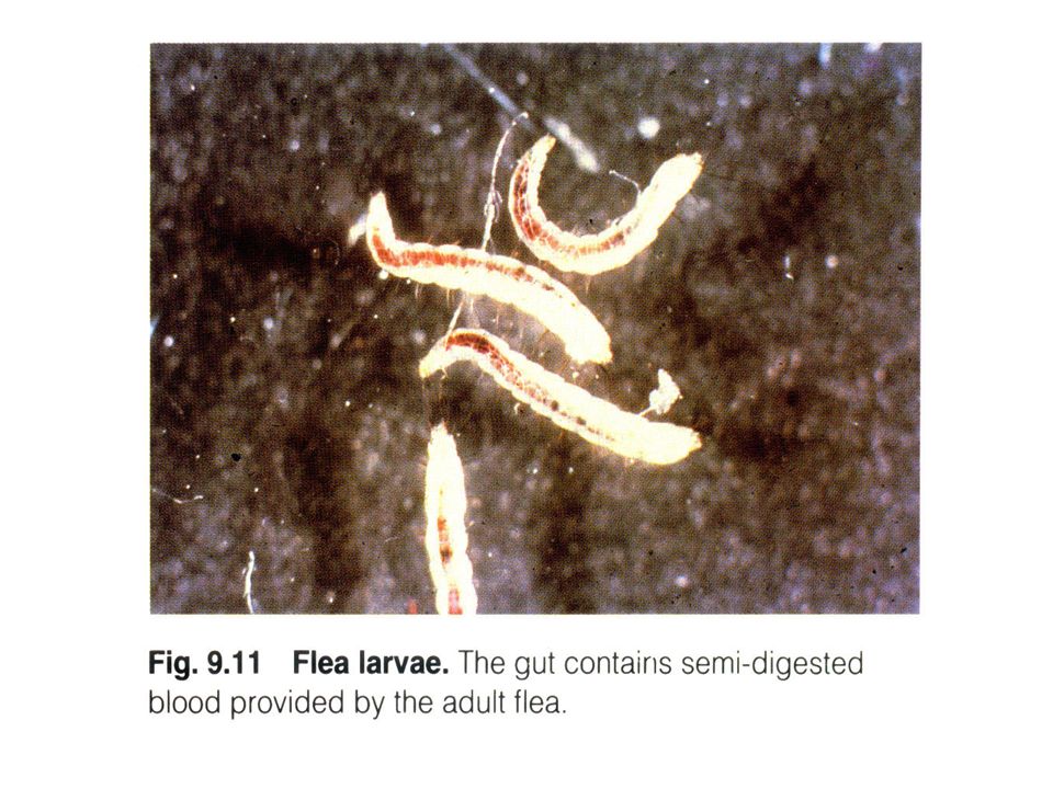
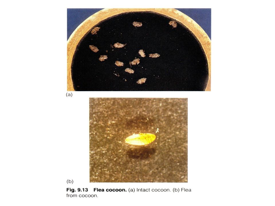
Flea Life Cycle (Ctenocephalides)
Adult life span: months Female produces avg. of eggs/day (up to 50) Egg production only after blood meal Incubation period: days Larval stage (needs humidity) feed on organic material and flea waste 2 molts, then the larva pupates (spins a cocoon) Lasts usually days Pupal stage Most resistant Lasts days to months dependent on environmental factors
 Egg production only after blood meal. Incubation period: 2-12 days. Larval stage (needs humidity) feed on organic material and flea waste. 2 molts, then the larva pupates (spins a cocoon) Lasts usually days. Pupal stage. Most resistant. Lasts days to months dependent on environmental factors.")
61
The Flea Fleas are very flat from side to
side. They have 3 pairs of legs. The most caudal pair is designed for jumping.

62
Flea Life Cycle

65
Fleas Ctenocephalides felis – The cat flea is the most commonly isolated flea from dogs and cats Flea saliva highly antigenic=> flea bite allergic dermatitis Clinical Signs Scratching/biting skin Alopecia: Dog - base of tail and caudal thighs initially then generalizes Cat- miliary dermatitis Flea dirt (flea feces) in hair coat Dx: Find fleas or flea dirt
 in hair coat. Dx: Find fleas or flea dirt.")
66
Miliary Dermatitis Common presentation of flea allergy dermatitis in cats

67
Flea allergy dermatitis
Common presentation of flea allergy dermatitis in the dog

68
Fleas Treatment Topicals: sprays, powders, dips, shampoos
Shampoos have no residual effect Systemic (spot-ons, injectable, tablets) Advantage (imidacloprid)- once a month spot-on; causes CNS paralysis and death of the flea Program: (Lufenuron -oral in dogs; injectable in cats); absorbed into fatty tissue and slowly distributed into bloodstream; Interferes with production of chitin (hard shell) Frontline (fipronil) - once a month spot-on; causes CNS paralysis or death Sentinel (Lufenuron + milbemycin) Once a month oral tablet (also heartworm preventive); inhibits chitin formation Revolution (Selamectin) – once a month spot-on; causes CNS paralysis and death; effective for heartworms, sarcoptes, ear mites, intestinal parasites in dogs and cats Comfortis (spinosad) - once a month ORAL tablet; causes CNS paralysis and death
 Advantage (imidacloprid)- once a month spot-on; causes CNS paralysis and death of the flea. Program: (Lufenuron -oral in dogs; injectable in cats); absorbed into fatty tissue and slowly distributed into bloodstream; Interferes with production of chitin (hard shell) Frontline (fipronil) - once a month spot-on; causes CNS paralysis or death. Sentinel (Lufenuron + milbemycin) Once a month oral tablet (also heartworm preventive); inhibits chitin formation. Revolution (Selamectin) – once a month spot-on; causes CNS paralysis and death; effective for heartworms, sarcoptes, ear mites, intestinal parasites in dogs and cats. Comfortis (spinosad) - once a month ORAL tablet; causes CNS paralysis and death.")
69
Fleas Client Info Treat environment and animal
Fleas spend most of time off host Foggers + vacuum well Prefer animals but will feed on humans All animals must be treated Skin damage/dermatitis can occur

70
Ticks: Brown Dog and Deer
Brown Dog Ticks: RMSF/Ehrlichia (Rhipicephalus sanguineus) A engorged female B female C male Deer Ticks: Lyme (Ixodes spp) D larvae E Nymphs F male G female H American Dog Tick (wood tick): RMSF (Dermacentor variabilis)
 A engorged female. B female. C male. Deer Ticks: Lyme. (Ixodes spp) D larvae. E Nymphs. F male. G female. H American Dog Tick (wood tick): RMSF. (Dermacentor variabilis)")
71
Hard ticks

72
Tick life cycle

73
Tick life cycle Female lays 1,000 to 10,000 eggs before dying

74
Ticks Irritation of the bite wound Vector for diseases
Neurotoxins in saliva of 12 different species Clinical signs vary with disease Dx: finding tick; hx of exposure to wooded and grassy areas

75
Brown Dog Tick The brown dog tick (Rhipicephalus sanguineus)
Transmits Ehrlichiosis in dogs Brown dog ticks are found throughout the world. Dogs are the primary host for the brown dog tick for each of its life stages.

76
Ticks Client Info Rx: Manually remove ticks
Grasp head parts close to skin with forceps (NOT HANDS) and pull backwards; no gasoline, cigarettes, etc. Topical treatments (sprays, shampoos, powder, dips) Collars Topical systemic products Client Info Routinely check pets for ticks, esp after walks in parks, etc Do not use bare hands to remove and kill ticks – blood may be infectious Ticks will feed on humans
 and pull backwards; no gasoline, cigarettes, etc. Topical treatments (sprays, shampoos, powder, dips) Collars. Topical systemic products. Client Info. Routinely check pets for ticks, esp after walks in parks, etc. Do not use bare hands to remove and kill ticks – blood may be infectious. Ticks will feed on humans.")
77
Tick paralysis Dermacentor variabilis
Neurotoxin in saliva of gravid female Dermacentor andersoni

78
The Mites Large variety of life styles among mites (free living vs parasitic), skin parasites vs respiratory parasites, etc. Life cycle involves egg, larva (6 legs), nymph and adult (8 legs).
, nymph and adult (8 legs).")
79
Burrowing Mites Family Sarcoptidae – Small, round, live in
skin tunnels. Short legs close to body. Cause mange. – Sarcoptes, Notoedres, Knemidocoptes Trixacarus Family Demodicidae – Live in hair follicles. – Adults cigar shaped. – Demodex

80
Mites (Demodectic mange)
")
81
Demodex Inhabits hair follicles, sebaceous glands or apocrine sweat glands D. canis Normal inhabitant in small numbers Some cats are asymptomatic carriers Spends entire life cycle on host Immune system controls infestation Genetic predisposition Localized or generalized Transmission Have not seen dog-to-dog or dog-to-human transmission

82
Demodex Demodex cati (right) Demodex canis (left)
 Demodex canis (left)")
83
Demodex D. cati D. gati - 2nd most common demodex mite on cat
Similar to D. canis – lives in hair follicles D. gati - 2nd most common demodex mite on cat Fat (broad, blunted abdomen) Lives more superficial in stratum corneum More pruritic Associated with immune suppression Similar lesions to dogs
 Lives more superficial in stratum corneum. More pruritic. Associated with immune suppression. Similar lesions to dogs.")
84
Demodex gati EPIDERMIS

85
DEMODEX

86
Demodex Clinical Signs Dx – skin scraping - deep Localized Generalized
Young dog (3m-1 y); Alopecia esp on face Erythema; crusty lesions Not pruritic unless secondary infections are present Generalized Often febrile Entire body surface involved Can result in protein loss through wounds Secondary bacterial infection - pustules Dx – skin scraping - deep
; Alopecia esp on face. Erythema; crusty lesions. Not pruritic unless secondary infections are present. Generalized. Often febrile. Entire body surface involved. Can result in protein loss through wounds. Secondary bacterial infection - pustules. Dx – skin scraping - deep.")
87
Demodex: Localized

88
Demodex: Generalized AUG

89
Demodex: Generalized

90
Demodex Tx Localized – Rotenone (Goodwinol) topical daily or mupiricin (Bactroban) Generalized Mitaban (amitraz) dips q 7 days x 3-6 treatments or 2 negative skin scrapings Side Effects – sedation for hrs (up to 72 hrs) Ivermectin: 0.3 mg/kg SQ repeat q 14 days OR PO SID x days extralabel use – client sign a release form – Not herding breeds Interceptor: 1/mo x 3 mo or more Oral antibiotics for secondary bacterial infections Client Info Many animals outgrow demodex as they age Not contagious to humans Tx does not completely remove mites If breeding dog, then do not treat and do not breed if does not clear on its own Strongly recommend OHE with treatment or will relapse with heat cycles Generalized form can be fatal.
 dips q 7 days x 3-6 treatments or 2 negative skin scrapings. Side Effects – sedation for hrs (up to 72 hrs) Ivermectin: 0.3 mg/kg SQ repeat q 14 days OR PO SID x days extralabel use – client sign a release form – Not herding breeds. Interceptor: 1/mo x 3 mo or more. Oral antibiotics for secondary bacterial infections. Client Info. Many animals outgrow demodex as they age. Not contagious to humans. Tx does not completely remove mites. If breeding dog, then do not treat and do not breed if does not clear on its own. Strongly recommend OHE with treatment or will relapse with heat cycles. Generalized form can be fatal.")
91
Demodex Rx Approved Not FDA approved Approved SQ or PO

92
Demodex Newer drugs and protocols Only APPROVED treatments
Ivermectin – lower initial dose due to side effects, then gradually increase dose over several days; µg/kg PO SID x 90 days SE: ataxia, bradycardia, mydriasis, resp arrest, salivation, stupor and tremors Milbemycin (Interceptor) – 2 mg/kg PO SID x days Fewer SE Expensive for large dogs Continue for 4 weeks post 2nd negative skin scraping – may be cost prohibitive moxidectin + Imidacloprid (Advantage Multi) Only APPROVED treatments Amitraz (Mitaban) Moxidectin + imidacloprid in topical formulation (Advantage Multi) Milbemycin oxime (Interceptor) orally
 – 2 mg/kg PO SID x days. Fewer SE. Expensive for large dogs. Continue for 4 weeks post 2nd negative skin scraping – may be cost prohibitive. moxidectin + Imidacloprid (Advantage Multi) Only APPROVED treatments. Amitraz (Mitaban) Moxidectin + imidacloprid in topical formulation (Advantage Multi) Milbemycin oxime (Interceptor) orally.")
93
Mites (Sarcoptic mange [Scabies])
![Mites (Sarcoptic mange [Scabies])](http://slideplayer.com/slide/6567389/23/images/93/Mites+%28Sarcoptic+mange+%5BScabies%5D%29.jpg "Mites (Sarcoptic mange [Scabies])")
94
Sarcoptic Mange Species specific Adults live 4-5 weeks
Egg-larva-nymph-adult cycle 17-21 days Entire life cycle on skin Infective in house environment for hrs Burrows under skin – stratum corneum (epidermis) Hypersensitivity reaction
 Hypersensitivity reaction.")
95
SARCOPTES

96
Sarcoptic mange Clincal Signs
Red crusty lesions on ears, elbows and trunk Intensely pruritic Progressively more severe DX – skin scraping; difficult to find mite Tx – easily killed Amitraz dip q 14 days Ivermectin SQ or PO q 14 days (extralabel use) Selamectin (revolution) – topically q 2 weeks x 3 treatments Client Info – highly contagious to other dogs and humans
 Selamectin (revolution) – topically q 2 weeks x 3 treatments. Client Info – highly contagious to other dogs and humans.")
97
Burrowing Mites Sarcoptes scabei

98
Scabies: Clinical signs

99
Sarcoptes Rx Approved Not FDA approved SQ or PO

100
Cheyletiellosis (Nonburrowing mites)
Cheyletiella spp

101
Cheyletiellosis “walking dandruff” Dogs, cats, rabbits, humans
Live in keratin layer of skin Feed on lymph Eggs deposited on hair shafts Eggs smaller than louse eggs Life cycle completed in 5-6 weeks Highly contagious among animals Direct contact and fomites

102
Cheyletiellosis Clinical Signs Severity of pruritis varies
Dry scales along back => entire body Patchy hair loss from scratching Dx: collect scales with clear tape, flea comb, skin scraping and ID with microscope Rx: Weekly baths with flea control products containing pyrethrins or permethrin and/or lime sulfur dip

103
Warbles (Cuterebra) Adult fly lays eggs in soil => larvae penetrates skin of animals=>matures=> leaves animal to become an adult fly Dx: swelling of skin behind ears with opening – can see larvae Tx: Surgery: open fistula and remove larvae Flush wound (betadine, Nolvasan) Oral antibiotics for skin infection Client Info: keep animals in fly-free environment
 Oral antibiotics for skin infection. Client Info: keep animals in fly-free environment.")
104
Warbles (Cuterebra)
")
105
Warbles (Cuterebra) Botfly, family cuterebra
 Botfly, family cuterebra")
106
“Education is a progressive discovery of our own ignorance.”
-Will Durant

107
Myiasis (Fly maggots)
")
108
Botfly ( Order Diptera)
")
109
Myiasis Many spp of flies lay eggs on wet, warm, damaged skin => larvae are maggots Dx: visually seen under matted hair with foul odor Rx: Clip hair Flush wound, clean daily Antibiotics Keep indoors to prevent re-infection Client info Disease of neglect More common in heavy coated animals in summer Do not use dips to remove maggots

110
Dog Lice (Linognathus setosus)
")
111
Dog Lice Rx Client Info Host specific; disease of neglect
Dx: lice cause intense itching Blood suckers => anemia if heavy infestation Presence of lice or nits or eggs diagnostic Rx Treat all animals in house with dip, shampoo or dust Topical insecticides used for fleas and ticks are adequate Wash bedding thoroughly Ivermectin orally (extra-label use) Client Info Humans don’t get lice from pets
 Client Info. Humans don’t get lice from pets.")
112
DOG LICE

113
Dermatophytes Microsporum canis

114
Dermatophytosis (Ringworm)
")
115
Dermatophytosis Superficial cutaneous infection
3 primary dermatophytes Microsporum canis Trichophyton mentagrophytes Microsporum gypseum Cat: 90% M. canis; Dog: all 3 Transmission Direct contact Contact with infected hair and scale Fomites Spores small and easily aerosolized Fleas Infected hairs – infective for up to 18 months Incubation period 1-3 weeks M. canis – cats Trichophyton – rodents or nests M. gypseum – geophilic (soil) More common in moist, warm environments
 More common in moist, warm environments.")
116
Dermatophytosis Clinical Signs Hair loss, scaling and crusting
+/- pruritus Cats Mimics other skin diseases Kittens – facial 1st Can form ulcerated dermal nodules Dog Focal or multifocal areas of hair loss Papules, scales and crusts Central area of hyperpigmentation

117
Dermatophytosis Dx: Wood’s Light – UV light Quick and easy screen
50% of M. canis fluoresce, rest do not Differentiate from scale, dust, dirt Lamp must warm up for 5 minutes prior to exam.

118
Wood’s lamp

119
Dermatophytosis Dx Tx Fungal culture – definitive
Color change to red in 1-3 weeks Confirm with microscopic exam Tx Usually self curing in healthy animal – may take 2-3 months, esp in kittens 3 elements Topical – reduces contamination on hair coat Systemic- reduces healing time Environmental-decreases contamination and spread

120
Microsporum canis

121
Trichophyton mentagrophytes

122
Microsporum gypseum

123
Dermatophyte Test Media - DTM

124
Dermatophytosis – Treatment
Clip haircoat, particularly long hairs Topicals Spot treatment may predispose to subclinical infections Whole body shampoos, dips, rinses twice weekly Lime-sulfur at 8oz/gal (4 oz not effective) Miconazole containing shampoos
 Miconazole containing shampoos.")
125
Dermatophytosis-Treatment
Systemic tx Griseofulvin (don’t use in pregnant animals: teratogenic effects) GI absorption variable Adverse effects Vomiting/diarrhea, anorexia Bone marrow suppression Neurologic signs Ketoconazole (Aplastic anemia in FIV + cats) Potential liver toxicity Intraconazole Less Side effects Lufenuron – inhibits chitin formation; chitin in outer wall of fungi Being studied Tx till resolution of signs and 2 negative cultures
 GI absorption variable. Adverse effects. Vomiting/diarrhea, anorexia. Bone marrow suppression. Neurologic signs. Ketoconazole (Aplastic anemia in FIV + cats) Potential liver toxicity. Intraconazole. Less Side effects. Lufenuron – inhibits chitin formation; chitin in outer wall of fungi. Being studied. Tx till resolution of signs and 2 negative cultures.")
126
Dermatophytosis – Treatment
Environment Remove or discard all bedding, brushes, fabric toys, etc Cheap vacuum to clean with, then discard Clean all surfaces; use bleach Vacuum daily Disinfect weekly Catteries – strict isolation

127
Dermatophytosis – Zoonosis
At risk populations: children, immune suppressed adults, older adults Vaccine – reduces severity but not occurrence of disease May not be available now Cats can be carriers w/o clinical signs

128
Ringworm (human)
")
129
Atopy (Atopic dermatitis)
Def: Allergy to inhaled environmental substances and manifested through irritation of skin and ears, usually starting at 1-2 yrs of age Allergens: Dust mites Pollens Feathers Molds Animal and human dander Tobacco smoke Clinical Signs: Pruritus Self-trauma to skin +/- secondary bacterial infection Staining of hair from saliva Licking/chewing feet Alopecia, scaling, hyperpigmentation

130
Atopy Dx Tx: No cure Accurate hx critical
R/o food allergies, flea allergy dermatitis, sarcoptic mange, contact dermatitis BEFORE diagnosis of atopy can be made Intradermal skin testing – most accurate to identify offending allergen Tx: No cure ID and eliminate cause best treatmetn ATOPICA – contains cyclosporin A (immunosuppressor), Anti-inflammatory and antipruritic Treat any bacterial or fungal infections before using immune suppressive drugs Medication (steroids, Abs) and allergy shots (desensitization) Omega 3 fatty acids Antihistamines-more effective if given before symptoms occur
, Anti-inflammatory and antipruritic. Treat any bacterial or fungal infections before using immune suppressive drugs. Medication (steroids, Abs) and allergy shots (desensitization) Omega 3 fatty acids. Antihistamines-more effective if given before symptoms occur.")
131
Atopy

132
Alopecia and erythema of the periocular skin and alopecia, erythema, and hyperpigmentation of the muzzle. Alopecia and hyperpigmentation of the muzzle.

133
Atopy: Appearance

134
Skin Testing Skin testing is performed to identify the allergens involved in allergic disease. Under profound sedation an area of hair on the chest is shaved and small injections of substances known to be possible allergens made. After minutes the reactions are recorded

135
Food Allergies 10% of all allergies Usually starts between 2-6 yrs
Processed foods increase likelihood of reaction Fillers, artificial colors, preservatives

136
Food allergies in dogs

137
Food Allergies Dx and Tx
Elimination diets: new source of protein for 3 mo. If improves, reintroduce original protein to see if symptoms recur – confirmation of dx Use new diet. May develop allergies to new diet later

138
Treatment

139
Food allergies

140
Treatment Foods can be obtained based on venision and potato, fish and potato, egg and rice, duck and pea, and even kangaroo. Generally recommends duck and potato based foods for dogs

141
How do you diagnose food intolerance (food allergy)?
The patient is fed a hypoallergenic diet for days. This allows the body to become desensitized to the offending allergens. When the previous diet is fed back to the pet, an acute hypersensitivity reaction may occur. This helps to identify that a food was the source of the allergic signs

142
Pruritits Algorithm Dogs Cats

143
Evaluating Feline Pruritus
Feline Food Allergy Year round pruritus Variable response to steroids 10-30% with concurrent GI symptoms Common offending proteins -Dairy, fish, beef, pork, chicken, rabbit, horse, lamb, eggs, clams, "commercial diets"

144
Managing Feline Pruritus
Diagnosing food allergy 10 week food trial Home cooked meat Potato/rice Canned food Innovative Veterinary Diets (IVD) Green pea & duck/rabbit Hills ZD???
 Green pea & duck/rabbit. Hills ZD")
145
Allergies

146
References Alleice Summers, Common Diseases of Companion Animals
Dr. Ralf S. Mueller, Dermatology for the Small Animal Practitioner

Similar presentations