Download presentation
Presentation is loading. Please wait.

2
1H15. Analyze the anatomy and physiology of the urinary system. Specific Objectives: 1H15.01Describe the structure of the urinary system. 1H15.02Analyze the function of the urinary system. 1H15.03Identify characteristics and treatment of common urinary disorders

3
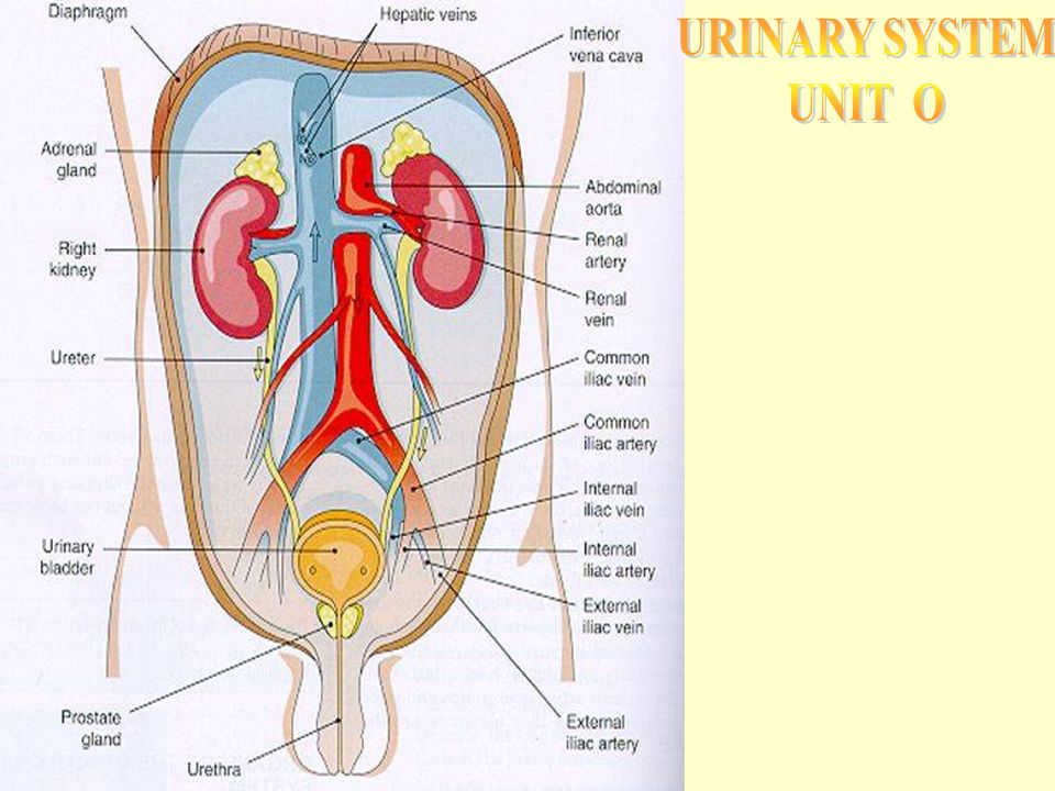
Kidney 1. Bean-shaped 2. Located between peritoneum and the back muscles (retroperitoneal) 3. Renal pelvis – funnel-shaped structure at the beginning of the ureter. 4. Medulla inner layer a. Inner, striated layer b. Striated cones are renal pyramids c. Base of pyramids empty into cuplike cavities called calyces 5.Cortex – composed of millions of microscopic nephrons.

4
Functions of Urinary System: 1. Excretion- removing nitrogenous wastes, (waste products) certain salts & excess water from blood. 2. Maintain acid-base balance 3. Secrete waste products in the form of urine. 4. Eliminate urine from bladder Toxic wastes would accumulate in the cells, and poison themselves if the kidneys were not functioning properly.
 certain salts & excess water from blood. 2. Maintain acid-base balance 3. Secrete waste products in the form of urine. 4. Eliminate urine from bladder Toxic wastes would accumulate in the cells, and poison themselves if the kidneys were not functioning properly..")
5
Nephron *Nephron –basic functional unit of the kidney. *Bowman’s capsule -double- walled capsule surrounding glomerulus. *Glomerulus-a cluster of capillaries that the Bowman’s capsule surrounds in the nephron that filters substances from the blood. *Proximal convoluted tubule *Loop of Henle *Distal convoluted tubule *Collecting tubule

6
Nephron Function Filtration 1. 1. First step in urine formation. 2. 2. Blood from renal artery enters glomerulus 3. 3. Blood pressure in glomerulus forces fluid (filtrate) to filter into Bowman’s capsule 4. 4. Filtrate does not contain plasma proteins or RBCs – they’re too big
 to filter into Bowman’s capsule Filtrate does not contain plasma proteins or RBCs – they’re too big.")
7
1. 1. Reabsorption-during the function of urine formation in the kidneys, useful substances filter out of the renal tubules & back into capillaries around tubules. a. a. Water (90%) and useful substances are reabsorbed b. b. If blood levels of certain substances are high (glucose, amino acids, vitamins, sodium) then those substances will NOT be reabsorbed
 and useful substances are reabsorbed b. b. If blood levels of certain substances are high (glucose, amino acids, vitamins, sodium) then those substances will NOT be reabsorbed.")
8
Pyramids The triangular-shaped divisions of the Medulla.

9
Path of urine formation! Glomerulus Bowman’s capsule Proximal convoluted tubule Loop of Henle Distal convoluted tubule Collecting tubule

10
Secretion Opposite of reabsorption Secretion transports substances from blood into collecting tubules Electrolytes are selectively secreted to maintain body’s acid- Base balance Ammonia is normally found in a urinalysis.

11
Urinary Output Average= 1500 ml/day Urinalysis- examination of urine to determine presence of blood cells, bacteria, acidity level, specific gravity and physical characteristics (color, clarity and odor)
")
12
Ureters The tubes connecting the kidneys & bladder. Carry urine from kidney to bladder Smooth muscle tube with mucous membrane lining Peristalsis pushes urine down ureters

13
Urethra The tube leading from the bladder to the outside of the body.

14
Urinary bladder Located in Pelvic cavity Stores urine – usually about 500 cc Elastic muscular organ that is capable of great expansion to store & aid in the elimination of urine. Emptying urine (voiding) is involuntary but controlled through nervous system (voluntary)
 is involuntary but controlled through nervous system (voluntary).")
15
Urinary Meatus The opening of the urethra to the outside of the body.

16
Control of Urinary Secretion Chemical control Production of urine controlled by ADH & Aldostrone hormones! Reabsorption of H2O in distal convoluted tubule controlled by ADH (antidiuretic hormone) Secretion and regulation of ADH controlled by hypothalamus Diuretics inhibit reabsorption of H2O
 Secretion and regulation of ADH controlled by hypothalamus Diuretics inhibit reabsorption of H2O.")
17
Nervous Control Direct Control through nerve impulses on kidney blood vessels. Indirect control through stimulation of endocrine glands.

18
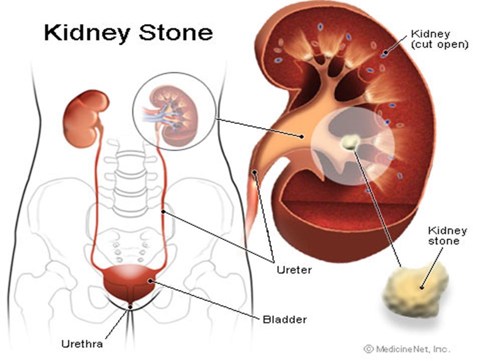
Renal calculi (kidney stones) Made of calcium and uric acid crystals Large stones can obstruct the flow of urine. Gradually they get larger until they block ureters First symptom – severe pain Other symptoms – nausea and vomiting, frequency, chills, fever, hematuria Diagnosis – by symptoms, ultrasound or x-ray Rx – increase fluids, medications, lithotripsy

20
Lithotripsy

21
Surgical procedure to remove kidney stones Shock waves hit dense stones and break them up. Done on outpatient basis

22
Nephritis Inflammation of the kidney (kidney infection)
")
23
Cystitis Bladder infection, most common cause is E. Coli bacteria! Symptoms – dysuria (painful urination) and frequency More often in females (shorter urethra) Rx - antibiotics
 and frequency More often in females (shorter urethra) Rx - antibiotics.")
24
Kidney Failure A early sign of acute kidney failure is Oliguria (absent or decreased production of urine)
")
25
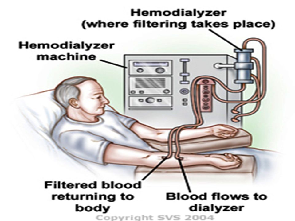
Dialysis (Hemodialysis) Used for kidney failure Involves the passage of blood through device with semipermeable membrane. Dialysis serves as substitute kidney Blood from patient flows through machine and if filtered. Can be done at home or in clinic Takes 2-4 hours, 2-3 times a week

27
Kidney Transplant As a last resort Involves donor organ from someone with a similar immune system Main complication- rejection

28
Incontinence (Involuntary urination)
")
29
Terminology Enuresis- bedwetting-from years 0-3! Gylcosuria- sugar in urine Nocturia- frequent urination at night Polyuria- large amounts of urine Anuria- no urine produced Hematuria- blood in urine Diuretic- drug or substance to increase urine production

Similar presentations


 Ca+, K+, Na+ -Regulate acid/base.>")