Download presentation
Presentation is loading. Please wait.

1
Eye care basics and optical options
Rick Smith & Sam Powdrill

2
Eye Anatomy

3
Visual Image

4
Components of Vision

5
The External Eye

6
The Skull

7
Bones of the Orbit Frontal Zygoma Ethmoid Maxilla Sphenoid

8
The Lacrimal System The tear film Lipid layer Aqueous layer
Mucus layer

9
The Eyelids

10
Vessels and Muscles of the Eyelids

11
Extrinsic Eye Muscles

12
Extrinsic muscles with innervation

13
The Globe Anterior chamber aqueous Iris Lens
Posterior chamber vitreous Optic nerve

14
Layers of the eye Protective Vascular Visual

15
Protective Layer Sclera Cornea

16
Vascular layer Iris Ciliary body Choroid

17
Angle of the Eye

18
Visual Layer The retina Optic nerve

19
Central Retina

20
Optic Nerve (II) Visual pathway
 Visual pathway")
21
Optic Nerve (II) – Visual pathway
 – Visual pathway")
22
Refraction

23
Prism

24
Converging

25
diverging

26
Lens Power Measured in diopters One diopter lens focuses at 1 meter
Lens power = cm focal length focal length = cm Lens power

27
Refractive errors Nearsighted – myopia Farsighted – hyperopia
Astigmatism Presbyopia

28
Refraction Convex lens Concave lens Accommodation Refractive error

29
Correction of Refractive Errors
Pinhole Myopia Hypermetropia Astigmatism

30
Exam of the Eye

31
Eye Exam

32
Pathways of Blindness Corneal Anterior chamber Lens Vitreous Retina
Optic nerve Occipital Functional

33
Instruments needed to examine the eye
Visual acuity chart Flashlight Ophthalmoscope Tonometer Simple loupe

34
The vital sign of the eye
Measure Visual acuity The vital sign of the eye Distance vision Near vision Peripheral vision Central vision Color vision

35
Snellen charts

36
Pinhole eliminates the refractive error

37
Visual acuity Cover the eye with the palm of the hand
Test the eyes separately then together Point to the letters clearly Use a random sequence

38
Recording Visual acuity
20/20 Upper number = distance in feet from the patient to the chart Lower number = distance in feet at which a person with excellent vision would see the same letter

39
Examples 20/40 20 /400 20/15 10/50 CF 10ft HM LP NLP

40
Central vision Test for loss of central vision - macula Amsler grid
Abnormal in macular degeneration

41
Amsler grid

42
Color vision Check for red / green color blindness Ishihara chart
Color comparison method with a red object Color blindness is an X linked genetic disorder that most often affects males.

43
Ishihara chart

44
Tonometry Schiotz Applanation Air puff tonopen Normal is 10 to 20 mmHg

45
Extraocular Muscles Six Cardinal eye movements
Corneal light reflex position Nystagmus Cover - uncover test Convergence

47
Testing the visual field
Peripheral vision Testing the visual field Confrontation method – compares the examiner’s visual field with the patient’s Automated – done in optometrist’s or ophthalmologist’s office Abnormal in glaucoma

48
Visual fields by confrontation

49
Eyelids Inspect Symmetry – skin creases Opening and closing completely
Eyelash position Ptosis Tremor or fasciculations Swelling or infection Flakiness

50
cataract Ophthlmoscope set on +4 Shadows against Red reflex

51
Direct light vs Coaxial light on the lens

52
Eyelids Palpate Eyelids for nodules Intraocular pressure
Evert upper and lower lids and inspect Color Discharge Lacrimal system

53
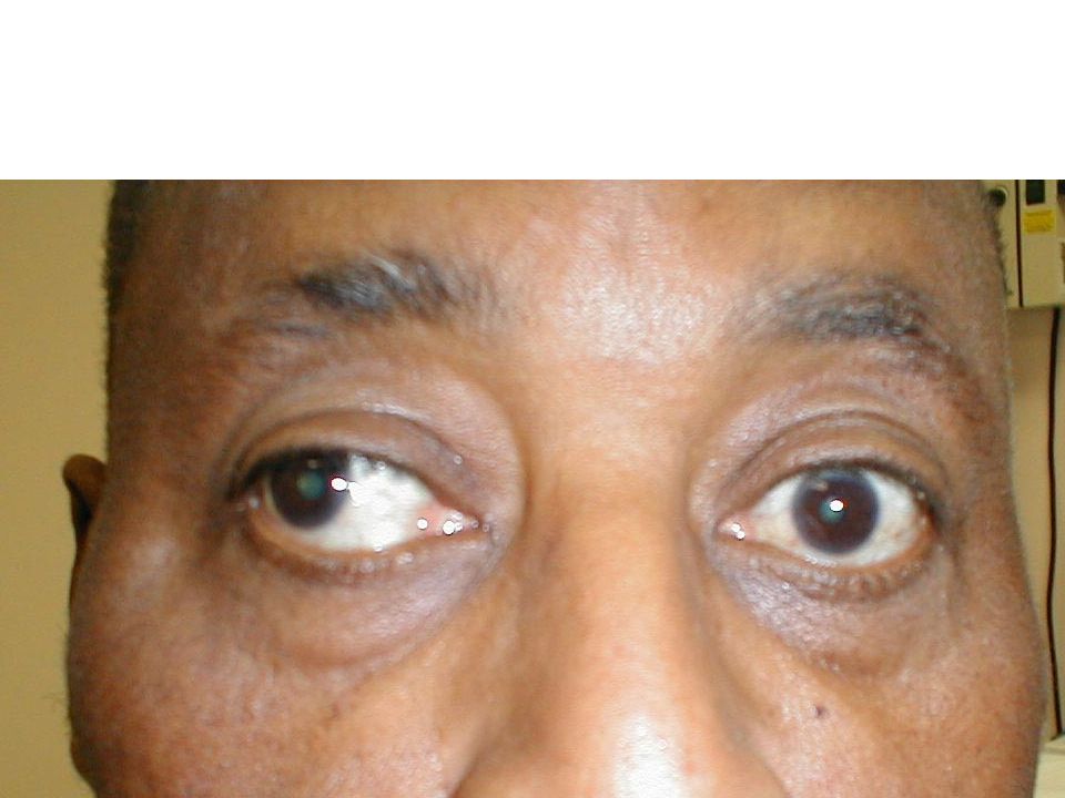
Conjunctiva and Sclera
Inspect Redness Vessels Growths or lesions, foreign bodies Proptosis enophthalmos

54
Cornea Inspect Clarity – scars obscure part of the iris.
Light reflex – reflex is scattered in edema Corneal sensitivity Arcus Depth of anterior chamber – iris shadow Abrasions or foreign bodies – use fluorescein stain with blue light to examine the cornea for defects

55
Iris and pupil Inspect Iris Color Angle lesions Inspect Pupil Size
Shape Reaction to light Reaction to near Consensual response PERRLA

56
Ophthalmoscope exam Lens clarity – lens opacity is a cataract
Red reflex – ophthlmoscope set on + 4 Vitreous clarity – floaters, sclerotic bands Retinal color and background Retinal vessels – hemhorrage, cotton wool spots, AV nicking, neovascularization, Optic disc – cup/disc ratio Optic nerve head – papilledema, cupping or atrophy Macula for drusen or changes

57
Retina

58
Retina

59
Retina

60
Retina

Similar presentations


 separated by the palpebral fissue Eyelashes Tarsal glands Lacrimal apparatus Vision Accessory structures.>")


 than.>")


 What are the 5 senses? 2.) T/F Of all the sensory receptors in the body, 70% are in the eyes. 3.) T/F Your eyebrows.>")
