Download presentation
Presentation is loading. Please wait.

1
LEPROSY

3
EPIDEMIOLOGY 8 MILLION PATIENTS 60,000 NEW CASES EACH YEAR WORLD WIDE
LEPROSY IS ENDEMIC IN ASIA & AFRICA APPOX. 80% CASES IN THESE AREAS IN THE WORLD

4
Global Leprosy Situation in 2005
WHO Region Point Prevalence Africa Americas East Mediterranean South East Asia Western Pacific World

5
PERVALENCE OF LEPROSY IN SEA REGION WHO 2003

6
DEFINITION LEPROSY IS A NONFATAL ,CHRONIC INFECTIOUS DISEASE CAUSED BY MYCOBACTRIUM LEPRAE (By G.A.Hansen 1873) INVOLVING - SKIN PERIPHERAL NERVOUS SYSTEM UPPER RESPIRATORY TRACT EYES TESTES M.LEPRAE HAS UNIQE TROPISM FOR PEIPHRAL NERVES REACTIONAL STATE RESPONSIBLE FOR MORBIDITY & DISEASE IF NOT TREATED LEADS TO CHARECTERSTIC DEFORMITY & PROFOUND SOCIAL STIGMA

7
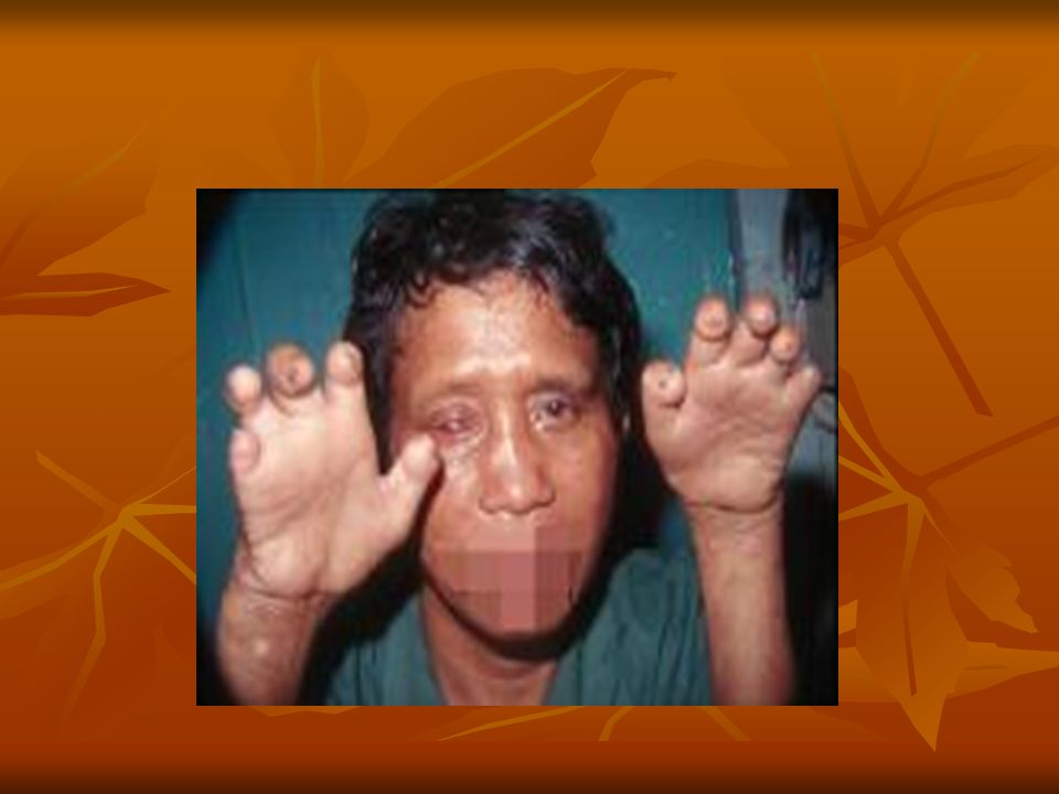
DEFORMITY

8
M. LEPRAE NONCULTIVABLE IN MEDIUM
FACULTATIVE OBLIAGTE INTRACELLULAR ORGANISM GRAM-POSITIVE ACID-FAST BACILUS PEPTIDOGLYCAN-BACK BONE ARABINOGALACTAN & MYCOLIC ACID PHENOLIC GLYCOLIPD-1 (PGL-1) ATTACHED TO LAMININ 2 OF SCHWANN CELLS MHC CLASS II FOR DISEASE EXPRESSION NOT FOR SUSCEPTIBILITY OF DISEASE SUSCEPTIBILTY GENE LOCUS ON CHROMOSOME-10P13
 ATTACHED TO LAMININ 2 OF SCHWANN CELLS. MHC CLASS II FOR DISEASE EXPRESSION NOT FOR SUSCEPTIBILITY OF DISEASE. SUSCEPTIBILTY GENE LOCUS ON CHROMOSOME-10P13.")
9
The route of transmission
Not been definitively established, Although human-to-human aerosol spread of nasal secretions is the most likely mode of transmission in most cases. The disease is not spread by touch, since the mycobacteria are incapable of crossing intact skin. Living near people with leprosy is associated with increased transmission. Among household contacts, the relative risk for leprosy is increased 8- to 10-fold in multibacillary and 2- to 4-fold in paucibacillary forms. Animal reservoirs do exist (armadillos, certain nonhuman primates), and cases of suspected zoonotic transmission have been reported.
, and cases of suspected zoonotic transmission have been reported.")
10
Two indices which depend on observation of M
Two indices which depend on observation of M. leprae in smears from skin or nasal smears are useful in assessing the amount of infection, the viability of the organisms the progress of the patient under treatment. They are - the morphological index (MI) the bacteriological index (BI).
 the bacteriological index (BI).")
11
1. The bacteriological index (BI)
This is an expression of the extent of bacterial loads. It is calculated by counting six to eight stained smears under the 100 x oil immersion lens. A smear stained by the Ziehl-Neelsen method and decolorized (but not completely) which 1% acid alcohol. The results are expressed on a logarithmic scale.
 which 1% acid alcohol. The results are expressed on a logarithmic scale.")
12
BACTERIOLOGICAL INDEX
LOGAITHMIC SCALE AS TO NUMBER OF BACILLI PER OIL IMMERSION FIELD(OIF) BI OF 6 IS 1000 OR MORE BACILLI/OIF BI OF 5 IS 100 TO 1000/OIF BI OF 4 IS 10 TO 100 BACILLI/OIF BI OF 3 IS 1 TO BACILLI/OIF BI OF 2 IS 1 BACILLUS/1 TO 10 OIFS BI OF 1 IS 1 BACILLUS/ 10 TO 100 OIFS BI OF 0 IS NO BACILLUS PER 100 OIFS
 BI OF 6 IS 1000 OR MORE BACILLI/OIF. BI OF 5 IS 100 TO 1000/OIF. BI OF 4 IS 10 TO 100 BACILLI/OIF. BI OF 3 IS 1 TO 10 BACILLI/OIF. BI OF 2 IS 1 BACILLUS/1 TO 10 OIFS. BI OF 1 IS 1 BACILLUS/ 10 TO 100 OIFS. BI OF 0 IS NO BACILLUS PER 100 OIFS.")
13
MORPHOLOGICAL INDEX PERCENTAGE OF SOLIDLY STAINED BACILLI IN STAINED SMEAR Only the solid-staining bacilli are viable. It is not unusual for solid-staining M. leprae to reappear for short periods in patients being successfully treated with drugs. It is important to recognize that measurement of MI is liable for observer variations and therefore not always reliable.

14
IS LEPROSY HOST IMMUNE DEPENDENT DISEASE?
YES…. CMI OR ANTIBODY MEDIATED ?

15
CLINICAL PRESENTATION
LEPROSY HAS SPECTRUM OF DISEASE Ridley-Jopling classification system TT BT BB BL LL INDICIES FOR SPECTRUM BACTERIOLOGICAL IMMUNOLOGICAL CLINICAL HISTOPATHOLOGICAL

16
WHO Classification system:
The WHO recommends classifying leprosy according to the number of lesions and the presence of bacilli on a skin smear. . Paucibacillary (PB) leprosy is characterized by 5 or fewer lesions with absence of organisms on smear. Includes the tuberculoid and borderline tuberculoid leprosy categories from the Ridley-Jopling system. Multibacillary (MB) leprosy is marked by 6 or more lesions with possible visualization of bacilli on smear. Includes Lepromatous, borderline lepromatous, and midborderline on the Ridley-Jopling scale .
 leprosy is characterized by 5 or fewer lesions with absence of organisms on smear. Includes the tuberculoid and borderline tuberculoid leprosy categories from the Ridley-Jopling system. Multibacillary (MB) leprosy is marked by 6 or more lesions with possible visualization of bacilli on smear. Includes Lepromatous, borderline lepromatous, and midborderline on the Ridley-Jopling scale .")
17
The cardinal signs of leprosy
Hypoesthesia, skin lesions, loss of hair and sweating & peripheral neuropathy. The first physical signs of leprosy are usually cutaneous. The subtype of leprosy often determines the degree of skin involvement.

18
Physical examination Evaluation of skin lesions
Careful sensory and motor examination Palpation of peripheral nerves for pain or enlargement. Particular attention should be paid to the following locations: Elbows - Ulnar nerve Wrist - Superficial radial cutaneous and median nerves Popliteal fossa - Common peroneal nerve Neck - Great auricular nerve

19
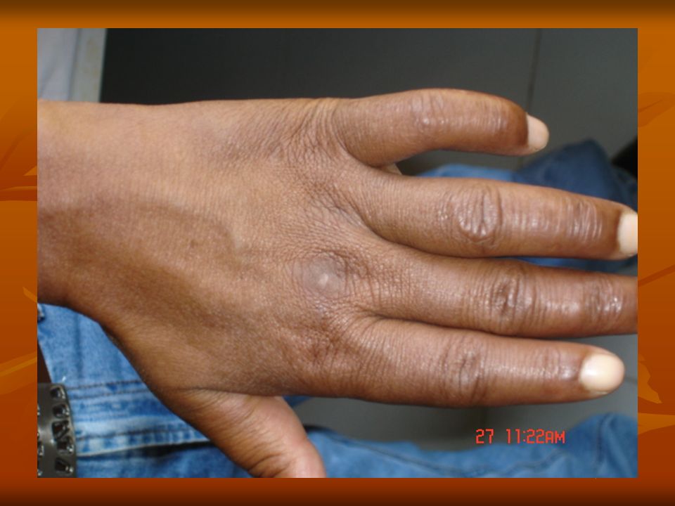
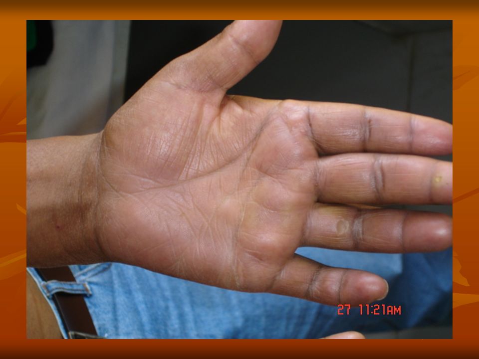
TUBERCULOID LEPROSY Single or few Lesions Erythematous plaques
Hypopigmanted Hypoasthatic Macular lesions Cutaneous nerve thickened

20
TUBERCULOID LEPROSY

21
TUBERCULOID LEPROSY

22
TUBERCULOID LEPROSY

23
BORDERLINE LEPROSY

24
BORDERLINE LEPROSY

25
POSTERIOR AURICULAR NERVE THICKNING

26
LEPROMATOUS LEPROSY

27
LEPROMATOUS LEPROSY

28
LEPROMATOUS LEPROSY

29
POSTERIOR AURICULR NERVE

30
NODULR LESION AT PINNA

31
NODULAR LESION AT PINNA

32
DIFFUSE INFILTRATION IN LEPROMATOUS LEPROSY

33
DIFFUSE INFILTRATION IN LEPROMATOUS LEPROSY

34
BORDERLINE LEPROMATOUS LEPROSY

35
LEFT ULNER NERVE PALSY

38
ANESTHATIC HAND

39
SKIN SMEAR FROM LESION SHOWS AFB++

40
HISTPATHOLOGY

41
Immunologic tests Lepromin skin test
Not diagnostic of exposure or infection with M leprae Assesses a patient's ability to mount a granulomatous response against a skin injection of killed M leprae. Patients with tuberculoid or borderline lepromatous leprosy typically have a positive response (>5 mm). Patients with lepromatous leprosy typically have no response.
. Patients with lepromatous leprosy typically have no response.")
42
PCR and recombinant DNA technology
Detection of antibodies to phenolic glycolipid-1 (PGL-1) This is a specific serologic test. This test has a sensitivity of 95% for the detection of lepromatous disease but only 30% for tuberculoid disease. PCR and recombinant DNA technology development of gene probes with M leprae–specific sequences. This technology can be used to identify the mycobacterium in biopsy samples, skin and nasal smears, and blood and tissue sections. Lymphocyte migration inhibition test (LMIT): As determined by a lymphocyte transformation and LMIT, cell-mediated immunity to M leprae is absent in the lepromatous form of disease but present in the tuberculoid form of disease. Contact or family screening for history of leprosy
 This is a specific serologic test. This test has a sensitivity of 95% for the detection of lepromatous disease but only 30% for tuberculoid disease. PCR and recombinant DNA technology. development of gene probes with M leprae–specific sequences. This technology can be used to identify the mycobacterium in biopsy samples, skin and nasal smears, and blood and tissue sections. Lymphocyte migration inhibition test (LMIT): As determined by a lymphocyte transformation and LMIT, cell-mediated immunity to M leprae is absent in the lepromatous form of disease but present in the tuberculoid form of disease. Contact or family screening for history of leprosy.")
43
REACTIONIONAL SATES IN LEPROSY

44
TYPE -1 REACTION OCCURS IN BORDERLINE CASES NOT IN POLAR LEPROSY
ACTIVATION OF PREVIOUSLY INVOLVED SKIN LESIONS PANFUL & TENDER NERVES DOWNGREADING REACTION - WHEN OCCURS BEFORE INITIATON OF CHEMOTHERAPY REVERSAL REACTION AFTER INITIATION OF CHEMOTHERAPY- TH1 RESPONSES WITH IFN-GAMMA,IL2 CORTICOSTEROID TREATMENT OF CHOICE

45
TYPE -1 REACTION

46
TYPE -2 REACTION OCCURS IN BL/LL FORM OF LEPROSY IN 50% OF CASES
IN 90% OF CASES MAY BE PRESENTING SYMPTOM OF DISEASE ENL- ERYTHEMA NODOSUM LEPROSUM FEVER ARTHRITIS UVEITIS ORCHITIS GLOMERULONEPHRITIS TNF- PLAYS CETERAL ROLE, IMMUNE COMPLEX DEPOSITION TH2 CYTOKINE PROFIL-IL6,IL8 THALIDOMIDE IS CHOICE OF TREATMENT

47
ERYTHEMA NODOSUM LEPROSUM

48
Erythema Nodosum

49
TREATMENT-MDT MULTI DRUG THERAPY

50
What is WHO MDT? Multi drug therapy (MDT) is a key element of the elimination strategy. MDT is available free of charge from WHO The drugs used in WHO-MDT are a combination of Rifampicin, clofazimine and Dapsone for MB leprosy patients and Rifampicin and Dapsone for PB leprosy patients. Treatment of leprosy with only one anti leprosy drug will always result in development of drug resistance. Treatment with Dapsone or any other anti leprosy drug used as monotherapy should be considered as unethical practice.

51
MULTI DRUG THERAPY Yes, it is the best combination available today, as proved by its successful application in leprosy control under varying conditions since 1982. The combination not only cures leprosy but is also highly cost-effective.

52
TREATMENT REGIMENS WHO
TUBERCULOID- PAUCIBACILLARY DAPSONE 100 mg/D UNSUPERVISED + RIFAMPIN 600mg/MONTH SUPERVISED FOR 6 MONTH MULTIBACILLARY DISEASE DAPSONE 100mg /D PLUS CLOFAZIMINE 50mg/D UNSUPERVISED PLUS RIFIAMPIN 600mg + CLOFAZIMNE 300mg MONTHLY SUPERVISED FOR 1 YEAR

53
Physiotherapy Reconstructive surgeries Rehabilitation programes Health education

54
THANK YOU…….

57
MDT Rifampicin: The drug is given once a month. No toxic effects have been reported in the case of monthly administration. The urine may be coloured slightly reddish for a few hours after its intake, this should be explained to the patient while starting MDT. Clofazimine: It is most active when administered daily. The drug is well tolerated and virtually non-toxic in the dosage used for MDT. The drug causes brownish black discoloration and dryness of skin. However, this disappears within few months after stopping treatment. This should be explained to patients starting MDT regimen for MB leprosy. Dapsone: The drug is very safe in the dosage used in MDT and side effects are rare. The main side effect is allergic reaction, causing itchy skin rashes and exfoliative dermatitis. Patients known to be allergic to any of the sulpha drugs should not be given dapsone.

Similar presentations


 is caused by the acid- fast bacillus Mycobacterium leprae.Unlike other mycobacteria, it does not grow in artificial.>")


>")


