Download presentation
Presentation is loading. Please wait.

1
Shujun Li Department of Forest Products Chemical Processing
WOOD CHEMISTRY Shujun Li Department of Forest Products Chemical Processing

2
Chapter 1 THE STRUCTURE OF WOOD
Trees belong to seed-bearing plants which are subdivided into gymnosperms (Gymnospermae) and angiosperms ( Angiospermae). Coniferous woods or softwoods belong to the first-mentioned category and hardwoods to the second group.
 and angiosperms ( Angiospermae). Coniferous woods or softwoods belong to the first-mentioned category and hardwoods to the second group.")
4
Chapter 1 THE STRUCTURE OF WOOD
Altogether 30,000 angiosperms and 520 coniferous tree species are known; most of the former grow in tropical forests. In North America the number of species is about 1200, while in Europe only 10 softwood and 51 hardwood species exist naturally. This limited number represents species surviving the period of glaciation, during which genera such as Sequoia and Pseudotsuga completely disappeared from Europe.

5
Chapter 1 THE STRUCTURE OF WOOD
1.1 The Macroscopic Structure of Wood Wood is composed of elongated cells, most of which are oriented in the longitudinal direction of the stem. They are connected with each other through openings, referred to as pits. These cells, varying in their shape according to their functions, provide the necessary mechanical strength to the tree and also perform the function of liquid transport as well as the storage of reserve food supplies.

7
Chapter 1 THE STRUCTURE OF WOOD
Figure 1-1 shows the macroscopic structure of wood as it appears to the naked eye. The centrally located pith is discernible as a dark stripe in the middle of the stem or branches. It represents the tissues formed during the first year of growth. The xylem or wood is organized in concentric growth rings (annual increments). It also contains rays in horizontal files, extending from the outer bark either to the pith(primary rays) or to an annual ring(secondary rays). Some softwoods also contain resin canals.
. It also contains rays in horizontal files, extending from the outer bark either to the pith(primary rays) or to an annual ring(secondary rays). Some softwoods also contain resin canals.")
8
Chapter 1 THE STRUCTURE OF WOOD
The inner part of a tree usually consists of dark-colored heartwood. The outer part, or sapwood, is lighter in color and conducts water from the roots to the foliage of the tree. The cambial zone is a very thin layer consisting of living cells between the wood (xylem) and the inner bark (phloem). The cell division and radial growth of the tree takes place in this region.
 and the inner bark (phloem). The cell division and radial growth of the tree takes place in this region.")
9
Chapter 1 THE STRUCTURE OF WOOD
1.2 The Living Tree The Growth of the Tree The tree grows through the division of the cells. The length of the growth period largely depends on the climate, but in many parts of North America and Scandinavia growth occurs from May to early September, and is most intensive in the spring. The majority of the cells develop into various permanent cells and only a very few are retained as growing cells capable of division.

10
Chapter 1 THE STRUCTURE OF WOOD
The growth of a tree is always continuous although it becomes slower in the course of time. Giant sequoias in California can be up to 4000 years old measuring 100 meters in height and 12 meters in diameter at the base. Longitudinal growth (primary growth), which takes place in the early season, proceeds at the end of the stem, branches and roots. The growth points are located inside the buds, which have been formed during the preceding autumn.
, which takes place in the early season, proceeds at the end of the stem, branches and roots. The growth points are located inside the buds, which have been formed during the preceding autumn.")
11
Chapter 1 THE STRUCTURE OF WOOD
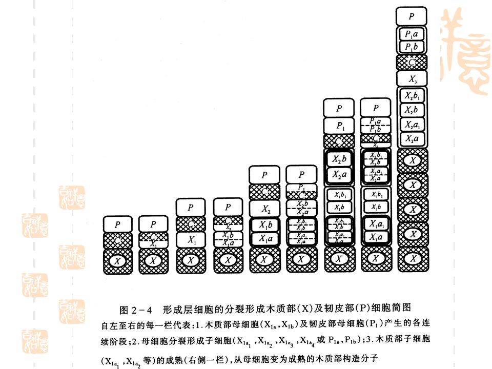
Radial growth begins in the cambium which is composed of a single layer of thin-walled living cells (initials) filled with protoplasm. The cambial zone consists of several rows of cells, which all possess the ability to divide. On division the initial cell produces a new initial and a xylem mother cell, which in its turn gives rise to two daughter cells; each of the latter is capable of further division. More cells are produced toward the xylem on the inside than toward the phloem on the outside; phloem cells divide less frequently than xylem cells. For these reasons, trees always contain much more wood than bark.
 filled with protoplasm. The cambial zone consists of several rows of cells, which all possess the ability to divide. On division the initial cell produces a new initial and a xylem mother cell, which in its turn gives rise to two daughter cells; each of the latter is capable of further division. More cells are produced toward the xylem on the inside than toward the phloem on the outside; phloem cells divide less frequently than xylem cells. For these reasons, trees always contain much more wood than bark.")
13
Chapter 1 THE STRUCTURE OF WOOD
1.2.2 Development of the cell When a cell divides, it first develops a cell plate, which is rich in pectic substances. Each of the two new cells subsequently encloses itself with a thin, extensible, primary wall, consisting of cellulose, hemicelluloses, pectin, and protein. During the following phase of differentiation, the cell first expands to its full final size, after which formation of the thick, secondary wall is initiated. At this stage, this wall consists of cellulose and hemicelluloses. Lignification begins while the secondary wall is still being formed. Figs.1-3 and 1-12 show the structures of a mature cell.

14
Fig.1-3. Development of the living cell to wood fiber(P34)
1. LIVING CELL; 2. NUCLEUS; 3. CYTOPLASM; 4. PRIMARY WALL; 5. GROWTH AND LIGNIFICATION OF THE CELL; 6. DEAD CELL; 7. LUMEN; 8. INNER LAYER S3; 9. MIDDLE LAYER S2; 10. OUTER LAYER S1; 11. SECONDARY WALL; 12. PRIMIARY WALL P; 13. WOOD FIBER

15
Chapter 1 THE STRUCTURE OF WOOD
Annual Rings At the beginning of the growth the tree requires an effective water transportation system. In softwoods thin-walled cells with large cavities are formed; in hardwoods special vessels take care of the liquid transportation. Comparatively light-colored and porous earlywood is thus formed. Later, the rate of growth decreases and latewood is produced. It consists of thick-walled fibers and gives mechanical strength to the stem and is darker and denser than the earlywood.

16
Chapter 1 THE STRUCTURE OF WOOD
The age of a tree can be calculated from the number of growth rings at the base of the stem. With a continuous growth period (tropical woods) regular annual rings are lacking. Alternation of wet and dry periods may, however, result in the formation of growth rings. The boundary between earlywood and latewood varies. It may be very sharp as in larch or nearly nonexisting (birch, aspen, and alder). Earlywood is weaker than the thick-walled latewood. Pulp fibers from earlywood and latewood also have different papermaking properties.
 regular annual rings are lacking. Alternation of wet and dry periods may, however, result in the formation of growth rings. The boundary between earlywood and latewood varies. It may be very sharp as in larch or nearly nonexisting (birch, aspen, and alder). Earlywood is weaker than the thick-walled latewood. Pulp fibers from earlywood and latewood also have different papermaking properties.")
18
Chapter 1 THE STRUCTURE OF WOOD
The width of the annual rings varies greatly depending on tree species and growth conditions. The variation limits for Scots pine in Scandinavia may be mm. For similar reasons the proportion of latewood may vary greatly. Typical percentages for the latewood in Scandinavia are 15-50% for pine and 10-40% for spruce; the values are higher in the northern than in the southern parts of these countries.

19
Chapter 1 THE STRUCTURE OF WOOD
1.2.4 Cell Types On the basis of their different shape wood cells can be divided into prosenchyma and parenchyma cells. The former are thin, long cells, narrower toward the ends; the latter are rectangular or round and are short cells. Depending on their functions, cells can be divided into three different groups: conducting cells, supporting cells, and storage cells.

20
Chapter 1 THE STRUCTURE OF WOOD
Conducting and supporting cells are dead cells containing cavities which are filled with water or air. In hardwoods the conducting cells consist of vessels and the supporting cells of fibers. In softwoods the tracheids perform both functions. The storage cells transport and store nutrients. They are thin-walled parenchyma cells which function as long as they remain in the sapwood.

21
Chapter 1 THE STRUCTURE OF WOOD
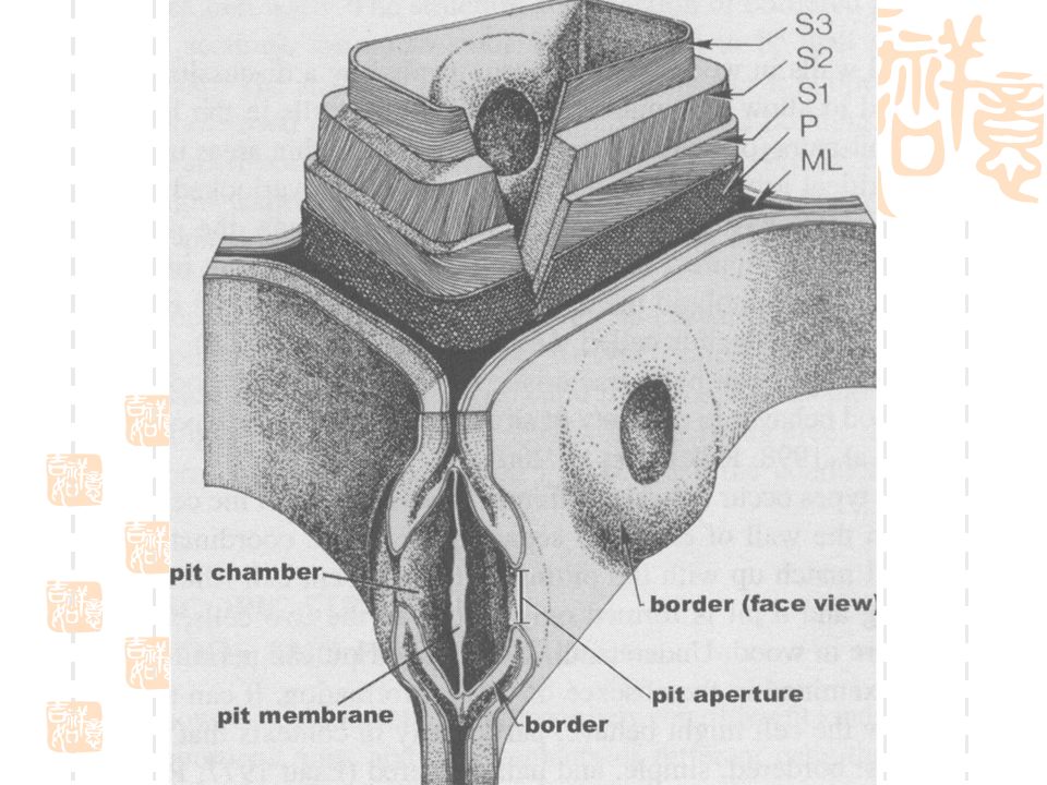
pits (P35) Water conduction in a tree is made possible by pits, which are recesses in the secondary wall between adjacent cells. Two complementary pits normally occur in neighboring cells thus forming a pit pair (Fig. 1-5). Water transport between adjacent cell lumina occurs through a pit membrane which consists of a primary wall and the middle lamella.
 Water conduction in a tree is made possible by pits, which are recesses in the secondary wall between adjacent cells. Two complementary pits normally occur in neighboring cells thus forming a pit pair (Fig. 1-5). Water transport between adjacent cell lumina occurs through a pit membrane which consists of a primary wall and the middle lamella.")
22
Fig.1-5 Types of pit pairs. A, bordered pit pair; B, half bordered pit pair; C, simple pit pair; M, middle lamella; P, primary wall; S, secondary wall.

24
Chapter 1 THE STRUCTURE OF WOOD
Bordered pit pairs are typical of softwood tracheids and hardwood fibers and vessels. In softwoods the pit membrane might be pressed against the pit border thus preventing water transport, since the torus is impermeable. The pits connecting tracheids, fibers and vessels with the ray parenchyma cells are half-bordered. Simple pits without any border connect the parenchyma cells with one another.

25
Chapter 1 THE STRUCTURE OF WOOD
The different shape of the pits are distinctive features in the microscopic identification of wood and fibers. Knowledge of the porous structure of wood is also of great importance for understanding the phenomena which are associated with the impregnation of wood.

26
Chapter 1 THE STRUCTURE OF WOOD
Softwood cells The wood substance in softwoods is composed of two different cells: tracheids (90-95%) and ray cells (5-10%). Tracheids give softwoods the mechanical strength required (especially the thick-walled latewood tracheids ) and provide for water transport, which occurs through the thin-walled early wood tracheids with their large cavities.
 and ray cells (5-10%). Tracheids give softwoods the mechanical strength required (especially the thick-walled latewood tracheids ) and provide for water transport, which occurs through the thin-walled early wood tracheids with their large cavities.")
27
Chapter 1 THE STRUCTURE OF WOOD
The liquid transport from one tracheid to another takes place through the bordered pits, their amount in earlywood tracheids is about 200 per tracheid, most of them located in the radial walls in one to four lines. Latewood tracheids have only 10 to 50 rather small bordered pits.

28
Fig. 1-6. Cells of coniferous woods
Fig Cells of coniferous woods. An earlywood (a) and a latewood (b) pine tracheid, an earlywood spruce tracheid (c), ray tracheid of spruce (d) and of pine (e), ray parenchyma cell of spruce (f) and pine (g) (P41)
 and a latewood (b) pine tracheid, an earlywood spruce tracheid (c), ray tracheid of spruce (d) and of pine (e), ray parenchyma cell of spruce (f) and pine (g) (P41)")
29
Fig.1-7. Radial section of a spruce ray (above) and radial and tangential section of a pine ray (below). (a) Longitudial tracheids. (b) Rows of ray tracheids (small bordered pits). (c) Rows of ray parenchyma. (d) Pits in the cross fields leading from ray parenchyma to longitudial tracheids. (e) A bordered pit pair between two tracheids. (f) A bordered pit pair between a longitudial and a ray tracheid
 and radial and tangential section of a pine ray (below). (a) Longitudial tracheids. (b) Rows of ray tracheids (small bordered pits). (c) Rows of ray parenchyma. (d) Pits in the cross fields leading from ray parenchyma to longitudial tracheids. (e) A bordered pit pair between two tracheids. (f) A bordered pit pair between a longitudial and a ray tracheid.")
30
Chapter 1 THE STRUCTURE OF WOOD
Liquids move from the tracheids to the ray parenchyma cells through half-bordered pits. The location and nature of these pits are characteristic and used for the identification of different wood species (compare the small elliptic pits in spruce with the large window pores in Scots pine, Figs. 1-6 and 1-7). As in other cells, the dimensions of the tracheids vary depending on genetic factors and growth conditions. Variations exist among different species and individuals as well as between different parts of the stem and within one and the same growth ring.
. As in other cells, the dimensions of the tracheids vary depending on genetic factors and growth conditions. Variations exist among different species and individuals as well as between different parts of the stem and within one and the same growth ring.")
31
Chapter 1 THE STRUCTURE OF WOOD
The fiber length in the stem increases from the pith toward the cambium and reaches a maximum at the middle of the bole. Tracheids in the latewood or in narrow, annual rings are usually longer and narrower than those formed more rapidly. The tangential width of the fibers varies only slightly but large differences exist in the radial direction between earlywood and latewood tracheids. The average length of Scandinavian softwood tracheids (Norway spruce and Scots pine) is 2-4 mm and the width in the tangential direction is mm (Fig 1-8). The thickness of earlywood and latewood tradteids is 2 -4 μm and 4-8 μm, respectively.
 is 2-4 mm and the width in the tangential direction is mm (Fig 1-8). The thickness of earlywood and latewood tradteids is 2 -4 μm and 4-8 μm, respectively.")
32
Chapter 1 THE STRUCTURE OF WOOD
The width of a ray usually corresponds to one cell. Several parenchyma cell files are placed on top of one another. Ray tracheids are often located at the upper and lower edges of this tier. (Fig. 1-7). Parenchyma cells are thin-walled, living cells. In Norway spruce and Scots pine their length and width vary between mm and 2-50μm, respectively. The ray tracheids are of the same size and also provide liquid transport in the radial direction. Rays in Scots pine, for example, contain ray tracheids per square millimeter in a tangential section.
. Parenchyma cells are thin-walled, living cells. In Norway spruce and Scots pine their length and width vary between mm and 2-50μm, respectively. The ray tracheids are of the same size and also provide liquid transport in the radial direction. Rays in Scots pine, for example, contain ray tracheids per square millimeter in a tangential section.")
33
Chapter 1 THE STRUCTURE OF WOOD
Resin canals are intercellular spaces building up a uniform channel network in the tree. Horizontal canals are always located inside the rays which appear together in several files (fusiform rays). The resin canals and lined by epithelial parenchyma cells, which secrete oleoresin into the canals. Pine wood contain more and larger resin canals than does spruce wood. In pine they are concentrated in the heartwood and root, whereas in spruce they are evenly distributed throughout the whole wood. The diameters of the resin canals in pine are on the average about 0.08 mm (vertical) and 0.03 mm (radial) .
. The resin canals and lined by epithelial parenchyma cells, which secrete oleoresin into the canals. Pine wood contain more and larger resin canals than does spruce wood. In pine they are concentrated in the heartwood and root, whereas in spruce they are evenly distributed throughout the whole wood. The diameters of the resin canals in pine are on the average about 0.08 mm (vertical) and 0.03 mm (radial) .")
34
Chapter 1 THE STRUCTURE OF WOOD
1.2.7 Hardwood Cells Hardwoods contain several cell types, specialized for different functions (Fig.1-9). The supporting tissue consists mainly of libriform cells, the conducting tissue of vessels with large cavities, and the storage tissue of ray parenchyma cells. In addition, hardwood contains hybrids of the above mentioned cells which are classified as fiber tracheids. Although the term fiber is frequently used for any kind of wood cells, it more specifically denotes the supporting tissue, including both libriform cells and fiber tracheids. In birch these cells constitute 65 to 70% of the stem volume.
. The supporting tissue consists mainly of libriform cells, the conducting tissue of vessels with large cavities, and the storage tissue of ray parenchyma cells. In addition, hardwood contains hybrids of the above mentioned cells which are classified as fiber tracheids. Although the term fiber is frequently used for any kind of wood cells, it more specifically denotes the supporting tissue, including both libriform cells and fiber tracheids. In birch these cells constitute 65 to 70% of the stem volume.")
35
Fig Hardwood cells. Vessel elements of birch (a), of aspen (b), and of oak in earlywood (c) and in latewood (c1), as well as a birch vessel (a1). Longitudinal parenchyma of oak (d) and ray parenchyma of aspen (e) and of birch (f). Tracheids of oak (g) and birch (h) and a birch libriform fiber (i).
, of aspen (b), and of oak in earlywood (c) and in latewood (c1), as well as a birch vessel (a1). Longitudinal parenchyma of oak (d) and ray parenchyma of aspen (e) and of birch (f). Tracheids of oak (g) and birch (h) and a birch libriform fiber (i)..")
36
Chapter 1 THE STRUCTURE OF WOOD
Libriform cells are elongated, thick-walled cells with small cavities containing some simple pits. The dimensions of birch libriform fibers are mm or on an average mm (length), 14-40μm (width), and 3-4μm cell wall thickness). In some tropical hardwood species the average length may reach 4 mm. Vessels are composed of thin-walled and rather short ( mm) and wide ( μm) elements, which are placed on top of one another to form a long tube. The ends have disappeared more or less completely.
, 14-40μm (width), and 3-4μm cell wall thickness). In some tropical hardwood species the average length may reach 4 mm. Vessels are composed of thin-walled and rather short ( mm) and wide ( μm) elements, which are placed on top of one another to form a long tube. The ends have disappeared more or less completely.")
37
Chapter 1 THE STRUCTURE OF WOOD
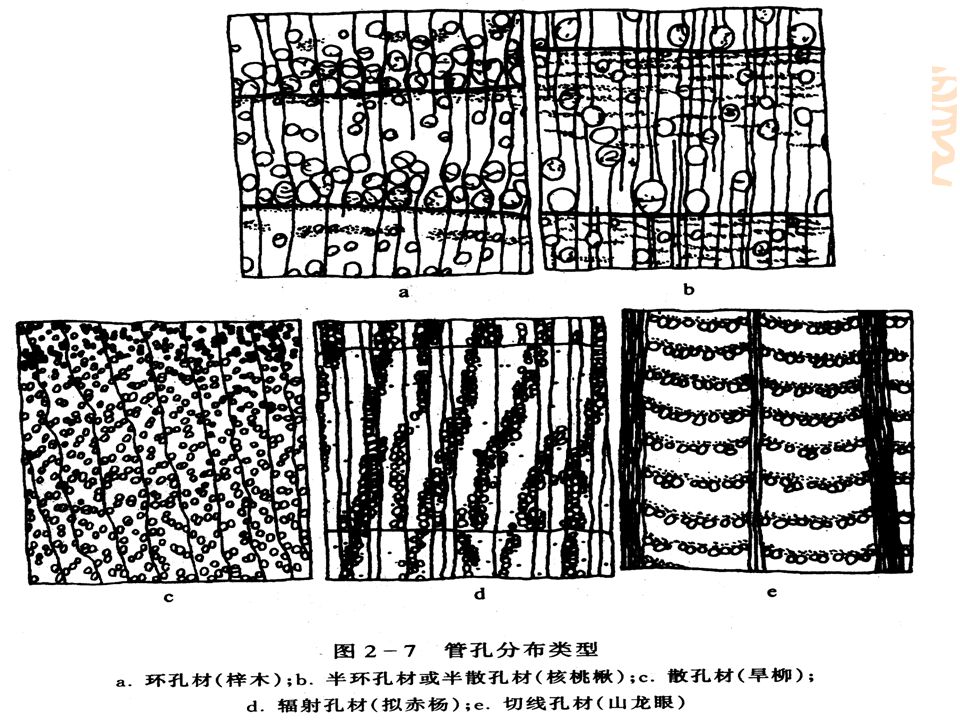
The channels thus formed, which might be several meters in length, are capable of a more effective water transport than the softwood tracheids. This is needed especially in the spring during the leafing. In diffuse-porous woods (aspen, birch, and maple ) the vessels are evenly distributed across the annual ring. The vessels are larger and more numerous in the earlywood portion in ring-porous woods, such as ash, elm, and oak. In birch and aspen the vessels amount to about 25% of the wood volume. Several different pores are present in the walls of the vessels.
 the vessels are evenly distributed across the annual ring. The vessels are larger and more numerous in the earlywood portion in ring-porous woods, such as ash, elm, and oak. In birch and aspen the vessels amount to about 25% of the wood volume. Several different pores are present in the walls of the vessels.")
38
Chapter 1 THE STRUCTURE OF WOOD
These differences together with other structural features are of great help in the identification of pulp fibers. Besides the usual vessels some hardwoods contain cells resembling softwood tracheids or small vessels. Their walls are rich in bordered pits. Hardwood rays consist exclusively of parenchyma cells. The ray width varies in the tangential direction. In aspen wood the rays form one row, in birch wood and oak wood 1-3 and 1-30 rows, respectively. The height varies from one up to several hundred tiers. The rays account for 5-30% of the stem volume.

39
Chapter 1 THE STRUCTURE OF WOOD
1.2.8 Sapwood and Heartwood At a certain age the inner wood of the stem of most trees begin to change to a completely dead heartwood and its proportion of the stem becomes successively larger as the tree grows. The dying parenchyma cells produce organic deposits such as resin, phenolic substances, pigments, etc. In softwoods the bordered pits are closed when the torus becomes pressed against either side of the border. In some hardwoods, such as oak or ash, the vessels are closed by tyloses, which enter the vessel from neighboring ray cells (Fig. 1-10).
.")
40
Fig. 1-10. Tyloses at the bud stage (A) and at a later stage filling the vessel cavity (B).
Wood with tyloses is impermeable to liquids and an excellent material for barrels. These anatomical and chemical changes often have a significant influence on the behavior of sapwood and heartwood during pulping.

41
Chapter 1 THE STRUCTURE OF WOOD
1.3 Wood Ultrastructure Building Elements The wood cell consists mainly of cellulose, hemicelluloses, and lignin. A simplified picture is that cellulose forms a skeleton which is surrounded by other substances functioning as matrix (hemicelluloses) and encrusting (lignin) materials. The length of a native cellulose molecule is at least 5000 nm corresponding to a chain with about 10,000 glucose units.
 and encrusting (lignin) materials. The length of a native cellulose molecule is at least 5000 nm corresponding to a chain with about 10,000 glucose units.")
42
Chapter 1 THE STRUCTURE OF WOOD
The smallest building element of the cellulose skeleton is considered by some to be an elementary fibril. This is a bundle of 16 parallel cellulose molecules which are held together by hydrogen bonds, but various opinions exist concerning this question. The cellulose molecules according to the "fringe micellar model" form completely ordered or crystalline regions, which without any distinctive boundary are changing into disordered or amorphous regions (Fig. 1-11). In native cellulose the length of the crystallites can be nm and the cross section, probably rectangular, is on an average 3 × 10 nm. According to this model the cellulose molecule continues through several crystallites.
. In native cellulose the length of the crystallites can be nm and the cross section, probably rectangular, is on an average 3 × 10 nm. According to this model the cellulose molecule continues through several crystallites.")
43
Fig. 1-11. Diagrammatic representation of fibrillar structure in the cell wall according to Mark
Heavy lines constitute the crystalline regions. The chain molecules may pass through one or more crystalline and amorphous regions.

44
Chapter 1 THE STRUCTURE OF WOOD
The microfibrils which are nm wide, are visible in the electron microscope without pretreatment. Microfibrils are combined to greater fibrils and lamellae, which can be separated from the fibers mechanically, although their dimensions greatly depend on the method used. Disordered cellulose molecules as well as hemicelluloses and lignin are located in the spaces between the microfibrils. The hemicelluloses are considered to be amorphous although they apparently are oriented in the same direction as the cellulose microfibrils. Lignin is both amorphous and isotropic.

45
Chapter 1 THE STRUCTURE OF WOOD
13.2 Cell Wail Layers The cell wall is built up by several layers, namely (Figs. 1-12) middle lamella (M), primary wall (P), outer layer of the secondary wall (S1), middle layer of the secondary wall (S2), inner layer of the secondary wall (S3), and warty layer (W). These layers differ from one another with respect to their structure as well as their chemical composition. The microfibrils wind around the cell axis in different directions either to the right (Z helix) or to the left (S helix). Deviations in the angular directions cause physical differences and the layers can be observed in a microscope under polarized light.
 middle lamella (M), primary wall (P), outer layer of the secondary wall (S1), middle layer of the secondary wall (S2), inner layer of the secondary wall (S3), and warty layer (W). These layers differ from one another with respect to their structure as well as their chemical composition. The microfibrils wind around the cell axis in different directions either to the right (Z helix) or to the left (S helix). Deviations in the angular directions cause physical differences and the layers can be observed in a microscope under polarized light.")
46
Fig Simplified stricture of a woody cell, showing the middle lamella (ML), the primary wall (P), the outer (S1), middle (S2), and inner (S3) layers of the secondary wall, and the warty layer (W).
, the primary wall (P), the outer (S1), middle (S2), and inner (S3) layers of the secondary wall, and the warty layer (W)..")
47
Chapter 1 THE STRUCTURE OF WOOD
The middle lamella is located between the cells and serves the function of binding the cells together. At an early stage of the growth it is mainly composed of pectic substances, but it eventually becomes highly lignified. Its thickness, except at the cell corners, is μm. The primary wall is a thin layer, μm thick, consisting of cellulose, hemicelluloses, pectin, and protein and completely embedded in lignin. The cellulose microfibrils form an irregular network in the outer portion of the primary wall; in the interior they are oriented nearly perpendicularly to the cell axis (Fig. 1-14).
.")
48
Bookp

49
Chapter 1 THE STRUCTURE OF WOOD
The secondary wall consists of three layers: thin outer and inner layers and a thick middle layer. These layers are built up by lamellae formed by almost parallel microfibrils between which lignin and hemicelluloses are located. The outer layer (S1) is μm thick and contains 3-4 lamellae where the microfibrils form either a Z helix or S helix. The microfibril angle of the crossed fibrillar network varies between 50 and 70º with respect to the fiber axis.
 is μm thick and contains 3-4 lamellae where the microfibrils form either a Z helix or S helix. The microfibril angle of the crossed fibrillar network varies between 50 and 70º with respect to the fiber axis.")
50
Chapter 1 THE STRUCTURE OF WOOD
The middle layer (S2) forms the main portion of the cell wall. Its thickness in softwood tracheids varies between 1 (earlywood) and 5 (latewood) μm and it may thus contain lamellae or more than 150 lamellae. The thickness naturally varies with the cell types. The microfibrillar angle varies between 10º (earlywood) and º (latewood). It decreases in a regular fashion with increasing fiber length. The characteristics of the S2 layer (thickness, microfibrillar angle, etc.) have a decisive influence on the fiber stiffness as well as on other papermaking properties.
 forms the main portion of the cell wall. Its thickness in softwood tracheids varies between 1 (earlywood) and 5 (latewood) μm and it may thus contain lamellae or more than 150 lamellae. The thickness naturally varies with the cell types. The microfibrillar angle varies between 10º (earlywood) and º (latewood). It decreases in a regular fashion with increasing fiber length. The characteristics of the S2 layer (thickness, microfibrillar angle, etc.) have a decisive influence on the fiber stiffness as well as on other papermaking properties.")
51
Chapter 1 THE STRUCTURE OF WOOD
The inner layer (S3) is a thin layer (ca. 0.1μm) consisting of several lamellae which contain microfbrils in both Z helices and S helices (50-90º angle). Great variations are noted among different wood species. The warty layer (W) is a thin amorphous membrane located in the inner surface of the cell wall in all conifers and in some hardwood, containing warty deposits of a still unknown composition. Each species has its own characteristic warty layer.
 is a thin layer (ca. 0.1μm) consisting of several lamellae which contain microfbrils in both Z helices and S helices (50-90º angle). Great variations are noted among different wood species. The warty layer (W) is a thin amorphous membrane located in the inner surface of the cell wall in all conifers and in some hardwood, containing warty deposits of a still unknown composition. Each species has its own characteristic warty layer.")
52
Chapter 1 THE STRUCTURE OF WOOD
1.3.3 Pits The normal structure of the cell wall is broken by pits. Changes appear already in the growth period of the cell. For instance, early stages of pit formation in softwoods are visible in the primary wall just before the cell reaches its final dimensions (primary pit fields). The microfibril network is loosened and new microfibrils are oriented around these points. The structure in the middle of the circles is tighten and the racially oriented microfibril bundles finally form a netlike membrane, permeable to liquids. The central, thickened portion of the pit membrane (torus) is formed after a secondary thickening of the primary wall. The torus is rich in pectic material and also contains cellulose in pine and spruce.
. The microfibril network is loosened and new microfibrils are oriented around these points. The structure in the middle of the circles is tighten and the racially oriented microfibril bundles finally form a netlike membrane, permeable to liquids. The central, thickened portion of the pit membrane (torus) is formed after a secondary thickening of the primary wall. The torus is rich in pectic material and also contains cellulose in pine and spruce.")
53
Chapter 1 THE STRUCTURE OF WOOD
1.4 Reaction Wood As a product of living organism the structure of wood fibers is so variable and complicated that a great number of details remains to be solved for understanding the anatomy and biology even of trees grown under normal conditions. When a tree is brought out of its natural, equilibrium position in space, for example by wind or by a landslide, the tree begins to produce a special tissue, referred to as reaction wood. The function of this type of wood is to restore the displaced stem or branch to its original position. In a leaning stem of a conifer, compression wood develops on the tower side.

54
Chapter 1 THE STRUCTURE OF WOOD
This wood expands longitudinally as it is being formed, and the pressure exerted along the grain forces the stem to bend upward. All movements of orientation in mature conifers are effected with the aid of appropriately located compression wood. In hardwoods, tension wood is fomred on the upper side of an inclined stem. This wood contracts as it is laid down and in this way forces the stem to bend upward. Compression wood can be said to push a stem or a branch up; tension wood pulls them up.

55
Chapter 1 THE STRUCTURE OF WOOD
Compression wood is heavier, harder, and denser than the normal wood. Its tracheids are short and thick-walled (even in earlywood) and in cross section rounded so that empty spaces remain between the cells. The S1 layer is thicker than in a normal wood while the S3 layer is absent. The S2 layer contains helical cavities that parallel the microfibrils and reach from the lumen deep into the S2. The cellulose content of compression wood is lower and the lignin content higher than for normal wood.
 and in cross section rounded so that empty spaces remain between the cells. The S1 layer is thicker than in a normal wood while the S3 layer is absent. The S2 layer contains helical cavities that parallel the microfibrils and reach from the lumen deep into the S2. The cellulose content of compression wood is lower and the lignin content higher than for normal wood.")
56
Chapter 1 THE STRUCTURE OF WOOD
Tension wood differs less from normal wood than compression wood. It contains thick-walled fibers, terminated towards the lumen by a gelatinous layer. This so-called G layer consists of pure and highly crystalline cellulose oriented in the same direction as the fiber axis. For this reason the cellulose content of tension wood is higher and the lignin content lower than in normal wood.

Similar presentations