Download presentation
Presentation is loading. Please wait.

0
Differentiation of pluripotent cells and transdifferentiation

1
What is differentiation of pluripotent cells?
Multipotent Differentiated cells Differentiation of ES/iPS cells creates specialized cells in vitro such as neurons, heart muscle cells, endothelial cells from blood vessels and insulin-secreting cells similar to those found in the pancreas, all of which can be used for cellular-based treatment or development of new therapies. Ectodermal cell brain ES/iPS cell Mesodermal cell heart • ES cells from the inner cell mass can give rise to all three germ layers and are pluripotent. • Cells in each germ layer retain the ability to proliferate and give rise to a more restricted spectrum of cells. Therefore, they are multipotent cells. • During embryonic development, proliferating precursors or progenitors eventually appear that have very limited fates and are unipotent, such as neurons or skin cells. • Stem cells or progenitors thus undergo successive steps of lineage restriction that limit the eventual cell types they can produce during development. Directed differentiation of ES cells in culture is the process of converting ES cells into specialized cells in vitro such as neurons, heart muscle cells, endothelial cells of blood vessels and insulin-secreting cells similar to those found in the pancreas. These cells can be used for cellular-based treatment or development of new therapies. Endodermal cell pancreas 1 1

2
Why do we care about directed differentiation of ES cells?
The differentiated cell types resulting from directed differentiation and lineage selection of pluripotent human ES cells can be applied to developmental biology, regenerative medicine and drug discovery. Mitochondrial heteroplasmy i.e. the process of transfering the mitochondria from one donor oocyte to another recepient oocyte (allogenic oocyte mitochondria and autogenic donor cell mitochondria) during the process of nuclear transfer, with the subsequent generation of ES cells from these oocytes, offers the possibility for deriving NTSC ES cell lines with mutations in mitochondrial DNA (mtDNA) or mitochondrial dysfunction in order to study these diseases. Autologous/heterologous differentiated cells derived from a patient’s hESCs can be either used as an in vitro model of that specific disease or, after repairing the abnormality, transferred back to the patient for treatment (with immunosuppressive treatments in the case of heterologous cells). In addition, hES cells, pES cells, NTSC (nuclear transfer-derived ES cells) or iPS-derived cells offer a more realistic material for drug discovery and toxicological screening. Differentiated embryonic stem cells (e.g. neural, cardiac or hepatic cells) can serve as a model preparation for scientists/clinicians and as a potential high-throughput system to test the pharmaceutical efficacy and toxicity of different drugs/chemicals. In this case, hESCs derived from different individuals or ethnic populations will open the possibility of determining whether genetic variants result in altered expression of the target gene or alter the responses of the gene product to therapeutic compounds. 2 2
 during the process of nuclear transfer, with the subsequent generation of ES cells from these oocytes, offers the possibility for deriving NTSC ES cell lines with mutations in mitochondrial DNA (mtDNA) or mitochondrial dysfunction in order to study these diseases. Autologous/heterologous differentiated cells derived from a patient’s hESCs can be either used as an in vitro model of that specific disease or, after repairing the abnormality, transferred back to the patient for treatment (with immunosuppressive treatments in the case of heterologous cells). In addition, hES cells, pES cells, NTSC (nuclear transfer-derived ES cells) or iPS-derived cells offer a more realistic material for drug discovery and toxicological screening. Differentiated embryonic stem cells (e.g. neural, cardiac or hepatic cells) can serve as a model preparation for scientists/clinicians and as a potential high-throughput system to test the pharmaceutical efficacy and toxicity of different drugs/chemicals. In this case, hESCs derived from different individuals or ethnic populations will open the possibility of determining whether genetic variants result in altered expression of the target gene or alter the responses of the gene product to therapeutic compounds")
3
Secreted factors keep ES cells pluripotent when cultured
Secreted factors (proteins): Cell feeder layer (fibroblasts) secretes proteins that interact with receptors in the ES cell membrane to maintain its pluripotency. LIF (Leukemia Inhibitory Factor) provided in the media binds LIF receptors in the ES cell membrane to maintain both mouse ES pluripotency and the rate of cell proliferation. Serum contains BMPs (bone morphogenetic proteins) that maintain pluripotency of mouse ES cells FGF-2 and TGFs maintain human ES cell pluripotency Feeders (MEFs) ES cells • ES cells are relatively easy to handle in culture. Several factors maintain the pluripotency of ES cells in culture. The factors can be divided in two groups: a) secreted factors, and b) intracellular factors. ES cells are cultured on a mitotically inactive mouse fibroblast “feeder” layer that maintains their pluripotency. Fibroblasts either promote self-renewal and/or suppress ES cell differentiation. LIF – leukemia inhibiting factor is another factor necessary to maintain pluripotency of mouse ES cells in culture. LIF is a member of the cytokine family of secreted proteins. It binds to a receptor on the ES cell surface and influences gene expression. LIF may influence: a) the rate of cell proliferation, b) cell cycle progression, c) activation of a signaling cascade involved in maintaining pluripotency. Serum contains bone morphogenetic proteins (BMPs) that are important for an ES cell’s self-renewing abilities. Human ES cells are maintained in a pluripotent state in culture in the presence of Fibroblast Growth Factor 2 (FGF-2). Mouse ES cells colonies in culture 3 3
: Cell feeder layer (fibroblasts) secretes proteins that interact with receptors in the ES cell membrane to maintain its pluripotency. LIF (Leukemia Inhibitory Factor) provided in the media binds LIF receptors in the ES cell membrane to maintain both mouse ES pluripotency and the rate of cell proliferation. Serum contains BMPs (bone morphogenetic proteins) that maintain pluripotency of mouse ES cells. FGF-2 and TGFs maintain human ES cell pluripotency. Feeders (MEFs) ES cells. • ES cells are relatively easy to handle in culture. Several factors maintain the pluripotency of ES cells in culture. The factors can be divided in two groups: a) secreted factors, and b) intracellular factors. ES cells are cultured on a mitotically inactive mouse fibroblast feeder layer that maintains their pluripotency. Fibroblasts either promote self-renewal and/or suppress ES cell differentiation. LIF – leukemia inhibiting factor is another factor necessary to maintain pluripotency of mouse ES cells in culture. LIF is a member of the cytokine family of secreted proteins. It binds to a receptor on the ES cell surface and influences gene expression. LIF may influence: a) the rate of cell proliferation, b) cell cycle progression, c) activation of a signaling cascade involved in maintaining pluripotency. Serum contains bone morphogenetic proteins (BMPs) that are important for an ES cell’s self-renewing abilities. Human ES cells are maintained in a pluripotent state in culture in the presence of Fibroblast Growth Factor 2 (FGF-2). Mouse ES cells colonies in culture")
4
(I) Directed differentiation of ES/iPS cells
Pluripotent Multipotent Differentiated cells ES/iPS cell Ectodermal cell brain ✗ ✗ • The most common approach to direct differentiation of ES cells in culture is to change their growth conditions in several specific ways: a) Remove the secreted factors that maintain ES cell pluripotency. For example, for mouse ES cells remove factors such as LIF or serum. b) Add growth factors to the culture medium that promote the differentiation into a specific lineage. These factors trigger the activation (or inactivation) of specific genes in ES cells. This initiates a series of molecular events that induces the cells to differentiate along a particular pathway. Change the chemical composition of the surface on which the ES cells are growing. For example, the plastic culture dishes used to grow both mouse and human ES cells can be treated with a variety of substances that allow cells either to adhere to the surface of the dish or to avoid adhesion and instead float in the culture medium. In general, an adherent substrate helps prevent ES cells from interacting with each other and thereby differentiating. In contrast, a nonadherent substrate allows the ES cells to aggregate and thereby interact with each other. These aggregates are called “embryoid bodies” (I.e. they mimic interactions that occur in the embryo in vivo). Cell-cell interactions are critical to normal embryonic development, so allowing some of these "natural" in vivo interactions to occur in the culture dish is a fundamental strategy for inducing mouse or human ES cell differentiation in vitro. ✗ ✗ Feeders Feeders 4 4
 Remove the secreted factors that maintain ES cell pluripotency. For example, for mouse ES cells remove factors such as LIF or serum. b) Add growth factors to the culture medium that promote the differentiation into a specific lineage. These factors trigger the activation (or inactivation) of specific genes in ES cells. This initiates a series of molecular events that induces the cells to differentiate along a particular pathway. Change the chemical composition of the surface on which the ES cells are growing. For example, the plastic culture dishes used to grow both mouse and human ES cells can be treated with a variety of substances that allow cells either to adhere to the surface of the dish or to avoid adhesion and instead float in the culture medium. In general, an adherent substrate helps prevent ES cells from interacting with each other and thereby differentiating. In contrast, a nonadherent substrate allows the ES cells to aggregate and thereby interact with each other. These aggregates are called embryoid bodies (I.e. they mimic interactions that occur in the embryo in vivo). Cell-cell interactions are critical to normal embryonic development, so allowing some of these natural in vivo interactions to occur in the culture dish is a fundamental strategy for inducing mouse or human ES cell differentiation in vitro. ✗ ✗ Feeders. Feeders")
5
(I) Directed differentiation of ES/iPS cells
Change growth conditions of ES cells: Remove secreted factors that maintain ES cell pluripotency from the media Add growth factors to the culture solution that trigger activation (or inactivation) of specific genes in ES cells, in order to promote differentiation into a specific lineage. Change the surface on which ES cells are growing: Grow ES cells on non-adherent substrates so that they aggregate with each other. These aggregates are called “embryoid bodies”. ES cells within aggregates will interact with each other. These cell-cell interactions mimic some of the interactions of ES cells in vivo that normally guide their differentiation. • 5 5
 of specific genes in ES cells, in order to promote differentiation into a specific lineage. Change the surface on which ES cells are growing: Grow ES cells on non-adherent substrates so that they aggregate with each other. These aggregates are called embryoid bodies . ES cells within aggregates will interact with each other. These cell-cell interactions mimic some of the interactions of ES cells in vivo that normally guide their differentiation. •")
6
Distinct signaling pathways specify discrete cell types during development
Cell signaling pathways Shh Activin/TGF- Erythropoietin (EPO) Patched/ Smoothened BMP-RI EPO receptor Progenitor cell Progenitor cell Progenitor cell • During development, immature cells or progenitors found at different locations in the developing embryo encounter distinct signals. Progenitors that will become motor neurons encounter a secreted molecule called Shh, which binds to receptors (Patched and Smoothened) in the plasma membrane of the progenitor. This triggers a signaling cascade inside the progenitors that promotes their differentiation into motor neurons. • Progenitors that will become heart muscle cells express a different receptor called BMPRI. This receptor interacts with the secreted molecules called Activin or TGF to trigger a signaling pathway that induces differentiation into heart muscle cells. Progenitors that become red blood cells express a third type of receptor that binds to a hormone named erythropoietin, to induce their differentiation into red blood cells. • The activation of different signaling pathways within distinct immature cells during development allows them to acquire different fates. Heart muscle cell (Cardiomyocyte) Motor neuron Red blood cells 6 6
 Patched/ Smoothened. BMP-RI. EPO receptor. Progenitor. cell. Progenitor. cell. Progenitor. cell. • During development, immature cells or progenitors found at different locations in the developing embryo encounter distinct signals. Progenitors that will become motor neurons encounter a secreted molecule called Shh, which binds to receptors (Patched and Smoothened) in the plasma membrane of the progenitor. This triggers a signaling cascade inside the progenitors that promotes their differentiation into motor neurons. • Progenitors that will become heart muscle cells express a different receptor called BMPRI. This receptor interacts with the secreted molecules called Activin or TGF to trigger a signaling pathway that induces differentiation into heart muscle cells. Progenitors that become red blood cells express a third type of receptor that binds to a hormone named erythropoietin, to induce their differentiation into red blood cells. • The activation of different signaling pathways within distinct immature cells during development allows them to acquire different fates. Heart muscle cell. (Cardiomyocyte) Motor neuron. Red blood cells")
7
(I) Induced differentiation of ES/iPS cells
Pluripotent Multipotent Differentiated cells ES/iPS cell Ectodermal cell brain Another way to direct differentiation of ES cells is to introduce foreign genes into the cells via transfection or other methods. The result of these strategies is to add an active gene to the ES cell genome, which then triggers the cells to differentiate along a particular pathway. The approach appears to be a precise way of regulating ES cell differentiation, but it will work only if we know which gene must be active at a particular stage of differentiation. Then, the gene must be activated at the right time — meaning during the correct stage of differentiation — and it must be inserted into the genome at the proper location. ✗ ✗ 7 7

8
(I) Induced differentiation of ES/iPS cells
Transfect ES cells with foreign genes: Adding an active gene or genes to the ES cell genome. The gene(s) trigger(s) ES cells to differentiate along a particular pathway. This approach is a precise way of regulating ES cell differentiation. Problems with this technology: It works ONLY if we know which gene(s) must be active at a particular stage of differentiation. The gene(s) must be activated at the right time, i.e. during the correct stage of differentiation The foreign gene(s) are often only required temporarily, but it is difficult to introduce them without permanently changing or “damaging” the genome. • 8 8
 trigger(s) ES cells to differentiate along a particular pathway. This approach is a precise way of regulating ES cell differentiation. Problems with this technology: It works ONLY if we know which gene(s) must be active at a particular stage of differentiation. The gene(s) must be activated at the right time, i.e. during the correct stage of differentiation. The foreign gene(s) are often only required temporarily, but it is difficult to introduce them without permanently changing or damaging the genome. •")
9
ICM cells form three germ layers during embryogenesis
Amnion Implantation Uterus Blastocyst Ectoderm Epithelial skin cells, inner ear, eye, mammary glands, nails, teeth, nervous system (spine and brain) Yolk sac • An early specialization during development is the segregation of the three basic germ layers that will each give rise to part of the adult animal • Ectoderm: nerve cells, skin cells, inner ear, eye, mammary glands, nails, teeth and the nervous system (spine and brain) • Mesoderm: blood, muscle, bone, heart, skeleton, gonads, urinary system, fat and spleen • Endoderm: gut, liver, pancreas, lungs, tonsils, pharynx and parathyroid glands • The future sperm and egg cells (reproductive cells) do not come from these three layers, but rather are partitioned early as primordial germ cells (not to be confused with germ layers) • Gastrulation also establishes the main axes of the embryo: anterior-posterior (head to toe), dorsal-ventral (back to front) and left-right Endoderm Stomach, gut, liver, pancreas, lungs, tonsils, pharynx, thyroid glands Mesoderm Blood, muscle, bones, heart, urinary system, spleen, fat 9 9
 Yolk sac. • An early specialization during development is the segregation of the three basic germ layers that will each give rise to part of the adult animal. • Ectoderm: nerve cells, skin cells, inner ear, eye, mammary glands, nails, teeth and the nervous system (spine and brain) • Mesoderm: blood, muscle, bone, heart, skeleton, gonads, urinary system, fat and spleen. • Endoderm: gut, liver, pancreas, lungs, tonsils, pharynx and parathyroid glands. • The future sperm and egg cells (reproductive cells) do not come from these three layers, but rather are partitioned early as primordial germ cells (not to be confused with germ layers) • Gastrulation also establishes the main axes of the embryo: anterior-posterior (head to toe), dorsal-ventral (back to front) and left-right. Endoderm. Stomach, gut, liver, pancreas, lungs, tonsils, pharynx, thyroid glands. Mesoderm. Blood, muscle, bones, heart, urinary system, spleen, fat")
10
Motor neurons and their diseases
One motor neuron per 106 cells in the body Reside in the ventral horn of the spinal cord Control movements of muscles Exist in various subtypes that control different muscle groups (limbs versus thoracic regions) Motor neuron diseases Paralysis from spinal cord trauma Spinal Muscular Atrophy (SMA) Amyotrophic Lateral Sclerosis (Lou Gehrig’s disease or ALS) A large number of research laboratories have focused their efforts in developing methods to direct differentiation of ES cells into motor neurons. Why? Motor neurons – one motor neuron per every million cells in the body – reside in the ventral horn of the spinal cord – control movement of muscles – different types of motor neurons are present in the body (limb versus thoracic areas) There are a number of diseases that affect motor neurons: – Paralysis from spinal cord trauma. Spinal cord injuries cause myelopathy, or damage to either nerve roots or myelinated fiber tracts that carry signals to and from the brain. Depending on its classification and severity, this type of traumatic injury can also damage the grey matter in the central part of the spinal cord, causing segmental losses of interneurons and motor neurons. – Spinal Muscular Atrophy (SMA) is a neuromuscular disease characterized by degeneration of motor neurons, resulting in progressive muscular atrophy (wasting away) and weakness. The clinical spectrum of SMA ranges from early infant death to normal adult life with only mild weakness. These patients often require comprehensive medical care that can be costly for society. The most common form of SMA is caused by mutation of the SMN (Survival of Motor Neurons) gene, and manifests over a wide range of severity affecting infants through adults. – Amyotrophic Lateral Sclerosis (ALS or Lou Gehrig’s disease) is a form of motor neuron disease. ALS is a progressive and fatal neurodegenerative disease caused by the loss of motor neurons. The condition is often called Lou Gehrig's disease in North America, after the famous New York Yankees baseball player who was diagnosed with the disease in There is no known cause for the disease in ~95% of cases, where the patient’s family history is unknown. There is a known hereditary factor in familial ALS (FALS), where the condition is known to run in families, although this only accounts for around 5% of all cases. An inherited genetic defect on chromosome 21 (coding for superoxide dismutase) is associated with approximately 20% of familial cases of ALS. This mutation is autosomal dominant. The most common ALS-causing SOD1 mutation in North America is A4V, characterized by an exceptionally rapid progression from onset to death. The children of those diagnosed with familial ALS have higher risk for developing the disease. 10
 Motor neuron diseases. Paralysis from spinal cord trauma. Spinal Muscular Atrophy (SMA) Amyotrophic Lateral Sclerosis (Lou Gehrig’s disease or ALS) A large number of research laboratories have focused their efforts in developing methods to direct differentiation of ES cells into motor neurons. Why Motor neurons. – one motor neuron per every million cells in the body. – reside in the ventral horn of the spinal cord. – control movement of muscles. – different types of motor neurons are present in the body (limb versus thoracic areas) There are a number of diseases that affect motor neurons: – Paralysis from spinal cord trauma. Spinal cord injuries cause myelopathy, or damage to either nerve roots or myelinated fiber tracts that carry signals to and from the brain. Depending on its classification and severity, this type of traumatic injury can also damage the grey matter in the central part of the spinal cord, causing segmental losses of interneurons and motor neurons. – Spinal Muscular Atrophy (SMA) is a neuromuscular disease characterized by degeneration of motor neurons, resulting in progressive muscular atrophy (wasting away) and weakness. The clinical spectrum of SMA ranges from early infant death to normal adult life with only mild weakness. These patients often require comprehensive medical care that can be costly for society. The most common form of SMA is caused by mutation of the SMN (Survival of Motor Neurons) gene, and manifests over a wide range of severity affecting infants through adults. – Amyotrophic Lateral Sclerosis (ALS or Lou Gehrig’s disease) is a form of motor neuron disease. ALS is a progressive and fatal neurodegenerative disease caused by the loss of motor neurons. The condition is often called Lou Gehrig s disease in North America, after the famous New York Yankees baseball player who was diagnosed with the disease in There is no known cause for the disease in ~95% of cases, where the patient’s family history is unknown. There is a known hereditary factor in familial ALS (FALS), where the condition is known to run in families, although this only accounts for around 5% of all cases. An inherited genetic defect on chromosome 21 (coding for superoxide dismutase) is associated with approximately 20% of familial cases of ALS. This mutation is autosomal dominant. The most common ALS-causing SOD1 mutation in North America is A4V, characterized by an exceptionally rapid progression from onset to death. The children of those diagnosed with familial ALS have higher risk for developing the disease. 10.")
11
Specification of motor neuron fate depends on nearby secreted signals
Hb9 Hb9::eGFP BMPs Wnts MNs Retinoic acid During development of the central nervous system, motor neurons are born in the ventral region of the posterior neural tube that will become the spinal cord in the adult organism. Progenitor cells in the neural tube encounter a variety of signaling molecules that specify their fates and direct their differentiation into various types of neurons. Progenitor cells in the ventral neural tube are specified into motor neurons by two signals: Sonic hedgehog (Shh) and Retinoic Acid (RA). Motor neurons are mature differentiated cells. They are characterized by the unique and early expression of a transcription factor called HB9. Motor neurons in the ventral neural tube can be visualized by expression of the Hb9 protein, as they are specified from immature progenitors in the neural tube and migrate laterally to occupy the ventral horns of the tissue. The Hb9 promoter and enhancer region (9 kb) can be used to drive expression of Green Fluorescent Protein in motor neurons and their axons as they exit the spinal cord and innervate peripheral muscle tissues to control their movement. This transgenic mouse has become a very valuable tool for researchers to understand the process of directed differentiation for ES cells into motor neurons in culture, and to test the ability of motor neurons that are generated in culture to innervate muscles after transplantation in vivo. Shh
 and Retinoic Acid (RA). Motor neurons are mature differentiated cells. They are characterized by the unique and early expression of a transcription factor called HB9. Motor neurons in the ventral neural tube can be visualized by expression of the Hb9 protein, as they are specified from immature progenitors in the neural tube and migrate laterally to occupy the ventral horns of the tissue. The Hb9 promoter and enhancer region (9 kb) can be used to drive expression of Green Fluorescent Protein in motor neurons and their axons as they exit the spinal cord and innervate peripheral muscle tissues to control their movement. This transgenic mouse has become a very valuable tool for researchers to understand the process of directed differentiation for ES cells into motor neurons in culture, and to test the ability of motor neurons that are generated in culture to innervate muscles after transplantation in vivo. Shh.")
12
Graded Shh signaling specifies ventral interneurons and motor neurons within the neural tube
Patched/ Smoothened Progenitor Cell During embryonic development of the neural tube, progenitor cells that will become motor neurons express two receptors on their surface (Patched and Smoothened). These receptors are activated by a secreted protein called Sonic Hedgehog (Shh), which is secreted from both a tube-like structure called the notochord and the region of the neural tube above the notochord called the floor plate. Shh mRNA can be detected in both of these tissues before motor neurons are generated. Shh diffuses away from the notochord and floor plate toward the neural tube and forms a gradient that is important for specifying the fates of several populations of neurons in the ventral spinal cord, including motor neurons. Motor neurons are generated at a very high efficiency from neural progenitors at a concentration of 3 nM. Motor neuron (HB9+)
. These receptors are activated by a secreted protein called Sonic Hedgehog (Shh), which is secreted from both a tube-like structure called the notochord and the region of the neural tube above the notochord called the floor plate. Shh mRNA can be detected in both of these tissues before motor neurons are generated. Shh diffuses away from the notochord and floor plate toward the neural tube and forms a gradient that is important for specifying the fates of several populations of neurons in the ventral spinal cord, including motor neurons. Motor neurons are generated at a very high efficiency from neural progenitors at a concentration of 3 nM. Motor neuron. (HB9+)")
13
Stem cell-based approaches to motor neuron diseases
Patients Pathways of degeneration iPS cells Astrocytes… Motor neurons Drug discovery ES cells Cell replacement therapy Animal models ES cells can be generated from animals models of motor neuron diseases such as SMN1/SMN2 mutant mice or SOD1G93A transgenic mice, which mimic the respective human diseases (SMA or ALS). iPS cells can also be generated from patients with human diseases such as SMA or ALS. Motor neurons or other CNS cells such as astrocytes can be derived from iPS cells or ES cells, and they can be used for a variety of scientific and clinical purposes: determine which cellular and molecular pathways are responsible for degeneration of motor neurons discover drugs using assays designed to allow isolation of compounds that improve the pathology of neurons c) cell replacement therapy 13
. iPS cells can also be generated from patients with human diseases such as SMA or ALS. Motor neurons or other CNS cells such as astrocytes can be derived from iPS cells or ES cells, and they can be used for a variety of scientific and clinical purposes: determine which cellular and molecular pathways are responsible for degeneration of motor neurons. discover drugs using assays designed to allow isolation of compounds that improve the pathology of neurons. c) cell replacement therapy. 13.")
14
Modeling ALS in a dish Skin cells from ALS patients ALS motor neurons
Dimos, JT et al. (2008). Induced Pluripotent Stem Cells Generated from Patients with ALS Can Be Differentiated into Motor Neurons. Science 321: ALS motor neurons Yamanaka method Oct4 Sox2 Klf4 C-Myc Motor neuron nuclei Axons ES cells may provide a new tool for studying disease mechanisms and for identifying drugs to slow amyotrophic lateral sclerosis (ALS), also known as Lou Gehrig’s disease. Two studies completed by researchers collaborating with Project A.L.S./Jenifer Estess Laboratory for Stem Cell Research, a privately funded laboratory focused exclusively on stem cells and ALS.The complementary studies, led by Kevin Eggan of the Harvard Stem Cell Institute and Serge Przedborsk, of Columbia University Medical Center, compellingly demonstrate that embryonic stem cells can be used to create an in vitro model of ALS. Until now, scientists have not known whether motor neurons in ALS die because of a problem within these cells or from outside the cell. The study by Eggan’s group describes successful use of a novel embryonic stem cell-based model for ALS that will help scientists to answer this and other questions. Dimos, JT et al., (2008). Induced Pluripotent Stem Cells Generated from Patients with ALS Can Be Differentiated into Motor Neurons. Science 321: Using this model, both Eggan’s and Przedborski’s groups observed that non-neuronal cells called astrocytes may have a toxic effect on motor neurons in ALS. The Columbia study provides further evidence that astrocytes are toxic to motor neurons in ALS. The Columbia team’s discovery of astrocytic, toxic mediators provides not only insight into how the damage associated with ALS occurs and a potential biomarker for early diagnosis, but also a target for potential new therapies aimed at slowing motor neuron degeneration in ALS. “That’s what happens when scientists from major institutions work together toward shared goals. Project A.L.S. prides itself on building productive research teams from elegant parts,” said Valerie Estess, director of research for Project ALS. iPS cells induced pluripotent stem cells 14
. Induced Pluripotent Stem Cells Generated. from Patients with ALS Can Be Differentiated into Motor Neurons. Science 321: ALS motor neurons. Yamanaka method. Oct4. Sox2. Klf4. C-Myc. Motor neuron nuclei. Axons. ES cells may provide a new tool for studying disease mechanisms and for identifying drugs to slow amyotrophic lateral sclerosis (ALS), also known as Lou Gehrig’s disease. Two studies completed by researchers collaborating with Project A.L.S./Jenifer Estess Laboratory for Stem Cell Research, a privately funded laboratory focused exclusively on stem cells and ALS.The complementary studies, led by Kevin Eggan of the Harvard Stem Cell Institute and Serge Przedborsk, of Columbia University Medical Center, compellingly demonstrate that embryonic stem cells can be used to create an in vitro model of ALS. Until now, scientists have not known whether motor neurons in ALS die because of a problem within these cells or from outside the cell. The study by Eggan’s group describes successful use of a novel embryonic stem cell-based model for ALS that will help scientists to answer this and other questions. Dimos, JT et al., (2008). Induced Pluripotent Stem Cells Generated from Patients with ALS Can Be Differentiated into Motor Neurons. Science 321: Using this model, both Eggan’s and Przedborski’s groups observed that non-neuronal cells called astrocytes may have a toxic effect on motor neurons in ALS. The Columbia study provides further evidence that astrocytes are toxic to motor neurons in ALS. The Columbia team’s discovery of astrocytic, toxic mediators provides not only insight into how the damage associated with ALS occurs and a potential biomarker for early diagnosis, but also a target for potential new therapies aimed at slowing motor neuron degeneration in ALS. That’s what happens when scientists from major institutions work together toward shared goals. Project A.L.S. prides itself on building productive research teams from elegant parts, said Valerie Estess, director of research for Project ALS. iPS cells. induced pluripotent stem cells. 14.")
15
Using motor neurons to screen drugs promoting their survival
Mouse disease models – creating ES cells from existing mouse model strains – genetic modification of existing ES cell lines Human disease models – genetically tested blastocysts from IVF clinics (SMA) – not applicable to ALS 15
 – not applicable to ALS. 15.")
16
Directed differentiation protocol for human ES cells into motor neurons
RA hES cells Day 10 primary neurectoderm (early rosettes) Day 14 secondary (late rosettes) Day 33 motor neurons Day 26 motor neuron progenitors RA/Shh Tubulin Hb9 Knowledge of the signaling molecules that specify the identity of motor neurons has also been used to guide differentiation of human ES cells into motor neurons in vitro. The directed differentiation of human ES cells into motor neurons requires the presence of both retinoic acid (RA) at 1 M and Shh, similar to the generation of motor neurons from mouse ES cells. Retinoic acid directs the specification of EBs into primary neuroectoderm, which is one of three germ layers present at early stages of embryonic development. The primary neuroectoderm then matures into a secondary neuroectoderm, which gives rise to motor neuron progenitors. This process takes 26 days for human ES cells. Motor neuron progenitors become motor neurons following incubation for 7 additional days with retinoic acid and Shh. The amount of Shh agonist required to specify motor neuron progenitors in culture is 1 M. However, the directed differentiation process for human ES cells requires a longer time. It takes 6 days to generate mouse motor neurons from ES cells, whereas to produce human motor neurons from ES cells requires 33 days. 10 days 4 days 12 days 7 days RA RA 1 M Shh agonist + RA 1 M Shh agonist + RA hES cells Early rosettes Late rosettes Motor neuron progenitors Motor neurons Li et al., Nature Neuroscience (2005) 16
 Day 14. secondary. (late rosettes) Day 33. motor neurons. Day 26. motor neuron. progenitors. RA/Shh. Tubulin. Hb9. Knowledge of the signaling molecules that specify the identity of motor neurons has also been used to guide differentiation of human ES cells into motor neurons in vitro. The directed differentiation of human ES cells into motor neurons requires the presence of both retinoic acid (RA) at 1 M and Shh, similar to the generation of motor neurons from mouse ES cells. Retinoic acid directs the specification of EBs into primary neuroectoderm, which is one of three germ layers present at early stages of embryonic development. The primary neuroectoderm then matures into a secondary neuroectoderm, which gives rise to motor neuron progenitors. This process takes 26 days for human ES cells. Motor neuron progenitors become motor neurons following incubation for 7 additional days with retinoic acid and Shh. The amount of Shh agonist required to specify motor neuron progenitors in culture is 1 M. However, the directed differentiation process for human ES cells requires a longer time. It takes 6 days to generate mouse motor neurons from ES cells, whereas to produce human motor neurons from ES cells requires 33 days. 10 days. 4 days. 12 days. 7 days. RA. RA. 1 M. Shh agonist. + RA. 1 M. Shh agonist. + RA. hES cells. Early. rosettes. Late. rosettes. Motor neuron. progenitors. Motor. neurons. Li et al., Nature Neuroscience (2005) 16.")
17
Dopaminergic neurons and their diseases
Neurons located in the midbrain that secrete dopamine - an important neurotransmitter in the brain These neurons degenerate in Parkinson’s disease, a movement disorder. Loss of these neurons is associated with muscle rigidity, tremor, posture and gait abnormalities as well as slowing or loss of physical movements. These neurons arise during development in response to two signals: Shh and FGF-8. A large number of research laboratories have focused their efforts at developing methods to direct differentiation of ES cells into dopaminergic neurons: These are neurons that are found in midbrain and secrete dopamine - an important neurotransmitter These neurons degenerate in Parkinson’s disease, a movement disorder. Loss of these neurons is associated with muscle rigidity, tremor, postural and gait abnormalities and slowing or loss of physical movements. These neurons are born during development in response to two signals: Shh and FGF-8. Dopaminergic neurons 17

18
Directed differentiation of ES cells into dopaminergic neurons
Dopaminergic neurons require Shh and FGF-8 Mouse EBs are grown in the absence of serum for 4 days on a non-adherent substrate. EBs are transferred to an adherent substrate and grown in a serum-free media that promotes survival of neuronal progenitors. After 6-10 days, neural progenitors are exposed to Shh and FGF-8 to induce differentiation into dopaminergic neurons. Differentiation of human ES cells into dopaminergic neurons takes a longer time. 1) Undifferentiated mouse ES cells are dissociated into single cells and plated at low density. They proliferate in plastic culture dishes coated with gelatin. The growth media containe LIF and fetal calf serum supplemented with amino acids. These conditions promote the proliferation of undifferentiated ES cells. 2) ES cells are induced to form embryoid bodies (EB) by dissociation with trypsin. They are then replated at higher density on a nonadherent surface. These conditions allow the cells to aggregate and begin the process of differentiation. 3) After 4 days, the EBs are replated on an adherent substrate in the original growth medium. 24 hours later, the growth medium is replaced with serum-free insulin/transferrin/selenium/fibronectin (ITSFn) medium. This switch to a serum-free medium causes many cells to die, but allows the survival of cells that express nestin. This intermediate filament protein is used as a marker to identify CNS stem cells and progenitors in vivo and in vitro. This process marks the formation of neuroectoderm. 4) After 6 to 10 days in medium that selects for nestin-positive progenitors, the cells are dissociated and induced to divide in a medium that contains N2 supplemented with laminin and basic FGF, a growth factor that induces proliferation. The progenitors are then exposed to Shh and FGF-8. These signals trigger the development of progenitors that give rise to dopaminergic and serotonergic neurons. This complex, multistage differentiation process yielded a high percentage (30%) of neurons that express tyrosine hydroxylase (TH, the rate-limiting enzyme for the synthesis of dopamine). These neurons secrete dopamine into the culture medium, show electrical activity typical of neurons and respond to the addition of a high concentration of potassium ions (via the addition of KCl) by releasing more dopamine, much as they would in vivo. A separate population of neurons in these mouse ES cell cultures stain positive for serotonin. 18
 Undifferentiated mouse ES cells are dissociated into single cells and plated at low density. They proliferate in plastic culture dishes coated with gelatin. The growth media containe LIF and fetal calf serum supplemented with amino acids. These conditions promote the proliferation of undifferentiated ES cells. 2) ES cells are induced to form embryoid bodies (EB) by dissociation with trypsin. They are then replated at higher density on a nonadherent surface. These conditions allow the cells to aggregate and begin the process of differentiation. 3) After 4 days, the EBs are replated on an adherent substrate in the original growth medium. 24 hours later, the growth medium is replaced with serum-free insulin/transferrin/selenium/fibronectin (ITSFn) medium. This switch to a serum-free medium causes many cells to die, but allows the survival of cells that express nestin. This intermediate filament protein is used as a marker to identify CNS stem cells and progenitors in vivo and in vitro. This process marks the formation of neuroectoderm. 4) After 6 to 10 days in medium that selects for nestin-positive progenitors, the cells are dissociated and induced to divide in a medium that contains N2 supplemented with laminin and basic FGF, a growth factor that induces proliferation. The progenitors are then exposed to Shh and FGF-8. These signals trigger the development of progenitors that give rise to dopaminergic and serotonergic neurons. This complex, multistage differentiation process yielded a high percentage (30%) of neurons that express tyrosine hydroxylase (TH, the rate-limiting enzyme for the synthesis of dopamine). These neurons secrete dopamine into the culture medium, show electrical activity typical of neurons and respond to the addition of a high concentration of potassium ions (via the addition of KCl) by releasing more dopamine, much as they would in vivo. A separate population of neurons in these mouse ES cell cultures stain positive for serotonin. 18.")
19
Transdifferentiation
Transdifferentiation, also known as lineage reprogramming, is a process where one mature somatic cell transforms into another mature somatic cell without undergoing an intermediate pluripotent state or progenitor cell type Transdifferentiation, also known as lineage reprogramming, is a process where one mature somatic cell transforms into another mature somatic cell without undergoing an intermediate pluripotent state or progenitor cell type 19 19

20
Induced Transdifferentiation
TRANSCRIPTION FACTORS! 20 20

21
Induced in vivo Transdifferentiation
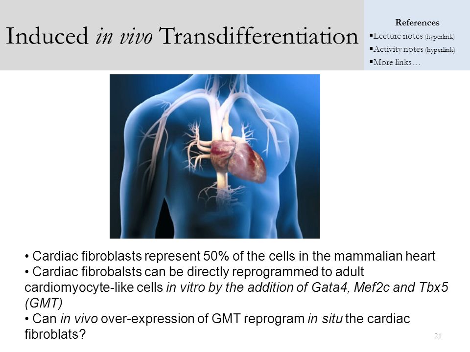
Cardiac fibroblasts represent 50% of the cells in the mammalian heart Cardiac fibrobalsts can be directly reprogrammed to adult cardiomyocyte-like cells in vitro by the addition of Gata4, Mef2c and Tbx5 (GMT) Can in vivo over-expression of GMT reprogram in situ the cardiac fibroblats? 21 21
 Can in vivo over-expression of GMT reprogram in situ the cardiac fibroblats")
22
Induced in vivo Transdifferentiation
22 22
23
Summary Directed differentiation of ES/iPS cells is the production of various mature cell types (e.g. motor neurons, dopaminergic neurons) using defined growth factors or cytokines. The defined growth factors are crucial for generating these cells during normal embryonic development. Induced differentiation of ES/iPS cells is the production of various mature cell types (e.g. motor neurons, dopaminergic neurons) using defined transcription factors. The defined transcription factors are overexpressed and are sufficient to promote the differentiation of the cells into the appropriate cell type Transdifferentiation is the production of various mature cell types (e.g. epithelial cells) from another mature cell type (e.g. blood cell). Induced Transdifferentiation is the production of various mature cell types (e.g. motor neurons, dopaminergic neurons) from another mature cell type (e.g. fibroblasts, epatocytes) using defined transcription factors. 23 23
 using defined growth factors or cytokines. The defined growth factors are crucial for generating these cells during normal embryonic development. Induced differentiation of ES/iPS cells is the production of various mature cell types (e.g. motor neurons, dopaminergic neurons) using defined transcription factors. The defined transcription factors are overexpressed and are sufficient to promote the differentiation of the cells into the appropriate cell type. Transdifferentiation is the production of various mature cell types (e.g. epithelial cells) from another mature cell type (e.g. blood cell). Induced Transdifferentiation is the production of various mature cell types (e.g. motor neurons, dopaminergic neurons) from another mature cell type (e.g. fibroblasts, epatocytes) using defined transcription factors")
Similar presentations


Default state is neural 2)Local secretion of BMPs by epidermis inhibits neural fate 3)Local secretion of noggin,>")


>")


>")

