Download presentation
Presentation is loading. Please wait.

1
The Upper Limb Viroj Mitranonda Department of Anatomy
Faculty of Science Mahidol University

2
Shoulder or Scapular region Arm Forearm Hand
Parts of the upper limb Shoulder or Scapular region Arm Forearm Hand

3
Bones Shoulder : Scapula, Clavicle Arm : Humerus
Forearm : Radius, Ulna Hand : 8 carpal bones, : 5 metacarpal bones, : 14 phalanges carpal bones : trapezium, trapezoid, capitate, hamate : scaphoid, lunate, triquetrum, pisiform

7
Superficial structures of the upper limb
Cutaneous nerves Superficial veins Lymph vessels

8
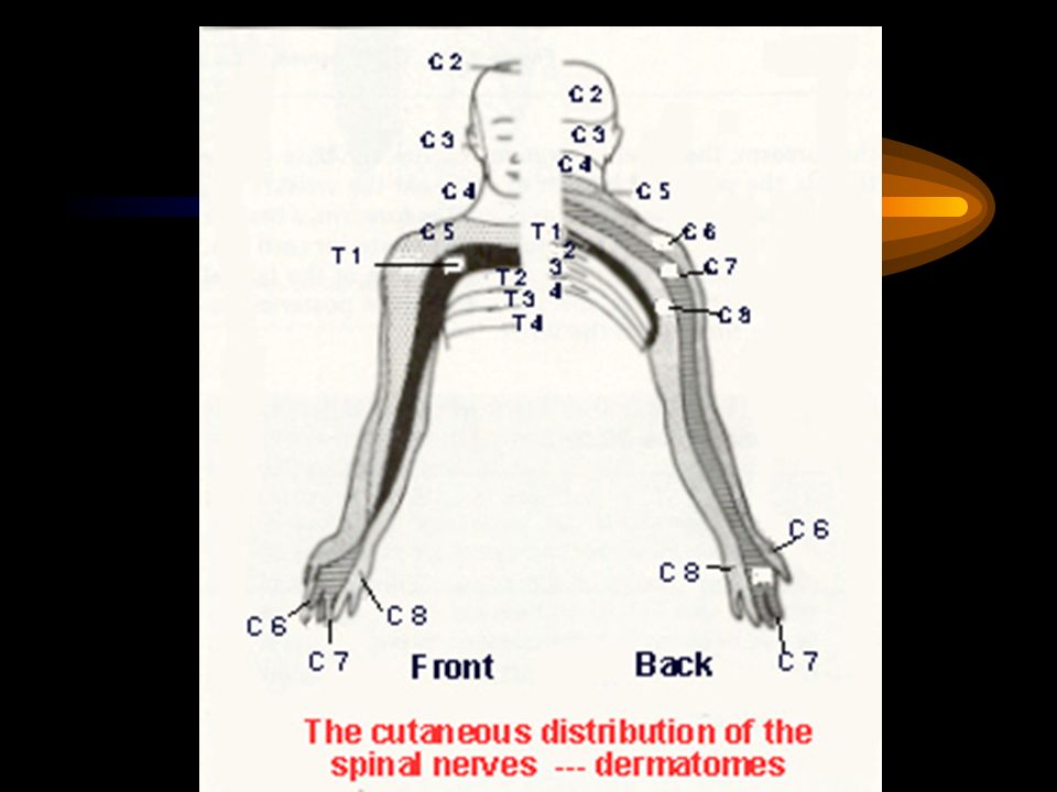
Cutaneous nerves of the upper limb
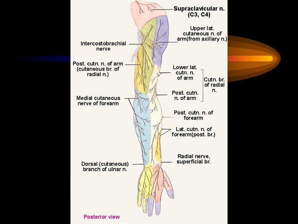
Shoulder : Lateral branches of the supraclavicular n.(c.3,4) Arm : Upper lateral cutaneous nerve of arm (axillary n.) : Lower lateral cutaneous nerve of arm (radial n.) : Medial cutaneous nerve of arm (medial cord) : Posterior cutaneous nerve of arm (radial n.) : Intercosto-brachial nerve (T.2)
 Arm. : Upper lateral cutaneous nerve of arm (axillary n.) : Lower lateral cutaneous nerve of arm (radial n.) : Medial cutaneous nerve of arm (medial cord) : Posterior cutaneous nerve of arm (radial n.) : Intercosto-brachial nerve (T.2)")
9
Cutaneous nerves of the upper limb (continued)
Forearm Lateral cutaneous nerve of forearm (from musculocutaneous n.) Medial cutaneous nerve of forearm (from medial cord) Posterior cutaneous nerve of forearm (from radial n.)
 Medial cutaneous nerve of forearm (from medial cord) Posterior cutaneous nerve of forearm (from radial n.)")
10
Cutaneous nerves of the upper limb (continued)
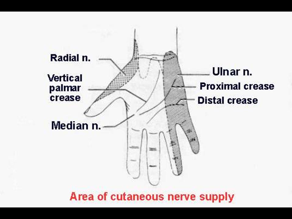
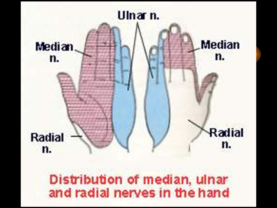
Hand Lateral 3 1/2 of the palm : Cutaneous branches of the median n. of the dorsum : Cutaneous branches of the radial n Medial 1 1/2 of the palm : Cutaneous branches of the ulnar n. of the dorsum : Cutaneous branches of the ulnar n. Distal part of the dorsum of the lateral 3 1/2 fingers : Cut. br. of the median n.

15
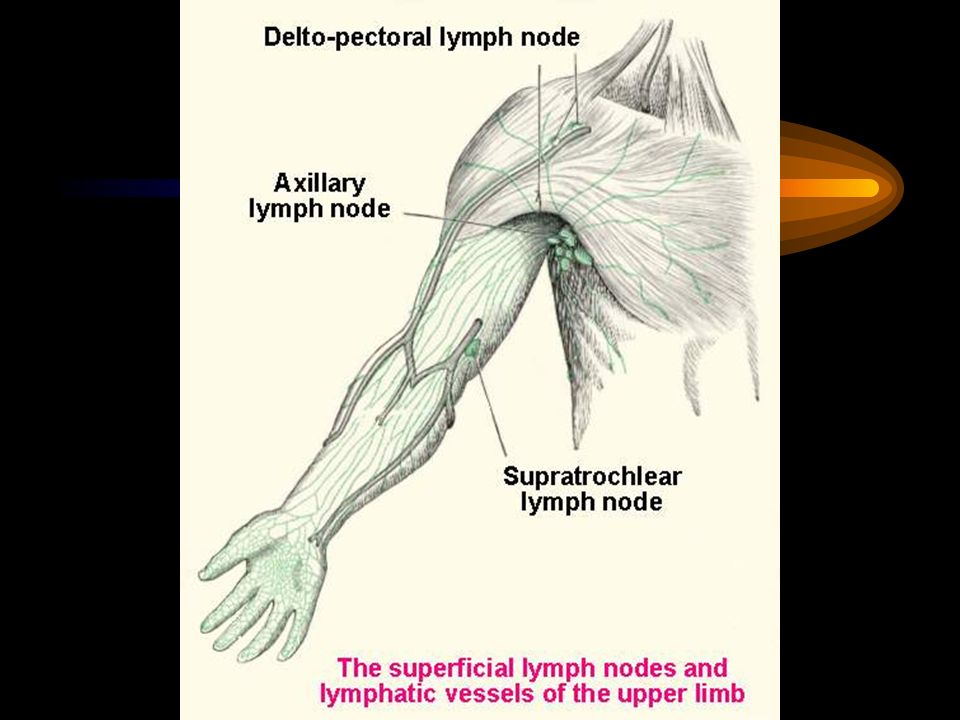
Superficial veins of the upper limb
Digital veins Dorsal venous arch Cephalic vein Basilic vein Median cubital vein Median veins of forearm (Median cephalic vein) (Median basilic vein)
 (Median basilic vein)")
19
The lymph vessels Accompanied the superficial veins The medial side
drains to the lateral group of the axillary node The lateral side drains to the delto-pectoral node

21
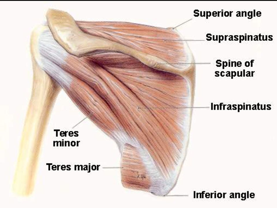
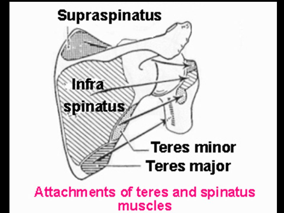
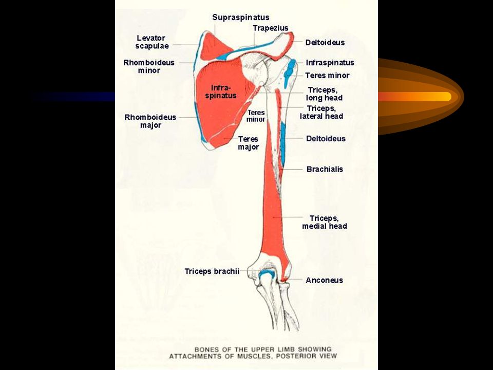
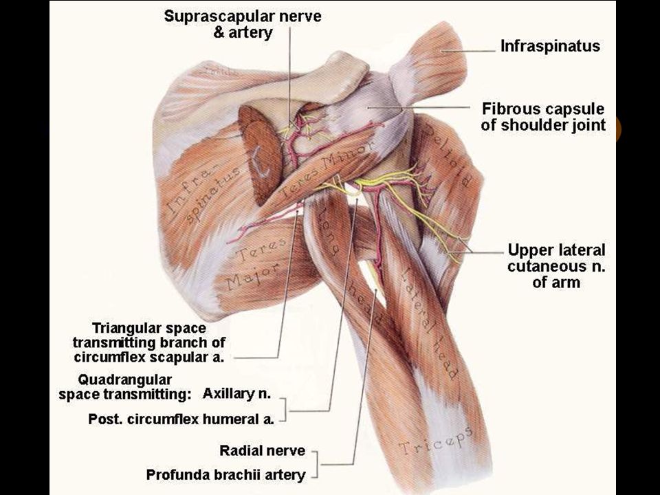
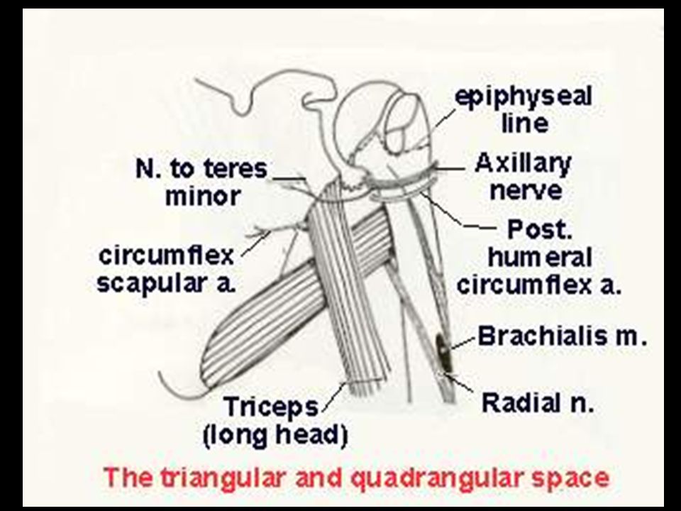
Muscles found in the shoulder (scapular) region
Axillary nerve Suprascapular nerve Upper and Lower subscapular nerves Lower subscapular nerve Deltoideus muscle Supraspinatus muscle Infraspinatus muscle Teres minor muscle Subscapularis muscle Teres major muscle

25
The rotator cuff muscles
Consists of : Supraspinatus m. Infraspinatus m. Teres minor m. Subscapularis m.

28
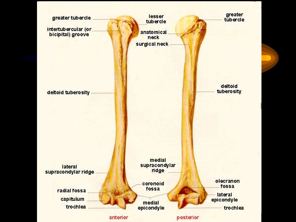
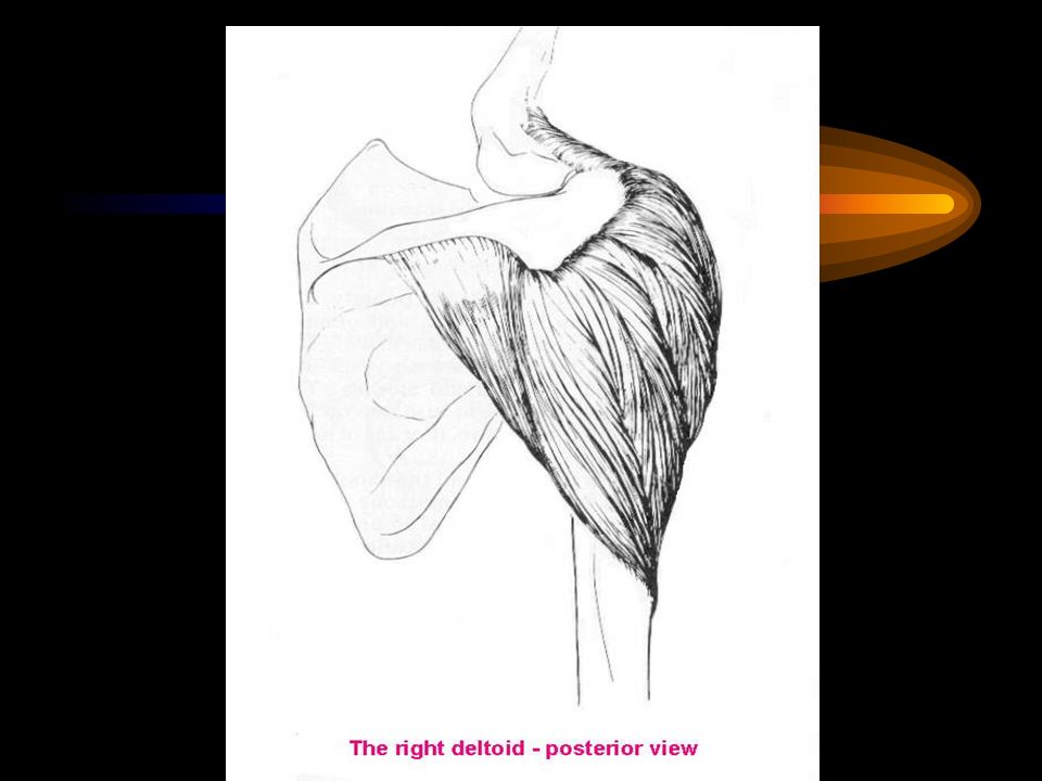
Deltoideus muscle Origin : Lateral 1/3 of clavicle,
: Acromion, Spine of scapula insertion : Humerus, deltoid tuberosity Action : Arm Flexion, medial rotation, Abduction, extension, lateral rotation nerve : Axillary nerve

30
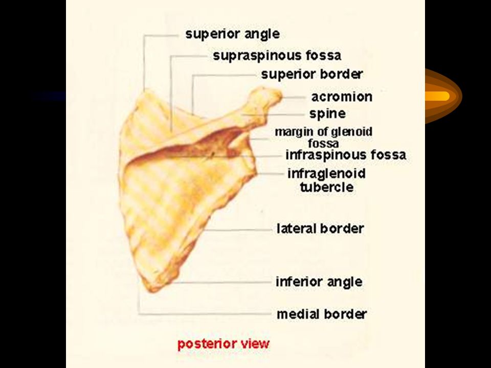
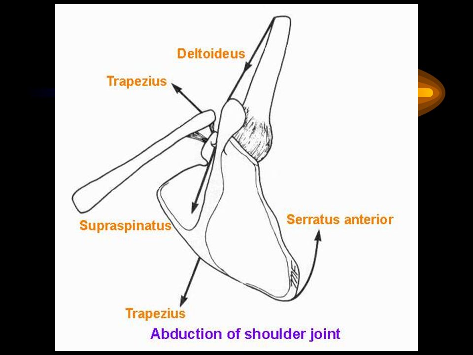
Supraspinatus muscle Origin : Scapula, supraspinous fossa
Insertion : Upper facet of greater tuberosity of Humerus Action : Abduction Nerve : Suprascapular nerve

31
Infraspinatus muscle Origin : Scapula, infraspinous fossa
Insertion : Middle facet of greater tuberosity of the Humerus Action : Lateral rotation Nerve : Suprascapular nerve

32
Teres minor muscle Origin : Scapula, superior 2/3 Lateral margin
Insertion : Lower facet of greater tuberosity of the Humerus Action : Lateral rotate nerve : Axillary nerve

36
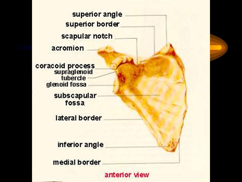
Subscapularis muscle Origin : Scapula, subscapular fossa
Insertion : Lesser tuberosity of the Humerus Action : Medial rotation Nerve : Upper and lower subscapular nerve

37
Teres major muscle Origin : Inf. 1/3 lat. margin of scapula
Insertion : Humerus, crest of lesser tubercle Action : Adduction,medial rotate, extension Nerve : Lower subscapular nerve

43
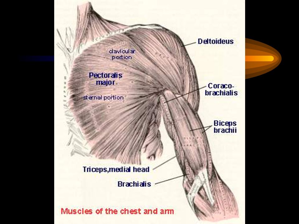
Flexor muscles of the arm
Musculocutaneous n. Musculocutaneous & radial nn. Coracobrachialis m. Biceps brachii m. Brachialis m. Extensor muscle of the arm Triceps brachii m. Radial n.

50
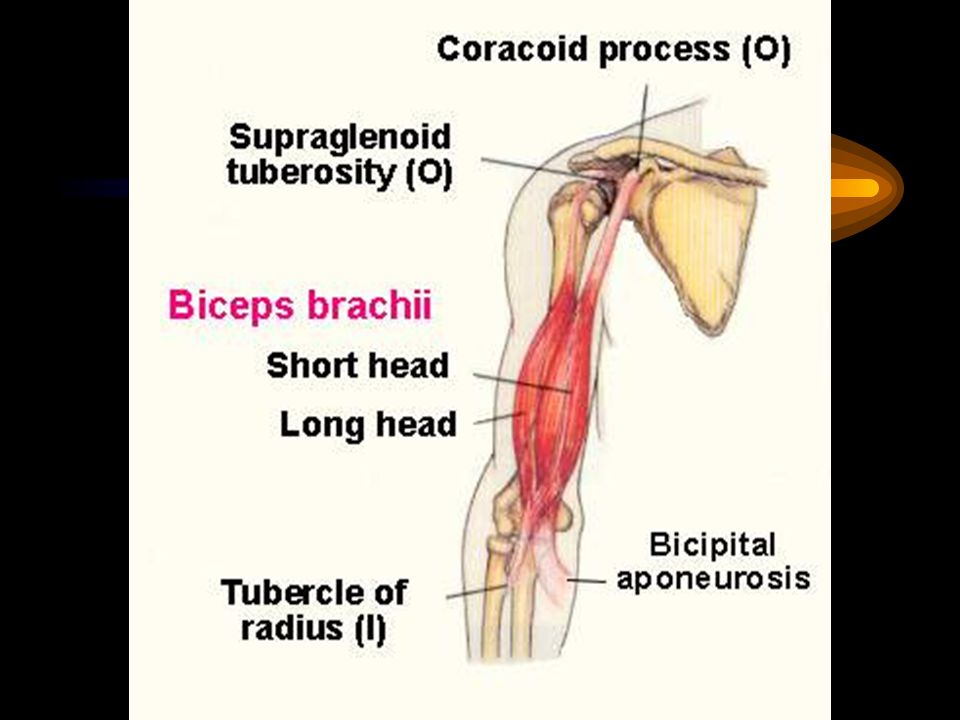
Biceps brachii muscle Origin : Short head; Coracoid process
: Long head; Supraglenoid tubercle Insertion : Radial tuberosity, bicipital aponeurosis Action : Supination, flexes elbow Nerve : Musculocutaneous nerve

52
Coracobrachialis muscle
Origin : Coracoid process Insertion : Humerus, middle of body,medial Action : Flexion (flexes shoulder) Nerve : Musculocutaneous nerve
 Nerve : Musculocutaneous nerve.")
54
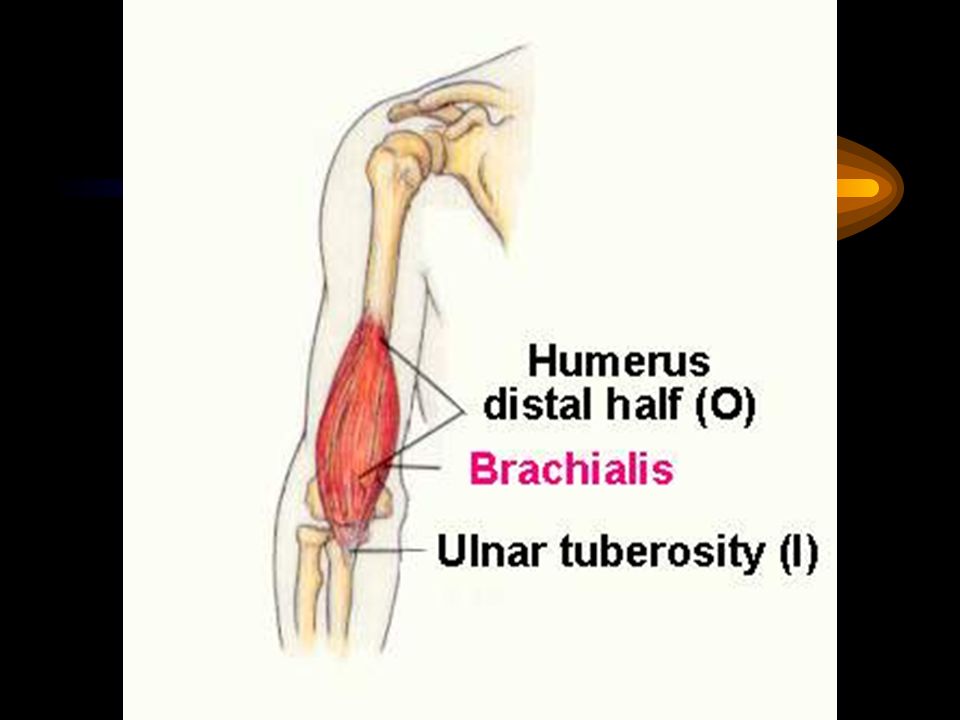
Brachialis muscle Origin : Humerus, ant. surface distal 1/2
Insertion : Ulna, coronoid process, ulnar tuberosity Action : Flexes elbow Nerve : Musculocutaneuos n. & radial n.

56
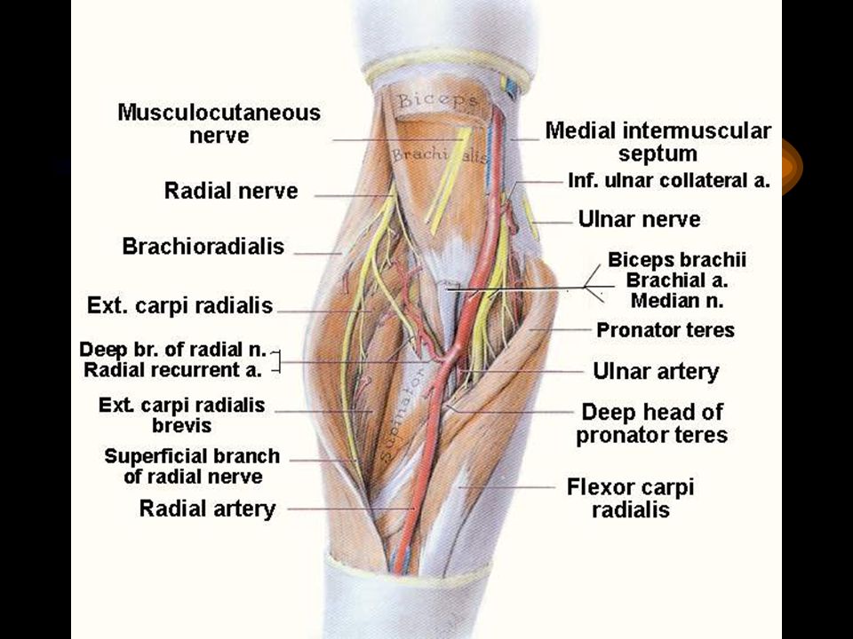
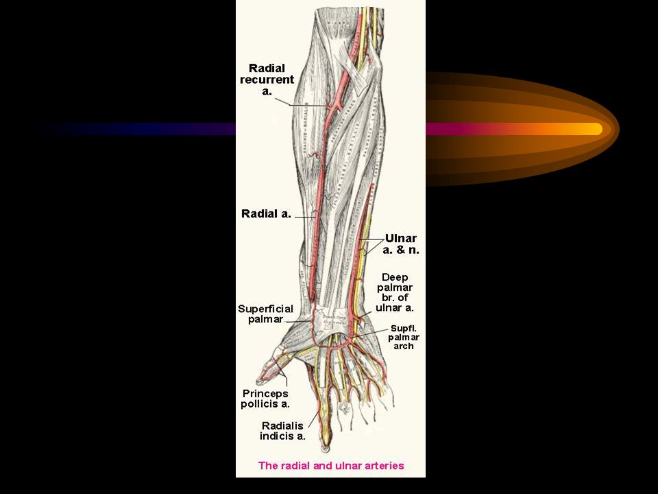
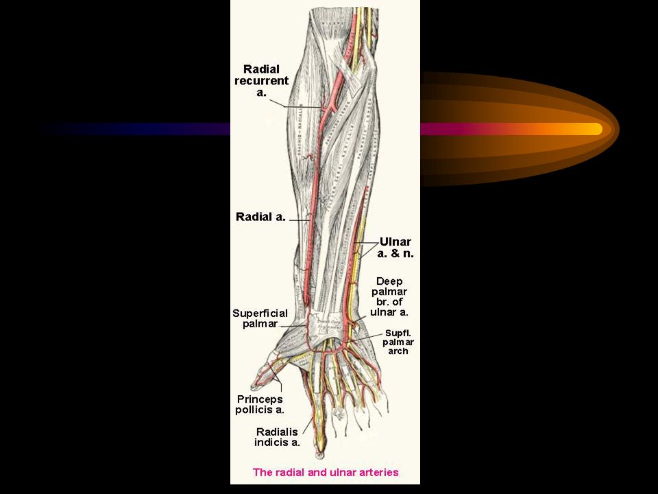
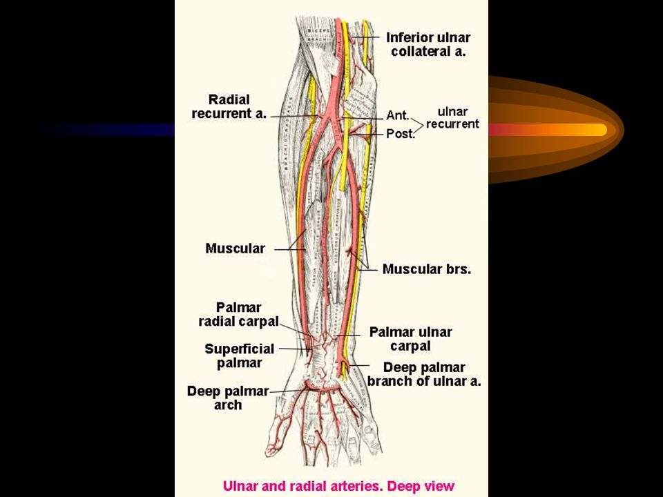
Cubital fossa Boundaries
Base : Imaginary line between the condyles of the humerus Laterally : Brachioradialis muscle Medially : Pronator teres muscle Apex : Brachioradialis m. overlapped Pronator teres m. Roof : Skin, superficial fascia & veins, bicipital aponeurosis Floor : Brachialis m.,Supinator m. Contents : Tendon of Biceps brachii m. Brachial a. and its’ terminal branches(radial a. and ulnar a.) Median n., fat
 Median n., fat.")
62
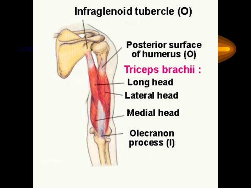
Triceps brachii muscle
Origin : Long head;Scapula, infraglenoid tubercle : Lateral head; Humerus, post. surface above groove for radial nerve : Medial head; below groove for radial n. Insertion : Ulna,olecranon process Action : Extends elbow Nerve : Radial nerve

63
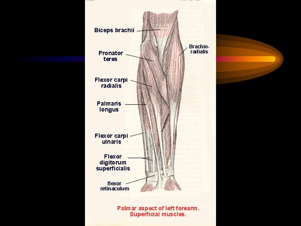
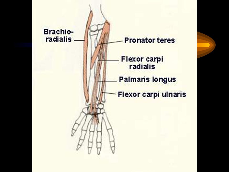
Flexor muscles of forearm
The forearm Flexor muscles of forearm Superficial layer Pronator teres muscle Flexor carpi radialis muscle Palmaris longus muscle Flexor carpi ulnaris muscle Median nerve Median nerve Ulnar nerve

65
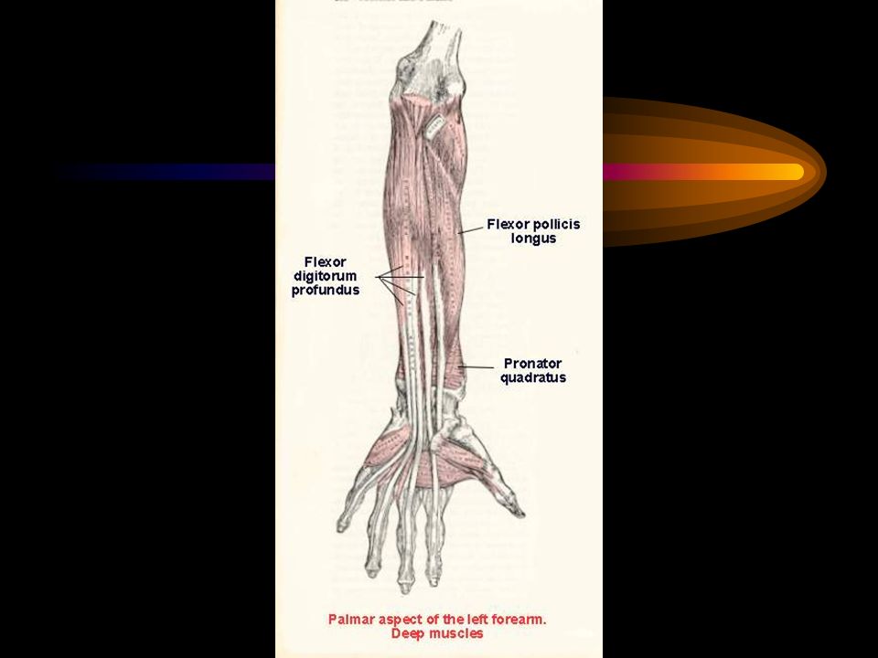
Flexor muscles of forearm (continued)
Intermediate layer Flexor digitorum superficialis (sublimis) muscle Median nerve Deep layer Flexor pollicis longus muscle Flexor digitorum profundus muscle Pronator quadratus muscle Median nerve Median&Ulnar nn.
 muscle. Median nerve. Deep layer. Flexor pollicis longus muscle. Flexor digitorum profundus muscle. Pronator quadratus muscle. Median nerve. Median&Ulnar nn.")
68
Pronator teres muscle Origin : Medial epicondyle of humerus,
: Coronoid process of ulna Insertion : Midlateral surface of radius Action : Pronation Nerve : Median nerve

69
Flexor carpi radialis muscle
Origin : Medial epicondyle of humerus Insertion : Base of 2nd and 3rd metacarpal bones Action : Flexes and abducts wrist (hand) Nerve : Median nerve
 Nerve : Median nerve.")
70
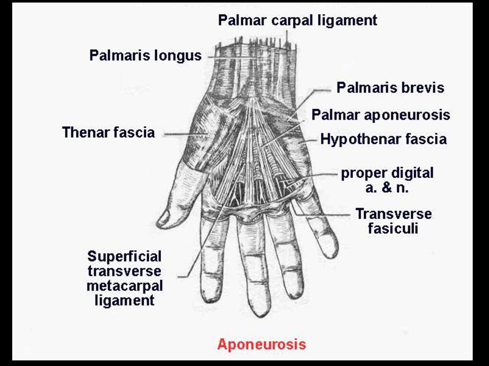
Palmaris longus muscle
Origin : Medial epicondyle of humerus Insertion : Palmar aponeurosis Action : Flexes wrist Nerve : Median nerve

71
Flexor carpi ulnaris muscle
Origin : Medial epicondyle of humerus, Upper dorsal border of ulna Insertion : Pisiform bone (& base of 5th metacarpal bone) Action : Flexes wrist, adduct wrist (hand) Nerve : Ulnar nerve
 Action : Flexes wrist, adduct wrist (hand) Nerve : Ulnar nerve.")
73
Flexor digitorum superficialis muscle
Origin : Medial epicondyle of humerus, Coronoid process of ulna, Oblique line of radius Insertion : Middle phalanges of medial four fingers Action : Flexes middle phalanges of each finger, Flexion of MP & PIP joints Nerve : Median nerve

75
Flexor pollicis longus muscle
Origin : Radius, ant. surface middle 1/2 Insertion : Distal phalanx of thumb Action : Flexes thumb Nerve : median nerve

76
Flexor digitorum profundus muscle
Origin : Ulna, prox. 2/3 ant. and medial surfaces Insertion : Bases of distal phalanges of medial four fingers Action : Flexes distal phalanges of each finger (DIP joints) Flexes MP & IP joints (PIP joints) Nerve : Median and ulnar nerves (lateral 1/2 : median nerve, medial 1/2 : ulnar nerve)
 Flexes MP & IP joints (PIP joints) Nerve : Median and ulnar nerves. (lateral 1/2 : median nerve, medial 1/2 : ulnar nerve)")
78
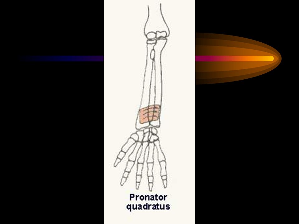
Pronator quadratus muscle
Origin : Ulna, ant. surface distal 1/4 Insertion : Radius, ant. surface distal 1/4 Action : Pronation. Holds radius to ulna Nerve : Median nerve

85
THE HAND Viroj Mitranonda Department of Anatomy Faculty of Science
Mahidol University

86
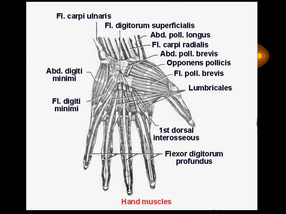
Muscles of the hand 3 Thenar muscles : Abductor pollicis brevis m.
Flexor pollicis brevis m. Opponens pollicis m. Median n. Adductor pollicis m. Ulnar n.

87
Muscles of the hand (continued)
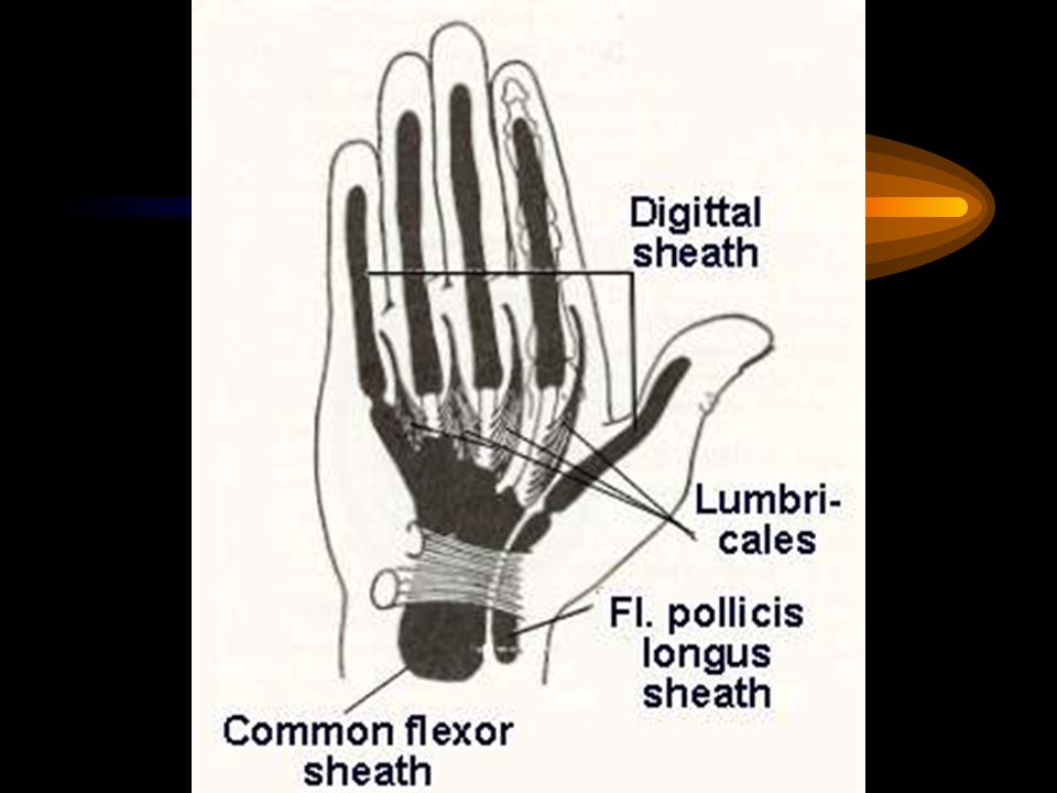
3 Hypothenar muscles : Abductor digiti minimi m. Flexor digiti minimi m. Opponens digiti minimi m. Ulnar n. 3 Palmar interossei mm. 4 Dorsal interossei mm. 4 Lumbricales mm. Ulnar n. 1st and 2nd Median n. 3rd and 4th Ulnar n. Palmaris brevis m. Ulnar n.

111
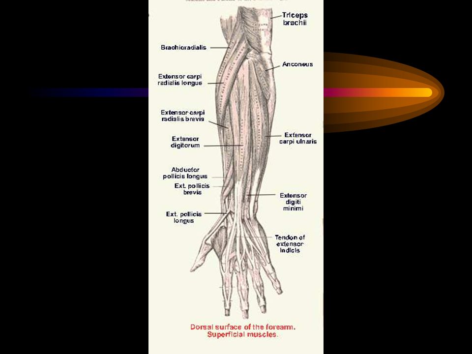
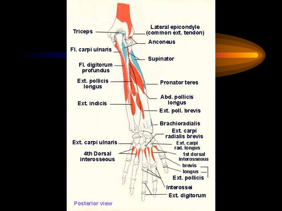
Extensor muscles of forearm
Superficial group Brachioradialis m.* Extensor carpi radialis longus m. * Extensor carpi radialis brevis m.** Laterally Posteriorly Extensor digitorum m.** Extensor digiti minimi m.** Extensor carpi ulnaris m.** Anconeus m.* * = Radial nerve ** = deep branch of radial nerve

113
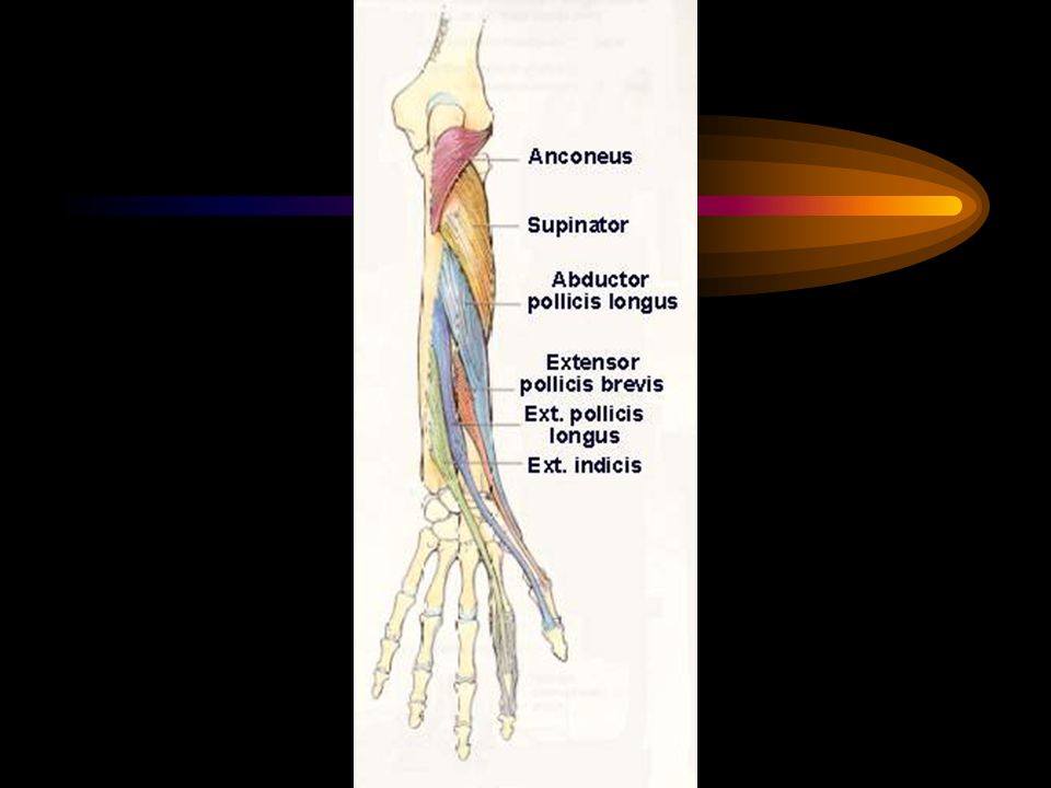
Extensor muscles of forearm (continued)
Deep group Supinator m. Abductor pollicis longus m. Extensor pollicis brevis m. Extensor pollicis longus m. Extensor indicis m. Nerve supply : Deep branch of radial nerve or posterior interosseous nerve

116
Brachioradialis muscle
Origin : Humerus, lateral supracondylar line Insertion : Radius, base of styloid process Action : Flexes elbow Nerve : Radial nerve

118
Extensor carpi radialis longus muscle
Origin : Humerus, lateral supracondylar line Insertion : Base of second metacarpal bone Action : Extends wrist, abduct hand Nerve : Radial nerve

119
Extensor carpi radialis brevis muscle
Origin : Humerus, lateral epicondyle Insertion : Base of third metacarpal bone Action : Extends wrist, abduct hand Nerve : Deep branch of the radial nerve

120
Extensor carpi ulnaris muscle
Origin : Lateral epicondyle of humerus, posterior border of ulna Insertion : Base of fifth metacarpal bone Action : Extends wrist, adduct hand Nerve : Deep branch of radial nerve

122
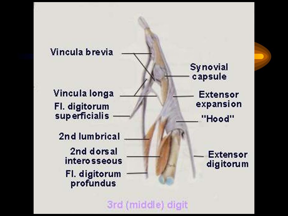
Extensor digitorum muscle
Origin : Humerus, lateral epicondyle Insertion : Middle and distal phalanges, extensor expansion Action : Extends phalanges Nerve : Deep branch of radial nerve

124
Extensor digiti minimi muscle
Origin : Humerus, lateral epicondyle Insertion : Extensor expansion of fifth digit Action : Extends fifth digit Nerve : Deep branch of radial nerve

126
Anconeus muscle Origin : Lateral epicondyle of humerus
Insertion : Proximal 1/3 lat. surface of ulna Action : Holds ulna to radius, extends elbow Nerve : Radial nerve

127
Supinator muscle Origin : Humerus, lateral epicondyle
: Ulna, supinator crest Insertion : Proximal 1/3 of radius Action : Supination Nerve : Deep branch of radial nerve

130
Abductor pollicis longus muscle
Origin : Radius and ulna distal to supinator m. Insertion : Base of first metacarpal bone Action : Abducts thumb Nerve : Deep branch of radial nerve (Posterior interosseous nerve)
")
131
Extensor pollicis brevis muscle
Origin : Radius, posterior surface Insertion : Base of proximal phalanx of thumb Action : Extends thumb Nerve : Deep branch of radial nerve (Posterior interosseous nerve)
")
132
Extensor pollicis longus muscle
Origin : Middle 1/3 post. surface of ulna Insertion : Distal phalanx of thumb Action : Extends thumb Nerve : Deep branch of radial nerve (Posterior interosseous nerve)
")
133
Extensor indicis muscle
Origin : Ulna, posterior surface Insertion : Extensor expansion Action : Extends index Nerve : Deep branch of radial nerve (Posterior interosseous nerve)
")
134
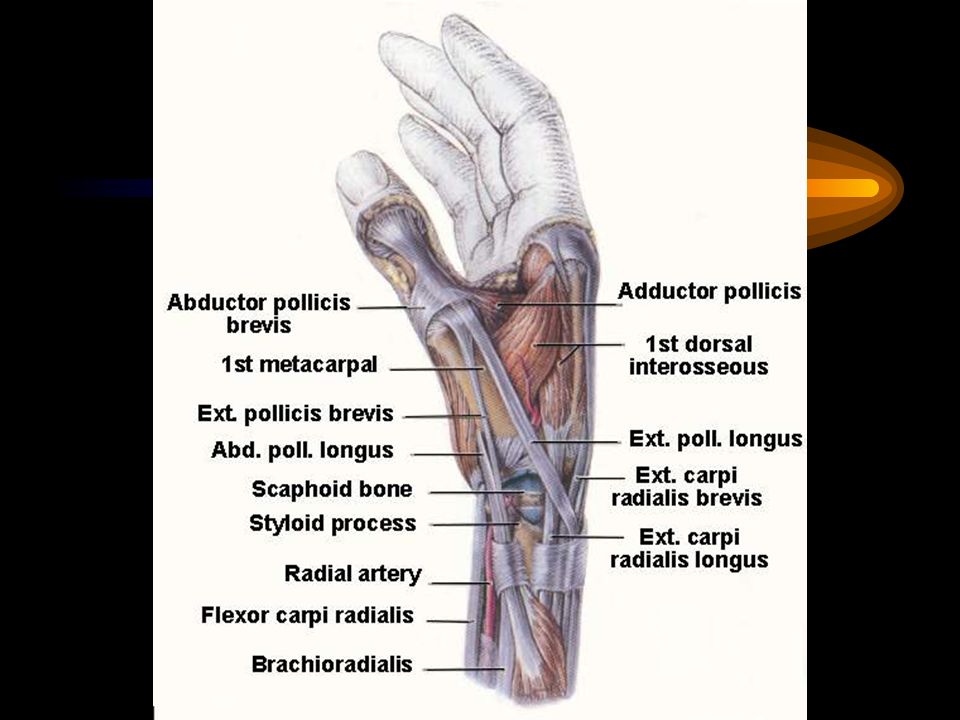
: Bounded the anatomical snuff box
Out cropping muscles Abductor pollicis longus m. Extensor pollicis brevis m. Extensor pollicis longus m. : Bounded the anatomical snuff box

140
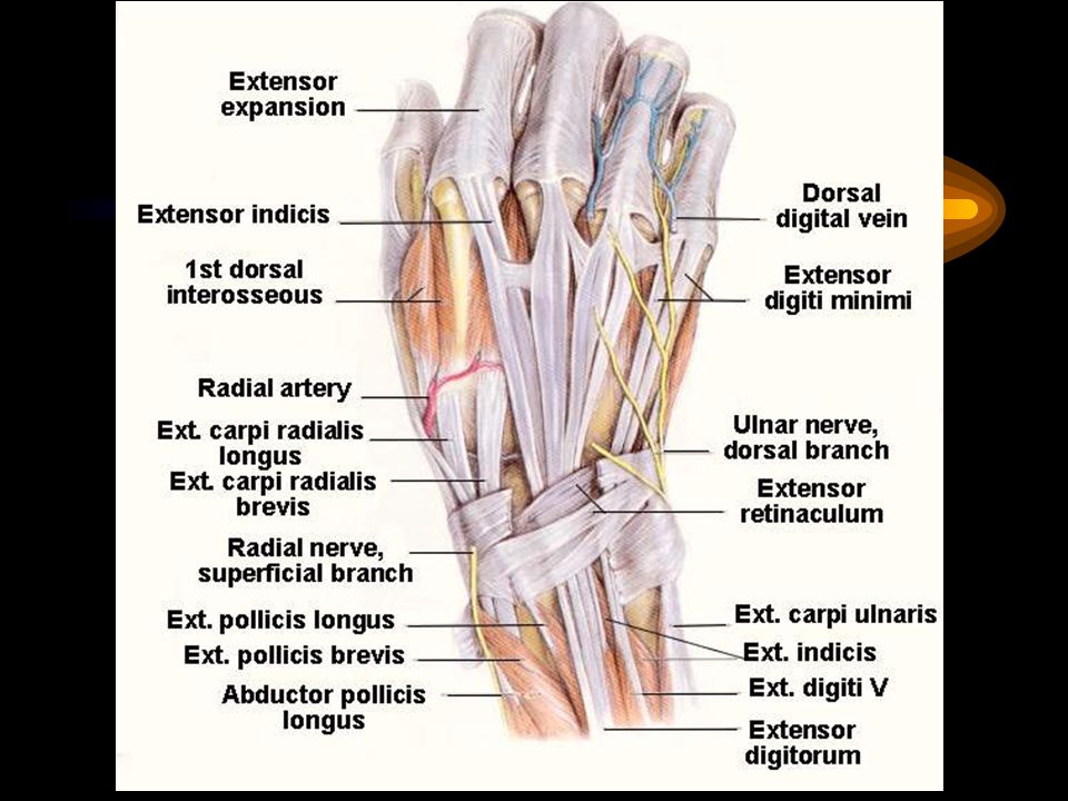
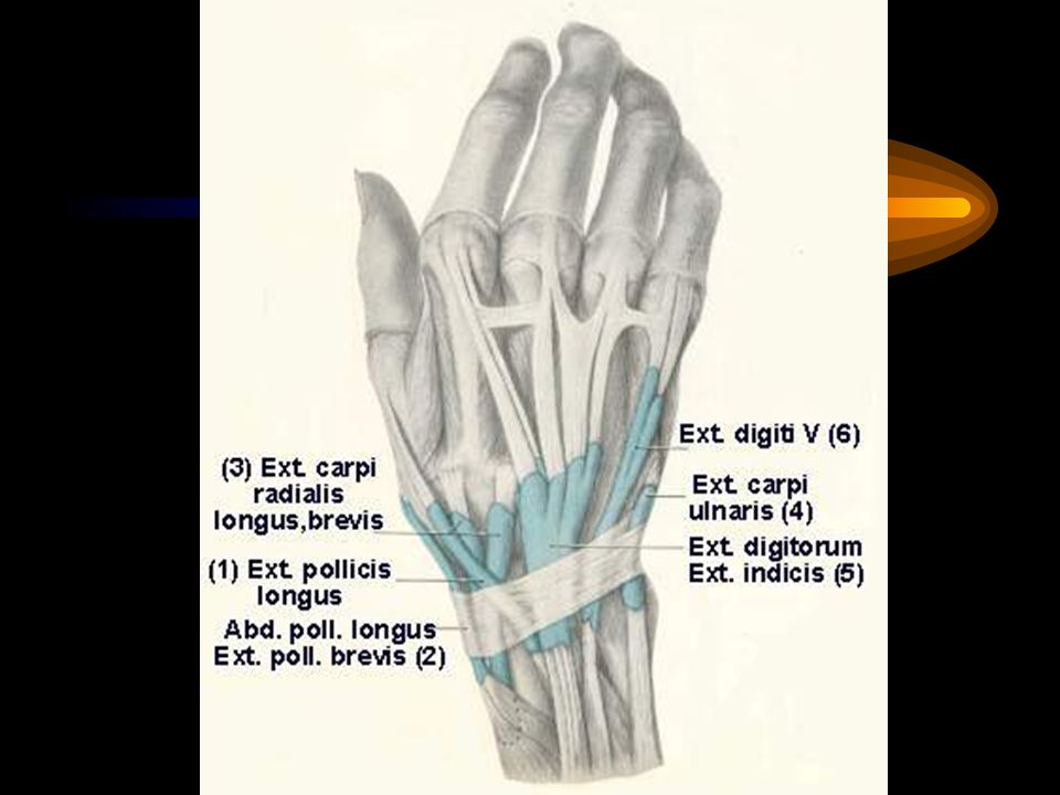
Tendon of the muscles in the fascial tunnels
2 = Abductor pollicis longus and extensor pollicis brevis 3 = Extensor carpi radialis longus and brevis 1 = Extensor pollicis longus 5 = Extensor digitorum and extensor indicis 6 = Extensor digiti minimi 4 = Extensor carpi ulnaris

142
Arthrology (Study of joint)
1. According to the movement 1.1 Immovable or synarthrosis eg. suture

143
1.2 Slightly movable or amphi(ar)throsis
Fibrous tissue lies between the bones (syndesmosis) eg. Intermediate radio-ulnar jt. Cartilage joins the bones (synchondrosis) eg. Pubic symphysis, intervertebral disc, sterno-costal joint
 eg. Intermediate radio-ulnar jt. Cartilage joins the bones (synchondrosis) eg. Pubic symphysis, intervertebral disc, sterno-costal joint.")
144
1.3 Freely movable (diarthrosis)
have joint cavity, joint capsule eg. Shoulder joint, elbow joint

145
2.According to the structures found in the joint
2.1. Fibrous joint there are fibrous tissue in the joint eg. Suture, radio-ulnar joint, tibio-fibular joint 2.2. Cartilagenous joint there are cartilage in the joint eg. Pubic symphysis, sterno-costal joint

146
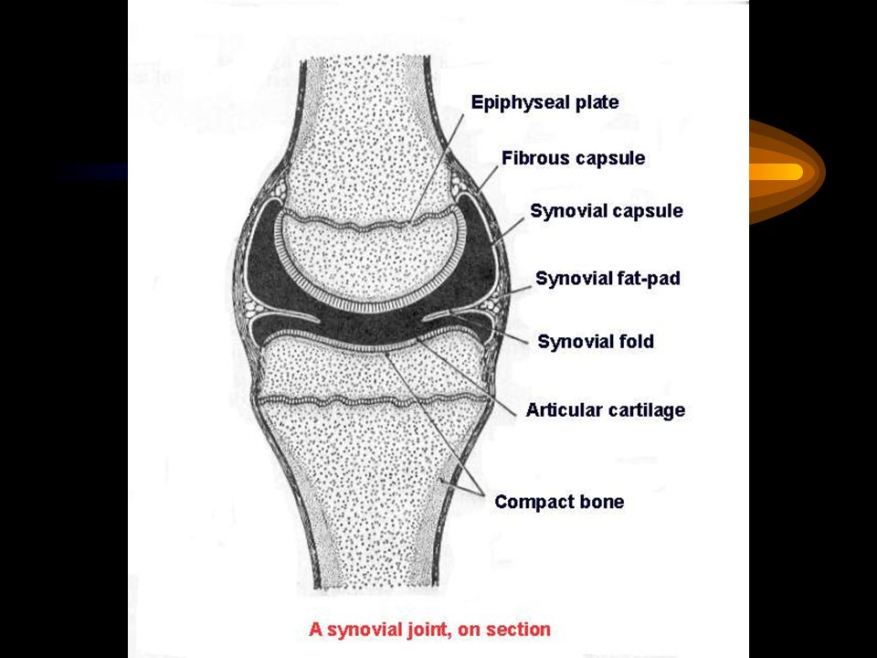
2.3. Synovial joint General patterns of synovial joint
There are at least 2 bones It’s articular surfaces lied with hyaline cartilage (articular cartilage) It has articular or joint capsule It has joint cavity Inner surface of the joint capsule lied with synovial membrane which secrete the synovial fluid or synovia
 It has articular or joint capsule. It has joint cavity. Inner surface of the joint capsule lied with synovial membrane which secrete the synovial fluid or synovia.")
148
Classification of the synovial joint
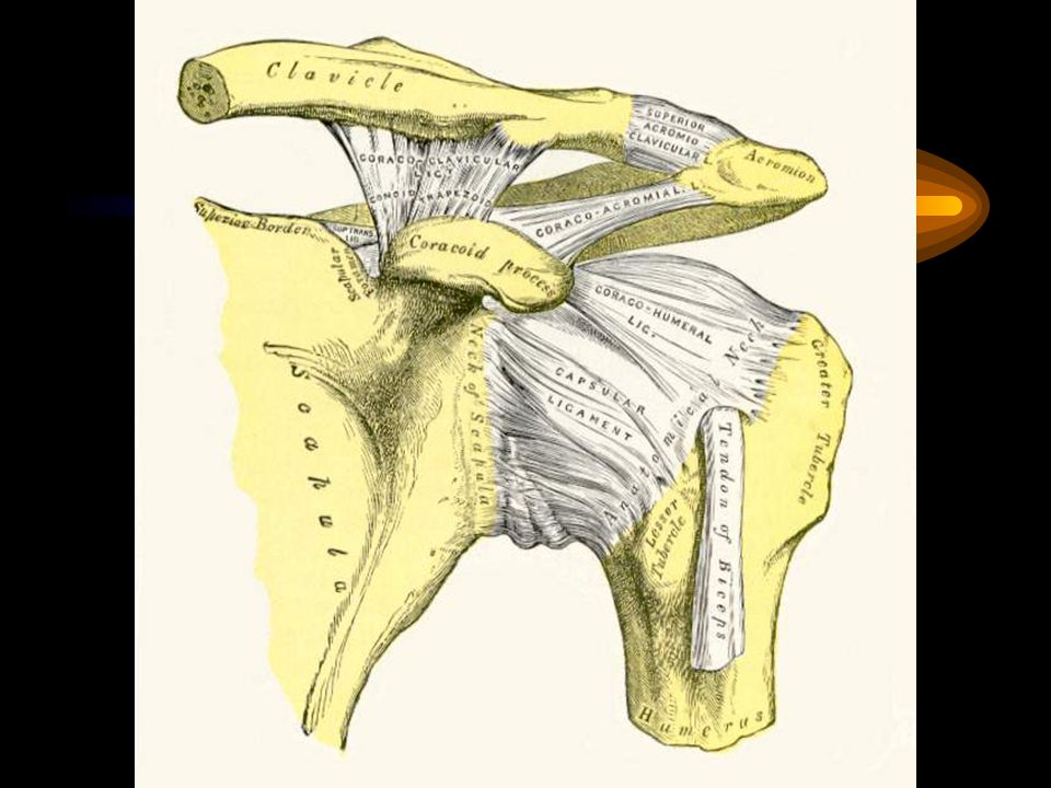
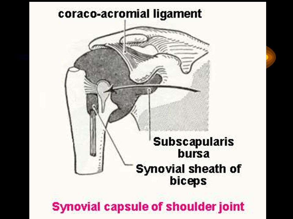
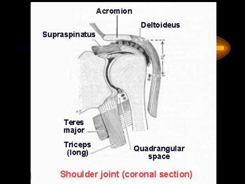
Classified into 7 types 1. The ball and socket type For example : shoulder joint Bones : humerus and scapula Ball : the head of the humerus Socket : primary socket is the glenoid cavity of the scapula : secondary socket is the coraco-acromial ligament

149
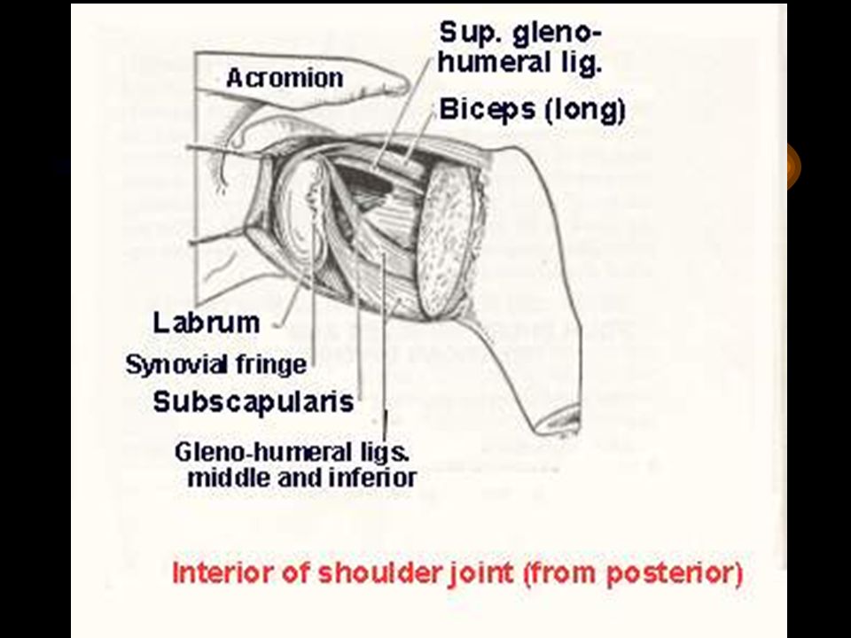
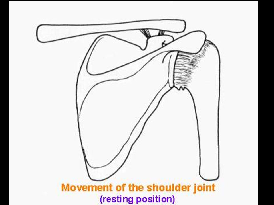
The ball and socket type (continue)
Movement : multiaxial - flex, extend, adduct,abduct and circumduct Joint capsule : thin fibrous membrane Ligaments : coraco-humeral, superior, middle and inferior gleno-humeral ligaments

160
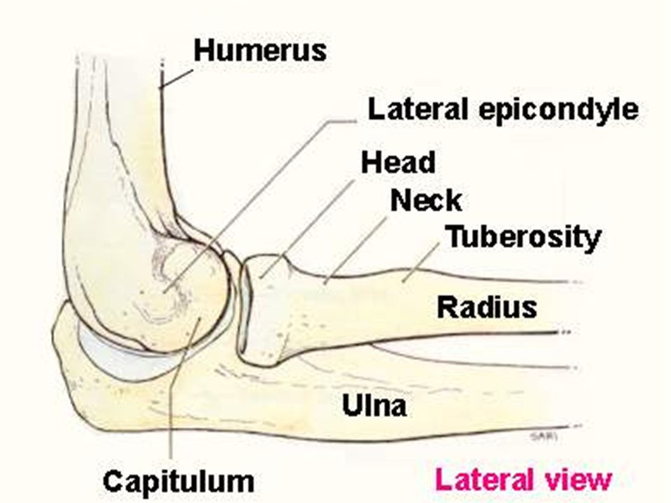
2. Hinge type For example : the elbow joint
Bones : humerus, ulna and radius Movement : around horizontal axis; flex and extend Joint capsule : thin at the anterior and posterior sides : thicken at medial and lateral sides Ligaments : medial or ulnar collateral ligament : lateral or radial collateral ligament

166
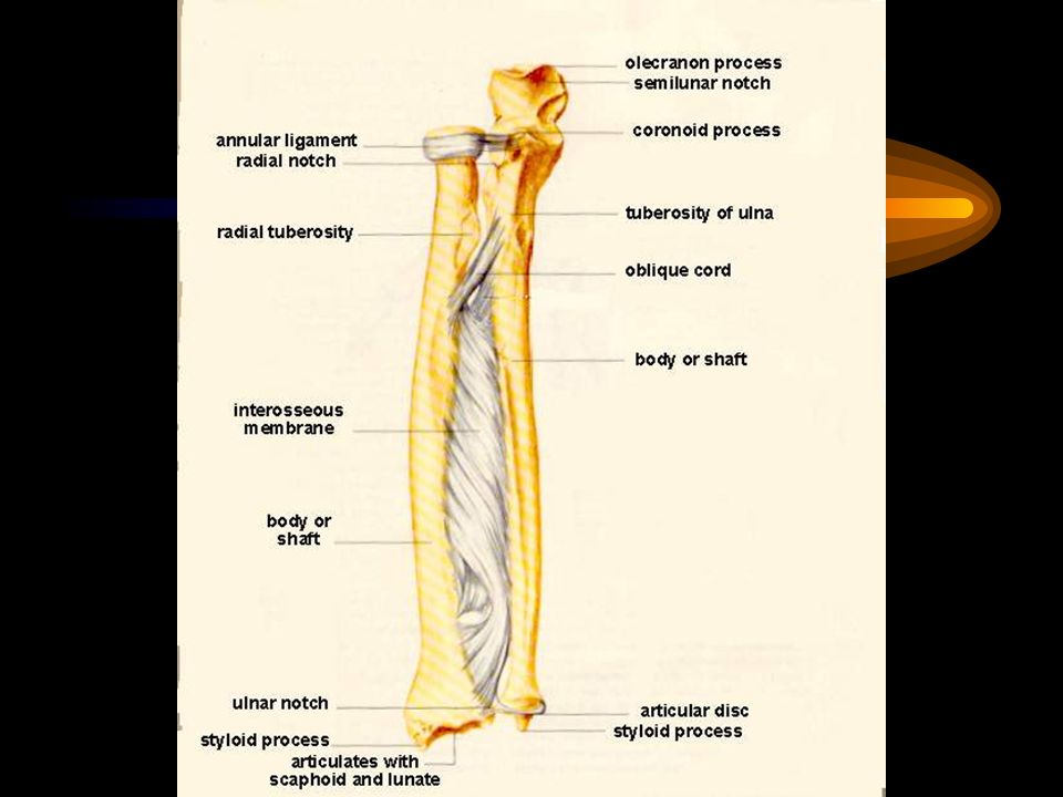
3. Pivot type For example : proximal radio-ulnar joint
Bones : radius and ulna Movement : around the vertical axis ; medial and lateral rotation Ligament : annular ligament

168
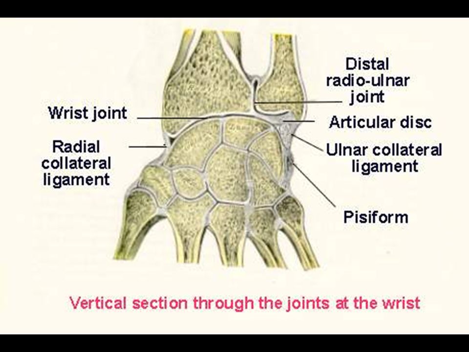
4. Ellipsoidal type For example : wrist or radio-carpal joint
Bones : distal end of radius, scaphoid, lunate and triquetrum Movement : modified hinge type; flex, extend, adduct and abduct Ligaments : medial or ulnar collateral ligament : lateral or radial collateral ligament

171
5. Plane or gliding type For example : intercarpal joints Bones : carpal bones Movement : gliding Ligaments : interosseous ligaments

172
6. Saddle type For example :1st carpometacarpal joint Bones : 1st metacarpal bone and trapezium Movement : flex, extend, adduct, abduct and circumduct

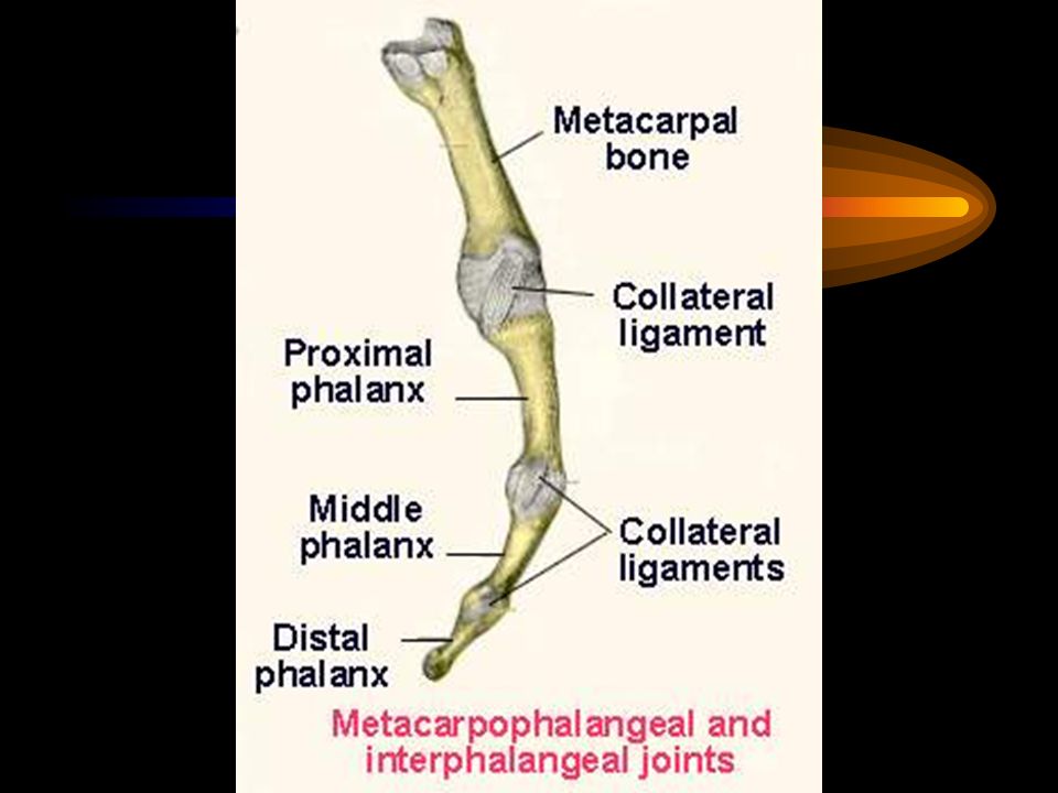
174
7. Condyloid type For example : metacarpophalangeal joint (MP) interphalangeal joint (IP) Bones : metacarpal bone, phalanges Movement : flex and extend, (adduct, abduct) Ligaments : medial and lateral collateral ligaments
 Ligaments : medial and lateral collateral ligaments.")
176
Joints of the upper limb
Sternno-clavicular joint Coraco-acromial joint Coraco-clavicular joint Acromio-clavicular joint Shoulder joint Elbow joint

177
Joints of the upper limb (continue)
Radio-ulnar joints (prox.,middle and distal) Wrist or radio-carpal joint Intercarpal joints Carpometacarpal joints Intermetacarpal joints Metacarpophalangeal joints (MP joint) Interphalangeal joints (IP joint)
 Wrist or radio-carpal joint. Intercarpal joints. Carpometacarpal joints. Intermetacarpal joints. Metacarpophalangeal joints (MP joint) Interphalangeal joints (IP joint)")
179
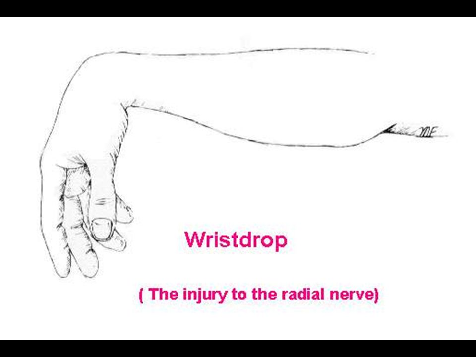
NERVE LESIONS OF UPPER LIMBS
BY VIROJ MITRANONDA Department of Anatomy Faculty of Science, Mahidol University

Similar presentations


>")


