Download presentation
Presentation is loading. Please wait.

1
JOINTS OF THE UPPER LIMB
Dr.Lubna Nazli

2
Objectives At the end of the lecture the students should be familiar with: Shoulder joint. Elbow joint. Radioulnar joints. Wrist joint. 5. Carpometacarpal joint of thumb.

3
Shoulder joint Bones forming the joint. Capsule Ligaments Relations
Movements Blood supply Nerve supply Applied anatomy

4
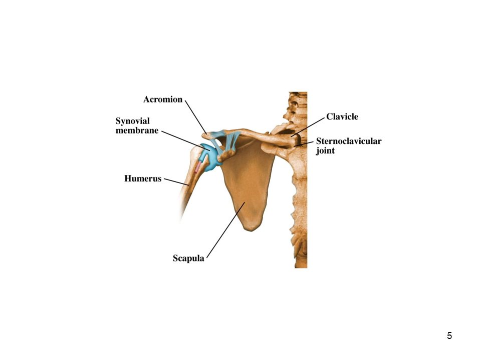
The shoulder-joint is a ball-and-socket type of synovial joint.
The bones entering into its formation are the hemispherical head of the humerus and the shallow glenoid cavity of the scapula. The joint is protected above by an arch, formed by the coracoid process, the acromion, and the coracoacromial ligament.

6
Articular cartilage The articular cartilage on the head of the humerus is thicker at the center than at the circumference, the reverse being the case with the articular cartilage of the glenoid cavity. Glenoid cavity is deepened by a rim of fibrocartilage called the glenoidal labrum.

8
Capsule The articular capsule completely encircles the joint.
It is attached, above, to the circumference of the glenoid cavity beyond the glenoidal labrum; below, to the anatomical neck of the humerus. It is thicker above and below.

10
Ligaments 1. Coracohumeral Ligament
This ligament is a broad band which strengthens the upper part of the capsule. It arises from the coracoid process, and passes to the greater tubercle of the humerus.

12
2. Glenohumeral ligaments:
There are 3 glenohumeral ligaments. Superior, middle & inferior. Superior ligament extends from the glenoid cavity to the upper part of the lesser tubercle of the humerus. Middle ligament passes from the glenoid cavity to the lower part of the lesser tubercle of the humerus.

13
Inferior ligament extends from glenoid cavity to the anatomical neck of the humerus.
3. Transverse Humeral Ligament It is a broad band passing from the lesser to the greater tubercle of the humerus.

14
Stability of the joint This is provided by rotator cuff of muscles or the musculotendinous cuff. What is this??

15
Synovial Membrane The synovial membrane is reflected from the margin of the glenoid cavity over the labrum; it is then reflected over the inner surface of the capsule, and covers the lower part and sides of the anatomical neck of the humerus as far as the articular cartilage on the head of the bone. The tendon of the long head of the Biceps brachii passes through the capsule and is enclosed in a tubular sheath of synovial membrane.

16
Relations Muscles in relation with the joint are:
Above: Supraspinatus. Below: long head of the Triceps brachii. In front: Subscapularis. Behind: Infraspinatus and Teres minor. Within: tendon of the long head of the Biceps brachii. Deltoid covers the joint in front, behind, and laterally.

17
Blood & nerve supply Arteries supplying the joint are anterior and posterior humeral circumflex, and transverse scapular arteries. Nerves are derived from the axillary and suprascapular nerves.

18
Movements The shoulder-joint has a variety of movements.
Flexion: by the Pectoralis major, anterior fibers of the Deltoid & Coracobrachialis. Extension: by Latissimus dorsi, Teres major, posterior fibers of the Deltoid.

19
Abduction: by the Deltoid and Supraspinatus
Adduction: by the Subscapularis, Pectoralis major, Latissimus dorsi, and Teres major. Medial rotation: by the Subscapularis, Latissimus dorsi, Teres major, Pectoralis major, and the anterior fibers of the Deltoid. Lateral rotation: by the Infraspinatus and Teres minor.

20
APPLIED ANATOMY Dislocations at the shoulder joint occur mostly anteriorly due to weakness of the anterior aspect of the joint. Anterior dislocations result from a fall on the outstretched arm. In frozen shoulder there is limited movement. The cause of limited movement may to some extent be anatomical/pathological. Anatomically the capsule may become adherent to itself, reducing the range of motion.

21
ELBOW JOINT Objectives: Bones forming the joint Capsule Ligaments
Muscles and movements Blood and nerve supply Applied anatomy

22
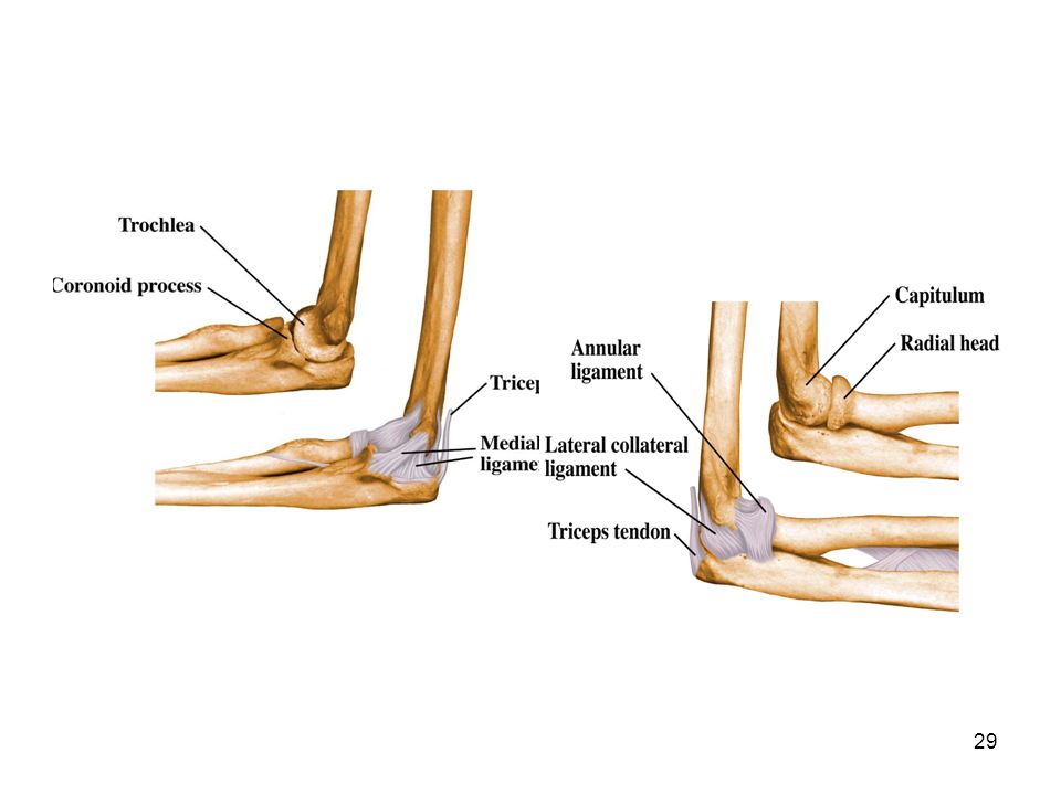
ELBOW JOINT The elbow-joint is a ginglymus or hinge-joint.
The trochlea of the humerus is received into the notch of the ulna, and the capitulum of the humerus articulates with the fovea on the head of the radius. CAPSULE: The articular surfaces are connected together by a capsule, which is thickened medially and laterally, and, to a less extent, in front and behind. These thickened portions are usually described as ligaments.

25
LIGAMENTS: The Anterior ligament. The Posterior ligament. The Ulnar Collateral. The Radial Collateral.

26
The Anterior Ligament The anterior ligament is a broad and thin fibrous layer covering the anterior surface of the joint. It is attached to the front of the medial epicondyle and to the front of the humerus. below to the anterior surface of the coronoid process of the ulna and to the annular ligament being continuous on either side with the collateral ligaments.

27
The Posterior Ligament
This posterior ligament is thin and membranous. Above, it is attached to the humerus immediately behind the capitulum and close to the medial margin of the trochlea, to the margins of the olecranon fossa, and to the back of the lateral epicondyle. Below, it is fixed to the upper and lateral margins of the olecranon, to the posterior part of the annular ligament, and to the ulna behind the radial notch.

28
The Ulnar Collateral Ligament
This ligament is a thick triangular band consisting of two portions, an anterior and posterior united by a thinner intermediate portion. The anterior portion, directed obliquely forward, is attached, above,to the front part of the medial epicondyle of the humerus; and, below, by its broad base to the coronoid process. The posterior portion, is attached, above, by its apex, to the back part of the medial epicondyle; below, to the medial margin of the olecranon. Between these two bands a transverse band bridges across the notch between the olecranon and the coronoid process.

30
The Radial Collateral Ligament
This ligament is a short and narrow fibrous band, attached, above, to the lateral epicondyle of the humerus; below, to the annular ligament.

31
Relations In front, the Brachialis. Behind , the Triceps and Anconeus.
Laterally , the Supinator, and the common tendon of origin of the Extensor muscles. Medially , the common tendon of origin of the Flexor muscles, and the Flexor carpi ulnaris.

32
Arteries supplying the joint are derived from the anastomosis between the profunda and the superior and inferior ulnar collateral branches of the brachial, with the anterior, posterior, and interosseous recurrent branches of the ulnar, and the recurrent branch of the radial. The nerves of the joint are from the ulnar and the median.

33
Movements The elbow-joint comprises three different portions
The joint between the ulna and humerus That between the head of the radius and the humerus. The proximal radioulnar articulation. All these articular surfaces are enveloped by a common synovial membrane.

34
Movements Flexion: Biceps brachii and Brachialis, assisted by the Brachioradialis. Extension: by the Triceps and Anconeus, assisted by the Extensors of the wrist. What is carrying angle?

35
Applied anatomy Supracondylar fracture of humerus,mostly seen in children. Dislocation of elbow seen in adults. Injury to lateral epicondyle produces increase in lateral deviation of forearm. This decreases carrying angle (cubitus valgus ) and thus ulnar nerve is stretched causing ulnar neuropathy.
 and thus ulnar nerve is stretched causing ulnar neuropathy.")
36
RADIO ULNAR JOINTS Superior radioulnar joint Middle radioulnar joint
Inferior radioulnar joint

37
Superior radioulnar joint
This articulation is a trochoid or pivot-joint between the circumference of the head of the radius and the ring formed by the radial notch of the ulna and the annular ligament. The Annular Ligament This ligament is a strong band of fibers, which encircles the head of the radius, and retains it in contact with the radial notch of the ulna.

38
Movements The movements are rotatory movements. Rotation forward being called pronation. Pronation is performed by the Pronator teres and Pronator quadratus. Rotation backward is supination. Supination is performed by the Biceps brachii and Supinator.

39
Middle Radioulnar joint
The shafts of the radius and ulna are connected by the Interosseous Membrane. The interosseous membrane is a broad and thin plane of fibrous tissue

40
Inferior radioulnar joint
This is a pivot-joint formed between the head of the ulna and the ulnar notch on the lower end of the radius. Ligaments: Anterior radioulnar ligament This ligament extends from the anterior margin of the ulnar notch of the radius to the front of the head of the ulna. Posterior radioulnar ligament

41
The Articular Disc The articular disc is triangular in shape, and is placed transversely beneath the head of the ulna. It is attached by its apex to the styloid process and the head of the ulna; and by its base to the radius, which separates the ulnar notch from the carpal articular surface.

42
Movements The movements consist of rotation of the lower end of the radius around an axis which passes through the center of the head of the ulna.

43
Applied anatomy Pulled elbow: It is seen in small children.
It is caused by dislocation of head of radius through the annular ligament due to pulling of hand in semi prone position.

44
Wrist joint The wrist-joint is a condyloid articulation.
The bones forming it are the lower end of the radius and under surface of the articular disc above and below are the carpal bones scaphoid,lunate and triquetral.

45
The joint is surrounded by a capsule.
It is strengthened by the ligaments. The anterior Radiocarpal. The posterior Radiocarpal. The Ulnar Collateral. The Radial Collateral.

46
The ulnar collateral ligament is a rounded cord, attached above to the end of the styloid process of the ulna & below to the carpal bones. The radial collateral ligament extends from the tip of the styloid process of the radius to the radial side of the navicular bone.

47
Movements The movements permitted in this joint are flexion, extension, abduction, adduction, and circumduction.

48
Carpometacarpal joint of the Thumb
It is saddle type of joint. The joint is surrounded by a capsule, which is thick but loose. It is lined by synovial membrane. Movements: Flexion & extension, adduction & abduction, circumduction and opposition.

Similar presentations


>")


 Lecture 3 Myology of the Elbow.>")


