Download presentation
Presentation is loading. Please wait.

1
Apheresis Matthew L. Paden, MD
Assistant Professor of Pediatric Critical Care Director, Pediatric ECMO

2
Disclosures Funded by NIH/FDA for CRRT/ECMO device development
Pending grant for pediatric apheresis device Much of this talk is stolen from others.

3
Objectives Review the technique of apheresis
Discuss common evidence based indications Few notes on technical aspects of concomitant ECMO/Plasma Exchange Things I have learned the hard way

4
Apheresis – what is it? Separation of blood into individual components based on density or molecular size Leukopheresis Erythrocytopheresis Plasmapheresis Plateletpheresis Common methods include Centrifugation Membrane filtration

5
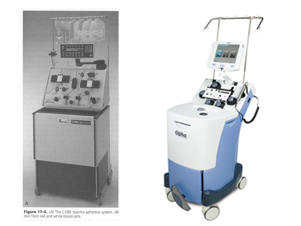
Apheresis Methods

6
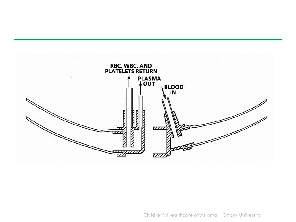
Separation by centrifugation
Milk separator Hand cranked Heavy milk goes to the side of the bowl Lighter cream stays in the middle Separate pathways for each to drain

9
Separation by density

11
Membrane Filtration Semi-porous membrane
Appropriate pore size for what you are trying to remove

12
The 5 “Whats” of Apheresis
What am I doing this for? What am I replacing with? What else am I removing? What is my anticoagulation? What is my extracorporeal volume?

13
What am I doing this for? Plasmapheresis / Plasma exchange
Most common apheresis procedure at our center Usually for removal of auto-antibodies (IgG) Only about 45% of your IgG is intravascular Need for repeated therapies One plasma volume (~45 mL/kg) removes about 63% of intravascular IgG
 Only about 45% of your IgG is intravascular. Need for repeated therapies. One plasma volume (~45 mL/kg) removes about 63% of intravascular IgG.")
14
What am I doing this for? Category I: primary/standard therapy
Category II: adjunctive therapy Category III: last-ditch effort (insufficient evidence to prove efficacy) Category IV: lack of efficacy in controlled trials
 Category IV: lack of efficacy in controlled trials.")
15
Description of the disease
Current management and treatment Rationale for therapeutic apheresis

16
Common Indications Category I Category II
Thrombotic thrombocytopenic purpura Guillian Barre Syndrome Wegener’s/Goodpasteur’s (dialysis dependence or pulmonary hemorrhage at presentation) Myasthenic crisis Category II Devic’s syndrome
 Myasthenic crisis. Category II. Devic’s syndrome.")
17
Common Indications Category III Category IV
Treatment of cardiac transplant antibody mediated rejection Sepsis with multiple organ failure Thyroid storm Category IV Diarrheal associated HUS SLE nephritis Schizophrenia

18
What am I replacing with?
Albumin or plasma? Depends on indication and patient condition Auto-antibody removal – almost always albumin Use FFP when you need replacement of factors Thrombotic thrombocytopenic purpura Liver failure Wegener’s granulomatosis with pulmonary hemorrhage Complication rate is higher with plasma Allergic, infectious, TRALI

19
What else am I removing? Coagulation factors ~25-50% Fibrinogen ~60%
Bilirubin ~45% Platelets ~30% Usually recover in 48 hours in HEALTHY patients Drugs – low volume of distribution, small molecular size

20
What is my anticoagulation?
Citrate Alkalosis – less than CRRT, because not continuous therapy Symptomatic hypocalcemia Serial monitoring of ionized calcium and patient symptoms If present, treat. Consider reduce citrate infusion rate, adding calcium drip, STOPPING THE PROCEDURE Hypomagesemia Some centers measure ionized magnesium levels as well Heparin rarely None

21
What is my extracorporeal volume?
Be aware of extracorporeal volume The disposables are made for adults not kids Current devices range from mL We blood prime if > 12% of TBV is extracorporeal Blood prime 125 mL pRBC 15 mL THAM 25 mL 25% Albumin 300 mg Calcium gluconate 10 mEq NaHCO3 50 units heparin

22
Erythrocytopheresis Removal/replacement of RBC
Commonly used for complications of sickle cell disease Acute stroke Acute chest syndrome Prevention of iron overload Rare other indications Babesiosis / Malaria Hereditary hemochromotosis Polycythemia vera

23
Leukopheresis Removal of WBC
Typically used for acute hematogenous cancers with evidence of end organ disease Thresholds are not well defined in pediatrics Range of WBC count in textbooks Differential range based on disease (AML, ALL, CML)
")
24
Photopheresis Remove buffy coat
Treat with a photoactive compound (psoralens) Expose to UVA light and reinfuse into patient Most commonly used with GVHD / T cell lymphoma Less commonly with Cardiac transplant rejection Pemphigus Nephrogenic systemic fibrosis
 Expose to UVA light and reinfuse into patient. Most commonly used with GVHD / T cell lymphoma. Less commonly with. Cardiac transplant rejection. Pemphigus. Nephrogenic systemic fibrosis.")
25
Lipopheresis Selective removal of lipoproteins in patients with familial hypercholesterolemia Common to have CAD by teenage years with AMI in 30’s Specific column Treatment for life or until liver transplant

26
Concomitant use with other extracorporeal therapies
ECMO Circuit is already anticoagulated with heparin Some devices still mandate citrate 10:1 is usual blood:citrate ratio Can increase to 50:1 Don’t need a calcium infusion Duration of procedure can be shortened

27
Things I have learned the hard way
People are scared of this Analogies to milk separation, platelet donation Usually an outpatient procedure Anaphylaxis kit Benadryl, Epinephrine, Steroids Calcium Need for central oversight Plasma exchange for autism?

Similar presentations


.>")


 –Formed elements 45%– rbc’s, wbc’s, platelets –Buffy coat – wbc and platelets.>")