Download presentation
Presentation is loading. Please wait.

1
Cell Organelles © J Beauchemin 2006
Use this presentation in conjunction with the Cell Organelle note-taking worksheet. Run through the entire presentation before using it in class so that you know what’s coming next! It helps to print the outline and notes to have with you while presenting so that there are no surprises. –JessB.org © J Beauchemin 2006

2
The flush of a toilet can send a spray 7 meters
The flush of a toilet can send a spray 7 meters. Where is your toothbrush??

3
What is a Cell? Red blood cells Cardiac muscle

4
Onion root Nerve cells (neurons)
")
5
Cell Theory -All living things are made of cells.
-Cells are the basic units of “structure and function” in organisms. (What does this mean?!!) -New cells are made from existing cells
 -New cells are made from existing cells.")
6
Handout

7
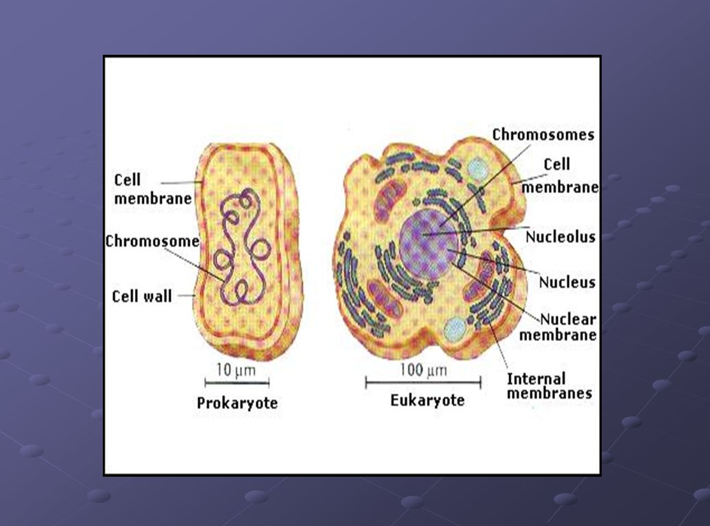
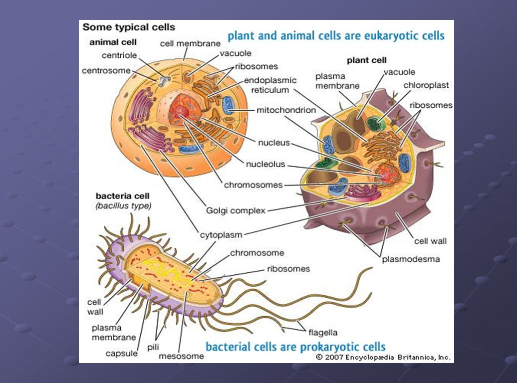
There are two main types of cells:
prokaryotic eukaryotic

9
Prokaryotic Eukaryotic cells: cells:
Genetic material is not in a nucleus. Usually smaller Simpler (fewer organelles with fewer functions) Bacteria Have a nucleus that contains genetic material. Generally larger Contain many organelles Found in all living things other than bacteria
 Bacteria. Have a nucleus that contains genetic material. Generally larger. Contain many organelles. Found in all living things other than bacteria.")
11
Cells are full of “things” called organelles.

12
Cell Organelles Organelle= “little organ”
Everything in a cell except the nucleus is cytoplasm You may or may not wish to distinguish between cytosol and cytoplasm. The correct use of each term is shown here. Most high school textbooks, however, use the word “cytoplasm” to mean “cytosol.”

13
Cell Membrane Boundary of the cell Emphasize word parts here:
phospho= phosphate head; lipid= fatty acid tail bi= 2

14
Cell Membrane Surrounds the cytoplasm and holds the shape of the cell
Protects the cell Allows certain material to pass through (selectively permeable)
")
15
Nucleus Control center of a eukaryotic cell Contains DNA and RNA
Surrounded by a double membrane Usually the easiest organelle to see under a microscope Usually one per cell Cells with more than one nucleus include muscle cells and liver cells, largely because of the massive volume of cytoplasm and number of organelles that need controlling.

16
Nucleolus Inside the nucleus is a small ball called the nucleolus.
Ribosomes are made here.

17
Cytoskeleton Acts as skeleton and muscle Provides shape and structure
Helps move organelles around the cell Actin, also found in muscle cells, mainly help maintain cell shape in their cytoskeletal role. Microtubules mostly move organelles around the cell. Intermediate filaments also provide structural support.

18
Endoplasmic Reticulum
“ER” Connected to nuclear membrane Highway of the cell Rough ER: studded with ribosomes; it makes proteins and delivers them to Golgi Apparatus Smooth ER: no ribosomes; it makes lipids; carries proteins It’s not necessary that the students can read the labels here; just point out the black dots are ribosomes.

19
Ribosome Where protein is made
Found attached to rough ER or floating free in cytoplasm Produced in a part of the nucleus called the nucleolus A polypeptide is a chain of amino acids. In this diagram, you can see the ribosome is making a polypeptide, also known as a protein.

20
Golgi Apparatus Looks like a stack of plates
Stores and packages proteins Carries particles in vesicles (little sacks or bubbles) Vesicles will pinch off the ends. AKA Golgi Complex. It is not necessary that the students read the labels, this diagram gives them a general idea of the Golgi’s shape.
 Vesicles will pinch off the ends. AKA Golgi Complex. It is not necessary that the students read the labels, this diagram gives them a general idea of the Golgi’s shape.")
21
Which organelles do lysosomes work with?
Garbage disposal of the cell (cannibals) Sack filled with enzymes that break down wastes Enzymes may destroy the cell, recycle material, fuse with food vacuoles to digest the food in single celled organisms. Students should recognize the shapes of the Golgi and ER even if they cannot read the captions. Which organelles do lysosomes work with?
 Sack filled with enzymes that break down wastes. Enzymes may destroy the cell, recycle material, fuse with food vacuoles to digest the food in single celled organisms. Students should recognize the shapes of the Golgi and ER even if they cannot read the captions. Which organelles do lysosomes work with")
22
Mitochondria “Powerhouse of the cell”
Cellular respiration occurs here to release energy for the cell to use Has its own strand of DNA Explain that this diagram shows the mitochondria cut open to reveal the internal membranes.

23
Chloroplast Found only in plant cells
Contains the green pigment chlorophyll Site of photosynthesis (glucose production) Chloroplasts absorb light, which is the catalyst for photosynthesis.
 Chloroplasts absorb light, which is the catalyst for photosynthesis.")
24
Cell Wall Found in plant, bacterial, fungi and some protista cells
Rigid, protective barrier Located outside of the cell membrane This is an actual microscopic image. Explain that the colors are added digitally to enhance the different parts.

25
What type of microscope may have been used to take this picture?
Vacuoles Large central vacuole usually in plant cells Many smaller vacuoles in animal cells Storage container for water, food, enzymes, wastes, pigments, etc. The image is 2D, so it must have been a light microscope or TEM. If the cell is very tiny, then a TEM was used. Otherwise, a strong light microscope could have captured this image. What type of microscope may have been used to take this picture?

26
Contractile Vacuole

27
Centriole Aids in cell division Found only in animal cells
Made of microtubules Microtubules are also part of the cytoskeleton.

28
Quick Review Which organelle is the control center of the cell?
Nucleus Which organelle holds the cell together? Cell membrane Which organelles are not found in animal cells? Cell wall, central vacuole, chloroplasts Which organelle helps plant cells make food? Chloroplasts What does E.R. stand for? Endoplasmic reticulum You may choose to delete the answers from the PowerPoint or change the animation so that they come in after all 5 questions are asked in case you want to quiz students individually at the end.

29
Video Review

Similar presentations