Download presentation
Presentation is loading. Please wait.

1
BASIC EMBRYOLOGY 1

2
It Literally means the study of embryos. It is the study of the origin and development of a single individual from the moment of its inception up to the time when it is born as an infant. WHAT IS EMBRYOLOGY ?

3
Every individual spends the first nine months(266 days or 38 weeks)of its life within the womb(uterus) of its mother. During this period it develops from a small one celled structure to an organism having billions of cells. WHAT IS EMBRYOLOGY?

4
It helps to tell us how a single cell develops into a newborn, containing numerous tissues and organs. Helps us understand many complicated facts of adult anatomy. Helps us understand why some children are born with organs that are abnormal. WHY EMBRYOLOGY?

5
A PERSON IS A PERSON NO MATTER HOW SMALL

6
Embryonic period – first 8 weeks ( first two months) Fetal period – remaining 30 weeks During the first 2 months we call the developing individual an EMBRYO. After that its called a FETUS. TESTES is male sex organ or gonad. OVARY is female sex organ or gonad, They produce GAMETES. PRENATAL PERIOD

7
Cells that carry out the special function of reproduction are called GAMETES. The development of a new individual begins when 1 male gamete(sperm or spermatozoa)meets and fuses with 1 female gamete(ovum or oocyte). The process of fusion of male and female gametes is called FERTILIZATION.
meets and fuses with 1 female gamete(ovum or oocyte). The process of fusion of male and female gametes is called FERTILIZATION..")
8
Male gametes produced by the testes are called SPERMATOZOA. The process is called SPERMATOGENESIS. Female gametes produced by ovary are called OVA. The process is called OOGENESIS. SPERMATOGENESIS and OOGENESIS are together called GAMETOGENESIS. Fertilization takes place when one spermatozoa enters an ovum. They fuse together to form a ZYGOTE. GAMATOGENESIS

9
Characters of the parents are transmitted to offspring through codes born on strands of DNA. GENES are made of such strands of DNA. They are located on chromosomes. A typical cell contains 46 chromosomes(DIPLOID number). The gamete contain 23 chromosomes(HAPLOID number). DNA
. The gamete contain 23 chromosomes(HAPLOID number). DNA.")
10
The diploid number of chromosomes is restored as a result of fertilization. Multiplication of the cell takes place by cell division. The usual method of cell division, seen in most tissues is called MITOSIS. Daughter cells resulting from a mitotic division are similar to the parent cell, and have the same number of chromosomes(46).
..")
11
A special kind of cell division takes place in the testis and ovary from formation of gametes. It is called MEIOSIS. The gametes resulting from meiosis have haploid number of chromosomes(23). The various gametes formed do not have the same genetic content.
. The various gametes formed do not have the same genetic content..")
12
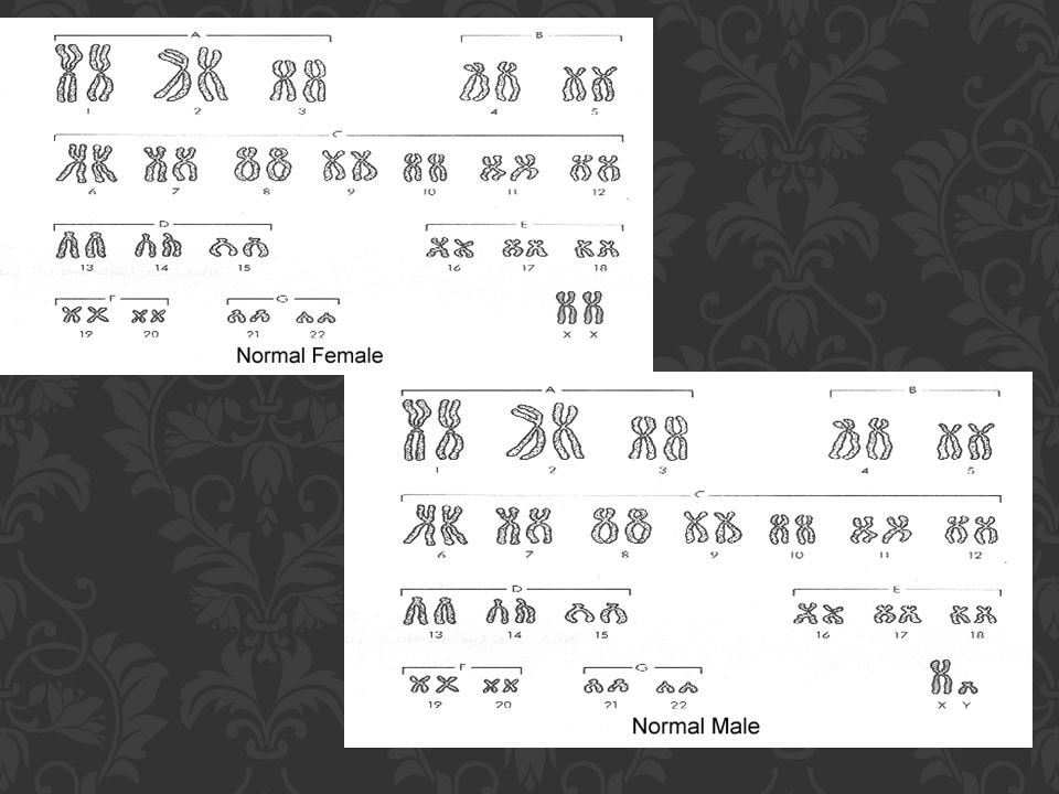
Haploid and Diploid Chromosomes The number of Chromosomes in each cell is fixed for a given species & in humans it is 46, referred as diploid. However, in spermatozoa & ova the number of Chromosomes is only half the diploid number i.e. 23,called haploid. So, after fertilization the resulting zygote has 23 Chromosomes from sperm & 23 Chromosomes from ovum, then the diploid number is restored. SOME FACTS ABOUT CHROMOSOMES

13
The 46 Chromosomes in each cell can be divided into 44 Autosomes & 2 sex Chromosomes. The sex Chromosome may be of 2 kinds ‘X’ &’Y’. Males have 44 autosomes & 1’X’ & 1‘Y’ chromosome. Females have 44 autosomes & 2 ‘X’ chromosomes. So there are 22 pairs of homologous Chromosomes which are exactly alike. In females, the 2 ‘X’ chromosomes form another such homologous pair. In males, this pair is represented by 1 ‘X’ & 1’Y’ chromosome. So, 1 Chromosome of each pair is derived from the mother & the other from the father. AUTOSOMES & SEX ‘C’

14
In resting cells, Chromosomes are not visible under a light microscope as chromatin material is dispersed. But during cell division the chromatin network in the nucleus becomes condensed into a number of Chromosomes. Its made up of 2 rod-shaped structures or CHROMATIDS placed more or less parallel to each other. They are united to each other at a light staining area called CENTROMERE(kinetochore) CHROMOSOME STRUCTURE
 CHROMOSOME STRUCTURE.")
15
Each chromatid has 2 arms 1 on either side of the centromere. The shorter of the two arms extending from the centromere is called the p arm; the longer is the q arm. Individual Chromosomes differ from one another in total length and some other characteristics, in order to be identified individually. Classification of Chromsomes in this way is called KARYOTYPING.

16
Each Chromosome bears on itself a very large number of structures called GENES. Genes are made up of nucleic acid called DNA. Genes are involved in synthesis of proteins. GENES Homologous Chromosomes

18
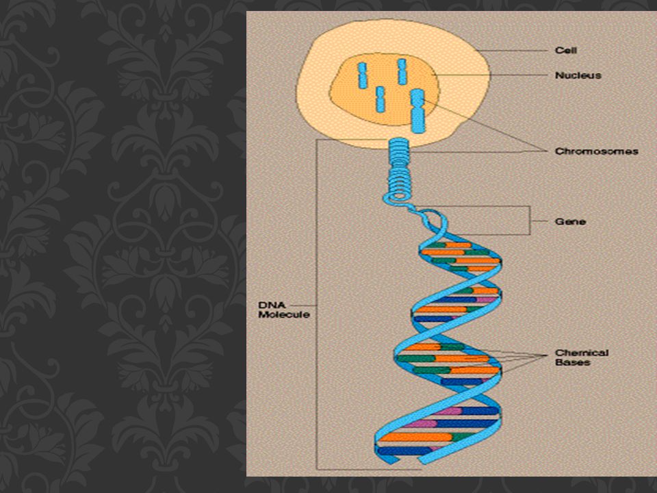
DNA, or deoxyribonucleic acid is a Double Helix structure. The information in DNA is stored as a code made up of four chemical bases: adenine (A), guanine (G), cytosine (C), and thymine (T). DNA bases pair up with each other, A with T and C with G, to form units called base pairs. An important property of DNA is that it can replicate, or make copies of itself. WHAT IS DNA?
, guanine (G), cytosine (C), and thymine (T). DNA bases pair up with each other, A with T and C with G, to form units called base pairs. An important property of DNA is that it can replicate, or make copies of itself. WHAT IS DNA .")
19
In the nucleus of each cell, the DNA molecule is packaged into thread-like structures called chromosomes. Each chromosome is made up of DNA tightly coiled many times around proteins called histones that support its structure.

21
A gene mutation is a permanent change in the DNA sequence that makes up a gene. Mutations range in size from a single DNA building block (DNA base) to a large segment of a chromosome. Gene mutations occur in two ways: they can be inherited from a parent or acquired during a person’s lifetime. GENE MUTATIONS
 to a large segment of a chromosome. Gene mutations occur in two ways: they can be inherited from a parent or acquired during a person’s lifetime. GENE MUTATIONS.")
22
Mutations that are passed from parent to child are called hereditary mutations or germline mutations (because they are present in the parent’s egg and sperm cells, which are also called germ cells). When an egg and a sperm cell unite, the resulting fertilized egg cell receives DNA from both parents. If this DNA has a mutation, the child that grows from the fertilized egg will have the mutation in each of his or her cells. HEREDITARY MUTATIONS OR GERMLINE MUTATIONS

23
Genetic changes that are described as de novo (new) mutations can be either hereditary or somatic. In some cases, the mutation occurs in a person’s egg or sperm cell but is not present in any of the person’s other cells. In other cases, the mutation occurs in the fertilized egg shortly after the egg and sperm cells unite. (It is often impossible to tell exactly when a de novo mutation happened.) As the fertilized egg divides, each resulting cell in the growing embryo will have the mutation. De novo mutations may explain genetic disorders in which an affected child has a mutation in every cell in the body but the parents do not, and there is no family history of the disorder. NEW (DE NOVO) MUTATIONS.
 As the fertilized egg divides, each resulting cell in the growing embryo will have the mutation. De novo mutations may explain genetic disorders in which an affected child has a mutation in every cell in the body but the parents do not, and there is no family history of the disorder. NEW (DE NOVO) MUTATIONS..")
24
They occur at some time during a person’s life and are present only in certain cells, not in every cell in the body. These changes can be caused by environmental factors such as ultraviolet radiation from the sun, or can occur if a mistake is made as DNA copies itself during cell division. Acquired mutations in somatic cells (cells other than sperm and egg cells) cannot be passed on to the next generation. ACQUIRED (OR SOMATIC) MUTATIONS
 cannot be passed on to the next generation. ACQUIRED (OR SOMATIC) MUTATIONS.")
25
Somatic mutations that happen in a single cell early in embryonic development can lead to a situation called mosaicism. These genetic changes are not present in a parent’s egg or sperm cells, or in the fertilized egg, but happen a bit later when the embryo includes several cells. As all the cells divide during growth and development, cells that arise from the cell with the altered gene will have the mutation, while other cells will not. Depending on the mutation and how many cells are affected, mosaicism may or may not cause health problems. Some genetic changes are very rare; others are common in the population. MOSAICISM

26
Genetic changes that occur in more than 1 percent of the population are called polymorphisms. They are common enough to be considered a normal variation in the DNA. Polymorphisms are responsible for many of the normal differences between people such as eye color, hair color, and blood type. Although many polymorphisms have no negative effects on a person’s health, some of these variations may influence the risk of developing certain disorders. POLYMORPHISMS

27
Gene therapy is an experimental technique that uses genes to treat or prevent disease. There are several approaches to gene therapy, including: 1.Replacing a mutated gene that causes disease with a healthy copy of the gene. 2.Inactivating, or “knocking out,” a mutated gene that is functioning improperly. 3.Introducing a new gene into the body to help fight a disease. Although gene therapy is a promising treatment option for a number of diseases (including inherited disorders, some types of cancer, and certain viral infections), the technique remains risky and is still under study to make sure that it will be safe and effective. Gene therapy is currently only being tested for the treatment of diseases that have no other cures. GENE THERAPY
, the technique remains risky and is still under study to make sure that it will be safe and effective. Gene therapy is currently only being tested for the treatment of diseases that have no other cures. GENE THERAPY.")
29
ANY QUESTIONS?

30
1.The Developing Human Clinically Oriented Embryology Moore Persaud 9 th Edition 2.Langman’s Medical Embryology T. W. Sadler 6 th Edition 3.Google images REFERENCES

Similar presentations

 and (b).>")
. -Histones - help maintain the shape of the.>")


