Download presentation
Presentation is loading. Please wait.

1
Nuclear Medicine in the Evaluation of Trauma
Materials for medical students Helena Balon, MD Wm. Beaumont Hospital Royal Oak, MI, USA Charles University 3rd School of Medicine Dept Nucl Med, Prague

3
Radionuclide methods in traumatology
Musculoskeletal trauma Bone scan Trauma to internal organs (hematoma, laceration, fracture, perforation, leaks) Renal scan Myocardial scan Hepatobiliary scan (Liver / spleen scan) - CT preferred (Testicular scan) - US preferred Head trauma CT preferred Cerebral perfusion scan - brain death Cisternography - CSF leak
 Renal scan. Myocardial scan. Hepatobiliary scan. (Liver / spleen scan) - CT preferred. (Testicular scan) - US preferred. Head trauma. CT preferred. Cerebral perfusion scan - brain death. Cisternography - CSF leak.")
4
Bone scan in trauma Very sensitive
Detects areas of abnormal bone turnover Shows areas that need further radiol.evaluation Provides objective evidence of disorder when X ray negative

5
Bone scan Tracers: diphosphonates (Tc-99m MDP, HDP) Dose: 500-900MBq
Tracer localization (chemisorption onto surface of bone trabeculae) depends on: blood flow capillary permeability bone metabolism (activity of osteoblasts, osteoclasts, new bone formation)
 depends on: blood flow. capillary permeability. bone metabolism (activity of osteoblasts, osteoclasts, new bone formation)")
6
Bone scan Patient preparation Pre-test: none
Post-injection: good oral hydration Frequent voiding Perchlorate p.o. preinj. to decrease rad. dose to thyroid

7
Bone scan Methods Regular - imaging @ 2-4 hrs post injection
3-phase (dynamic angiogram + blood pool + delay) Planar or SPECT Whole body ANT & POST, additional views (lat.,oblique) Parallel hole or pinhole collimator (for small structures)
 Planar or SPECT. Whole body ANT & POST, additional views (lat.,oblique) Parallel hole or pinhole collimator (for small structures)")
8
Bone Scan in Trauma Fractures & occult fx
Child abuse (except skull fx) Stress fractures (insufficiency fx, fatigue fx) Avulsion injuries Shin splints Bone bruises (contusion) RSD (reflex sympathetic dystrophy) Osteochondral lesions
 Stress fractures (insufficiency fx, fatigue fx) Avulsion injuries. Shin splints. Bone bruises (contusion) RSD (reflex sympathetic dystrophy) Osteochondral lesions.")
9
Diagnosis of Fractures
Plain X ray, X ray tomography - if neg >>> Bone scan if neg >>> stop work-up if diagnostic >>> treat if more information needed >>> CT (subtle changes) or MRI (subtle changes, soft tissue trauma, bone bruise, precise dx of limited area)
 or. MRI (subtle changes, soft tissue trauma, bone bruise, precise dx of limited area)")
10
Fractures on Bone scan Acute fx
Positive on all 3 phases Positive immediately after trauma in most pts 90% sensitivity if imaged in < 48 hrs If scan neg. in pts > 75y >>> repeat scan in 3-7 d Bone scan remains positive for 6-24 mo (healing fx)
")
11
Acute compression fractures
80 y/o F w osteopenia fell 6 wks prior

12
Rib fractures

13
Multiple fx’s 59 F w breast ca MVA 10 d ago

14
Osteogenesis imperfecta

15
Bone Bruise Direct trauma with disruption of trabecular bone but not cortical bone X ray - negative Bone scan - 3-phase positivity MRI - bone marrow involvement (hemorrhage)
")
16
Leg & Foot Trauma

17
Shin / thigh splints Continuous spectrum from shin splint to stress fx
Stress related periostitis along muscle insertion sites (soleus, tibialis posterior, adductor longus/brevis, gluteus max) X ray - negative Bone scan Flow, blood pool - normal Delay- vertical, linear uptake along posteromedial tibial cortex (mid- or distal 1/3) medial or lateral femoral cortex (proximal 1/3)
 X ray - negative. Bone scan. Flow, blood pool - normal. Delay- vertical, linear uptake along posteromedial tibial cortex (mid- or distal 1/3) medial or lateral femoral cortex (proximal 1/3)")
18
Shin Splints

19
Shin splints, thigh splints

20
Thigh splints - mechanism

21
Stress Fractures Fatigue fractures Abnormal stress on normal bone
(jogging, gymnastics, skating, military) Insufficiency fractures Normal stress on abnormal bone (osteoporosis, osteomalacia, RA, HPT, steroids, radiation Rx)
 Insufficiency fractures. Normal stress on abnormal bone. (osteoporosis, osteomalacia, RA, HPT, steroids, radiation Rx)")
22
Stress fractures Pathophysiology - repetitive microtrauma (athletes)
Symptoms - pain, swelling Common locations: Tibia - proximal or distal 1/3 Fibula - distal 1/3 Metatarsals (2nd, 3rd) Tarsal bones (calcaneus, navicular) Femoral neck Inferior pubic ramus Lower lumbar spine (spondylolysis)
 Tarsal bones (calcaneus, navicular) Femoral neck. Inferior pubic ramus. Lower lumbar spine (spondylolysis)")
23
Stress fractures X ray may be initially negative (2-4 wks)
Bone scan, MRI – positive earlier Bone scan 3-phase positivity Flow + for ~ 1 mo Blood pool + for ~ 2 mo Delay + for ~ 9-12 mo Rx - restrict sports for 4-6 wks

24
Stress fx ?

25
Stress fractures

26
Metatarsal stress fracture

27
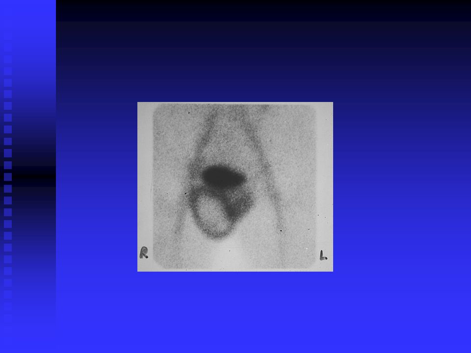
Metatarsal stress fracture

28
Metatarsal stress fx

29
Plantar fasciitis Heel pain
Post-traumatic inflammation of plantar ligament due to athletic overuse prolonged standing walking on hard surface Bone scan Focal blood pool + delayed uptake in inferior posterior calcaneus

30
Plantar fasciitis

31
Achilles tendonitis

32
Impingement syndromes
Posterior impingement sy (os trigonum sy) Excessive repeat plantar flexion (compression between posterior calcaneus & posterior tibia) Ballet dancers, gymnasts Anterior impingement sy Excessive repeat dorsal flexion >>> hypertrophic spur on dorsum (talus & anterior tibia) Ballet dancers, gymnasts, high jumping
 Excessive repeat plantar flexion (compression between posterior calcaneus & posterior tibia) Ballet dancers, gymnasts. Anterior impingement sy. Excessive repeat dorsal flexion >>> hypertrophic spur on dorsum (talus & anterior tibia) Ballet dancers, gymnasts, high jumping.")
33
Posterior impingement syndrome (os trigonum stress fx)

34
Hip & Pelvis Trauma

35
Femoral neck stress fracture
Thigh or groin pain in athletes Must distinguish femoral neck stress fx from pubic ramus stress fx Must treat / immobilize early to prevent complete fx, AVN

36
Femoral neck Fx 76F w L groin pain X ray neg

37
X ray 2 weeks later

38
Intertrochanteric fracture
93 F, fall 6 days ago, Rt hip pain

39
IT fx

40
Avascular necrosis (AVN)
Etiology trauma (fx) steroids, alcohol abuse pancreatitis, fat embolism vasculitis, SS disease idiopathic Pathophysiology: bone ischemia Diagnosis MRI most sensitive bone scan useful
 steroids, alcohol abuse. pancreatitis, fat embolism. vasculitis, SS disease. idiopathic. Pathophysiology: bone ischemia. Diagnosis. MRI most sensitive. bone scan useful.")
41
AVN Common locations Bone scan Femoral head (Legg-Perthes in children)
Carpal (scaphoid, lunate), tarsal (talus) Long bones, ribs in SS Bone scan Initially “cold” Revascularization starts in 1-3 wks, from periphery, diffusely “hot”, lasts for months
, tarsal (talus) Long bones, ribs in SS. Bone scan. Initially cold Revascularization starts in 1-3 wks, from periphery, diffusely hot , lasts for months.")
42
IT Fx + AVN 50 M w fall a few weeks ago

43
IT fx + AVN MRI

44
Sacrococcygeal Fx ANT POST

45
Sacral insufficiency fx
ANT POST 79 F fell 1 mo ago (“Honda” sign)
")
46
Pelvic fractures 4 days post fall 1 month later

48
Spine trauma

49
Spondylolysis Stress fx of posterior vertebral elements (pars interarticularis) due to repetitive trauma Teenagers, young adults Hyperextension sports (gymnastics, diving, weight lifting, soccer,hockey) Genetic predisposition? L5 > L4 > L3 Frequently bilateral >>> spondylolisthesis
 Genetic predisposition L5 > L4 > L3. Frequently bilateral >>> spondylolisthesis.")
51
Spondylolysis X ray Normal or sclerosis, later lucency 2º fx
Bone scan increased uptake in pars interarticularis SPECT better than planar Rx – discontinue activity

52
Pars interarticularis defect
14 y/o F basketball player trauma 1 mo prior

53
Pars defect

54
Transverse process fracture
planar SPECT CNM 2001:863

55
Hand & Wrist Trauma

56
Wrist fractures Scaphoid fx - most common Hook of hamate fx
70-80% carpal fx Fall on outstretched hand Common complications - AVN, non-union Hook of hamate fx Direct injury from handles (tennis, golf, baseball) Radial / ulnar styloid fx
 Radial / ulnar styloid fx.")
57
fall, injured Rt wrist

58
Fracture of radius + scaphoid
S/P fall, suspect scaphoid fx X ray neg.

59
Scaphoid Fx 14 y/o M fell 6 wks ago, X ray negative

60
Hook of the hamate fracture
R wrist pain

61
Hook of the hamate injury - mechanism

62
Reflex Sympathetic Dystrophy (Sudeck’s atrophy, Shoulder-hand sy, Causalgia, Chronic regional pain sy) Sympathetically mediated disorder (vasomotor instability) Etiology Trauma (blunt, fracture) MI Stroke/CVA Infection Idiopathic Symptoms: exquisite pain, tenderness, edema, skin changes, locally warm or cold UE or LE
 Etiology. Trauma (blunt, fracture) MI. Stroke/CVA. Infection. Idiopathic. Symptoms: exquisite pain, tenderness, edema, skin changes, locally warm or cold UE or LE.")
63
Reflex Sympathetic Dystrophy (RSD)
Bone scan Early stage: 3-phase positive Later stage (> 6 mo): only delayed phase posit. Delayed phase MDP: diffuse increased uptake in entire limb, “periarticular accentuation” in small joints Children: often all 3 phases or Sensitivity: 60-95% X ray Periarticular ST edema Late changes- bone resorption, osteopenia
: only delayed phase posit. Delayed phase MDP: diffuse increased uptake in entire limb, periarticular accentuation in small joints. Children: often all 3 phases or. Sensitivity: 60-95% X ray. Periarticular ST edema. Late changes- bone resorption, osteopenia.")
64
Reflex sympathetic dystrophy
(RSD) 73 F w Rt hand/wrist pain no trauma
 73 F w Rt hand/wrist pain no trauma.")
65
Non-accidental injury
1 mo old baby w intracranial hemorrhage, Lt parietal fx

67
Muscle trauma (Rhabdomyolysis)
MDP weight lifting CNM 2001: 344

68
Muscle uptake (Rhabdomyolysis)
pt w Ewing sarcoma, s/p BKA, walking on crutches

69
Trauma to internal organs

70
Hepatobiliary Scan Tc-99m IDA (disofenin, mebrofenin)
dose ~ MBq i.v. imaging of liver, abdomen, pelvis over 1 hr delayed images if 1st hr negative Bile leak - activity anywhere in peritoneal cavity Common after laparoscopic cholecystectomy Usually seals off spontaneously Leak clin. more significant if no transit into bowel seen (needs surgical intervention)
")
71
Bile leak

72
Liver - Spleen Scan Tc-99m sulfur colloid Parenchymal defects
dose ~ MBq i.v. SPECT imaging better than planar Parenchymal defects laceration, rupture, hematoma Splenosis splenic implants on peritoneum following spleen rupture

73
Splenosis MVA 30 y ago, S/P splenectomy Tc-99m S.C.

74
Pleuroperitoneal leak
Rt LAT ANT Pt. on peritoneal dialysis

75
Renal Scans Tc-99m MAG3 or DTPA Tc-99m DMSA ~ 100-300 MBq
Dynamic images over min Assessment of perfusion, function, leaks Tc-99m DMSA ~ MBq Static 2-4 hrs post injection High resolution needed for renal morphology pinhole, SPECT Parenchymal defects - laceration, rupture, hematoma Extrinsic defects - perinephric / retroperiton. hematoma

76
Urine leak CNM 2001:724

77
Testicular scan Indications: Acute torsion Delayed torsion
Epidymitis / orchitis Tc-99m pertechnetate Flow + immediate static images “Donut sign” Late torsion Abscess Trauma (hematoma) Tumor
 Tumor.")
79
Cisternography In-111 DTPA intrathecally
CSF leak - paraspinal (meningeal tears) CSF rhinorrhea, otorrhea imaging counting nasal pledgets for radioactivity pledget / plasma ratio
 CSF rhinorrhea, otorrhea. imaging. counting nasal pledgets for radioactivity. pledget / plasma ratio.")
80
Cerebral perfusion Tc-99m HMPAO or ECD dose ~ 800 MBq
Post-traumatic perfusion defects Assessment of brain death - role of NM complementary no flow no parenchymal uptake

81
Head Trauma ? Brain death?
15 y/o F with intracranial bleed

82
Brain death

83
Radionuclide synovectomy (radiosynoviorthesis, RSO)
Intraarticular treatment using beta rays Goal is to destroy inflammed synovia Alternative to surgical synovectomy Mostly in out-patients More than one joint treatment possiblity Repeated treatment More than 40 years experience

84
Indications Patients resistent to steroid injection
Rheumatoid arthritis Repetitive idiopatic swelling Repetitive decompensated arthrosis Psoriatic arthropathy Haemophillic arthropathy

85
Contraindications Pregnancy, breast feeding
Septic arthritis, infection around the joint Tumor of the joint Children (relative) Massive haemarthros Popliteal cyst rupture There are no side effects
 Massive haemarthros. Popliteal cyst rupture. There are no side effects.")
86
Radiopharmaceuticals
Sterile colloidal suspension Beta radiation (or mixed – imaging) Large joints (knee) Y-90 citrate, silikate Middle joints (shoulder, elbow, wrist, hip) Re -186 sulphate Small joints (hands) Er-169 citrate
 Large joints (knee) Y-90 citrate, silikate. Middle joints (shoulder, elbow, wrist, hip) Re -186 sulphate. Small joints (hands) Er-169 citrate.")
87
How it works Colloidal particles Small enough to be phagocytozed
Large enough to remain within the joint cavity Should be biodegradable Energy sufficient to penetrate and ablate the synovial tissue Not to damage underlying articular cartilage or overlying skin

88
Side effects Radiation synovitis
Reduced with simultaneous steroids injection Infection Less frequent than in steroids alone Tissue necrosis Fistula around injection, paraarticular injection Thrombosis Due to join fixation

89
Procedure Joint puncture Withdrawing of the fluid Injection of the RP
Injection of the steroid Joint fixation for 24 to 72 hours

90
Effectiveness Local hyperthermia and swelling decrease within 3-4 months. > 75% patients significant decrease in pain and swelling in rheumatoid arthritis, > 90% in hemophiliac joints reduction of bleeding episodes 52 ± 24% effect in osteoarthritis

91
Effectiveness depends on
Precise diagnosis Stage of the disease Correct indication Correct application Joint fixation Another injection could be performed 6 moths later Better repetitive injection of less radioactivity

92
Conclusion RSO is cost effective, safe and improves quality of life in patients with disabling arthritis The fear of developing cancer has been conclusively ruled out in extensive studies

Similar presentations