Download presentation
Presentation is loading. Please wait.

1
Liaoning Medical University Affiliated First Hospital
Electrocardiogram ECG Liaoning Medical University Affiliated First Hospital He Xin

2
一、Basic knowledge of ECG

3
Content 1、Electrophysiology 2、ECG Waveforms andintervals
3、ECG Lead system

4
1、Electrophysiology ---Depolarization and repolarization ---Vector and vectorcardiogram

5
Pacing and conducting system of the heart
2、ECG Waveforms and intervals Pacing and conducting system of the heart Sinus node Internodal tracts AV node right bundle branch (RBB) Purkinje fibers Bundle of His anterior fascicle left bundle branch (LBB) Purkinje fibers posterior fascicle
 Purkinje fibers. Bundle of His. anterior fascicle. left bundle branch (LBB) Purkinje fibers. posterior fascicle.")
6
Componcents of the conduction system
SA Node Bundle of AV junction AV Node Bundle of AV Left BundleBranch Right BundleBranch

7
3、ECG Lead system --bipolar leads(Standard leads)I, II, III --unpolar leads: aVR, aVL, aVF
I, II, III --unpolar leads: aVR, aVL, aVF")
8
3、ECG Lead system --Chest leads: V1, V2, V3, V4, V5, V6

9
二、 Measurement and Normal ECG

10
1. Measurement of ECG ECG paper Paper speed:25mm/s;Standard calibration: 1mV=10mm

11
(1) Measuring heart rate (HR)
60 60 =HR =75/min R-R (S) 0.80S ---Measuring heart rate (HR) = 60/R-R (bpm) ---300/the number of large time units between R-R /the number of small time units between R-R
 0.80S. ---Measuring heart rate (HR) = 60/R-R (bpm) /the number of large time units between R-R /the number of small time units between R-R.")
12
(2) Amplitude of waves or segment: P, QRS, S-T, T, U
 Amplitude of waves or segment: P, QRS, S-T, T, U")
13
(3) Width or duration of waves: P, QRS, T, U
(3) Width or duration of waves: P, QRS, T, U * Duration of intervals: P-R, Q-T * Shape of waves: P, QRS, T
 Width or duration of waves: P, QRS, T, U * Duration of intervals: P-R, Q-T * Shape of waves: P, QRS, T")
14
(4) Mean QRS axis ---normal cardiac axis range from -3090 ---significant left deviation: -30-90 ---right deviation: 90180 ---significant right deviation: 180-90

15
(5) Clockwise and counterclockwise rotation ---Clockwise rotation
V1V2 V3V4 V5V6 Clockwise rotation normal Counterclockwise rotation (5) Clockwise and counterclockwise rotation Clockwise rotation ---Counterclockwise rotation
 Clockwise and counterclockwise rotation. ---Clockwise rotation. ---Counterclockwise rotation")
16
2、 Normal ECG (1) P wave: atrial depolarization ---Amplitude 0.20 mv ---Duration 0.11 sec ---Positive in I, II, aVF, V4-V6; Negative in aVR (2) PR interval: the time for intraatrial, --- AV nodal, and His-Purkinje conduction, --- Duration: 0.12 ~ 0.20 sec
 P wave: atrial depolarization ---Amplitude 0.20 mv ---Duration 0.11 sec ---Positive in I, II, aVF, V4-V6; Negative in aVR (2) PR interval: the time for intraatrial, --- AV nodal, and His-Purkinje conduction, --- Duration: 0.12 ~ 0.20 sec")
17
(3) QRS complex: ventricular depolarization ---The width: 0. 060
(3) QRS complex: ventricular depolarization ---The width: 0.060.10 sec, 0.11 sec. ---From V1 to V6, the R waves gets bigger and bigger,the S waves gets smaller and smaller. --R/S < l in V1, but R/S > l in V5 --R in V5 and V6 < 2.5 mv, R in V1 < 1.0 mv ---R in aVR < 0.5 mv, R in aVL < 1.2 mv and R in aVF < 2.0 mv R in I < 1.5 mv ---Q < 0.04 sec in width, < 1/4 R in the same lead.
 QRS complex: ventricular depolarization ---The width: 0.060.10 sec, 0.11 sec. ---From V1 to V6, the R waves gets bigger and bigger,the S waves gets smaller and smaller. --R/S < l in V1, but R/S > l in V5 --R in V5 and V6 < 2.5 mv, R in V1 < 1.0 mv ---R in aVR < 0.5 mv, R in aVL < 1.2 mv and R in aVF < 2.0 mv R in I < 1.5 mv ---Q < 0.04 sec in width, < 1/4 R in the same lead.")
19
(4) ST segment: it reflects Phase 2 of the action potential
(4) ST segment: it reflects Phase 2 of the action potential. ---ST elevation < 0.3 mV in V1、V2; < 0.5 mV in V3;< 0.10 mV in V4 V6 ---ST depression < 0.05 mV in any leads (5) T wave: repolarization of ventricles ---It is upright in all the unipolar leads except aVR, and occasionally V1. ---T wave > 1/10 R in the same lead, maybe < 1.21.5 mV in the precordial leads
 ST segment: it reflects Phase 2 of the action potential. ---ST elevation < 0.3 mV in V1、V2; < 0.5 mV in V3;< 0.10 mV in V4 V6 ---ST depression < 0.05 mV in any leads (5) T wave: repolarization of ventricles ---It is upright in all the unipolar leads except aVR, and occasionally V1. ---T wave > 1/10 R in the same lead, maybe < 1.21.5 mV in the precordial leads")
20
(6) QT interval: the duration of depolarization and repolarizaion of ventricles The normal range is 0.320.44 sec (7) U wave: the wave following the T wave and is usually very smal ---Its cause is not completely understood ---Elevated U wave: low K+ in plasma
 QT interval: the duration of depolarization and repolarizaion of ventricles The normal range is 0.320.44 sec (7) U wave: the wave following the T wave and is usually very smal ---Its cause is not completely understood ---Elevated U wave: low K+ in plasma")
21
三、Atrial Enlargement and Ventricular Hypertrophy

23
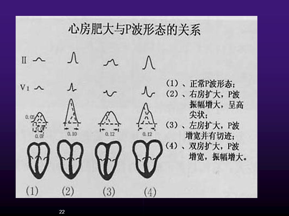
1、Atrial Enlargement (1) Right Atrial Enlargement Lead II
---P wave is peaked (P "pulmonale"); ---Amplitude of P wave ≥0.25 mV in limb leads. Lead V1 ---upright and amplitude ≥0.15 mV; ---biphasic and amplitude ≥0.20 mV
; ---Amplitude of P wave ≥0.25 mV in limb leads. Lead V1. ---upright and amplitude ≥0.15 mV; ---biphasic and amplitude ≥0.20 mV.")
26
(2) Left Atrial Enlargement
Lead II --- Duration of P wave ≥0.12 sec ---P wave become bifid (P "mitrale") ---The distance of two peak ≥ 0.04sec Lead V1 ---P wave become biphasic ---Ptfv1 mm·sec
 ---The distance of two peak ≥ 0.04sec. Lead V1. ---P wave become biphasic. ---Ptfv1 mm·sec.")
29
(3) Biatrial Enlargement
Lead II P wave duration and amplitude both increased

30
2、Ventricular Hypertrophy
(1) Left Ventricular Hypertrophy A. Increased voltage ---Rv5 or Rv6 > 2.5 mV SV1 + R V5 >3.5mV (female) or > 4.0mV (male) ---RI >1.5mV;RaVL >1.2mV;RaVF >2.0 mV RI + SIII >2.5 mV B. Left axis deviation C. longer duration of QRS ( s) D. ST depression and T inversion in V5-6
 Left Ventricular Hypertrophy. A. Increased voltage. ---Rv5 or Rv6 > 2.5 mV. SV1 + R V5 >3.5mV (female) or > 4.0mV (male) ---RI >1.5mV;RaVL >1.2mV;RaVF >2.0 mV. RI + SIII >2.5 mV. B. Left axis deviation. C. longer duration of QRS ( s) D. ST depression and T inversion in V5-6.")
33
(2) Right Ventricular Hypertrophy
A. Increased voltage (adults over 30) ---R/S ratio in V1 ≥ 1.0; R /Rs ---R/S ratio in V5 or V6 ≤ 1.0 ---R/q or R/S ratio in aVR≥1 --R V1+ S V5 >1.05mV (severe>1.2mV) --RaVR>0.5mV B. Right axis deviation ≥ (severe > +1100) C. ST depression and T inversion in V1-2
 ---R/S ratio in V1 ≥ 1.0; R /Rs. ---R/S ratio in V5 or V6 ≤ R/q or R/S ratio in aVR≥1. --R V1+ S V5 >1.05mV (severe>1.2mV) --RaVR>0.5mV. B. Right axis deviation ≥ +900 (severe > +1100) C. ST depression and T inversion in V1-2.")
36
(3) Biventricular Hypertrophy
---Normal ECG. ---One ventricular hypertrophy. ---Biventricular Hypertrophy.

37
四、Myocardial Ischemia and Myocardial infarction

38
1、Myocardial Ischemia Subendocardial: Upright T wave
Subepicardial: Inverted, diphasic, low T wave

39
1、Myocardial Ischemia Subendocardial: ST segment depression
Subepicardial: ST segment elevation( coronary spasm)
")
43
Summary ---ST segment depression ---ST segment elevation
---T wave tall positive ---T wave inversion These changes are transitory and mostly synchronous with symptoms

44
2、Myocardial infarction
(1) Basic changes ---Ischemic T Waves. Tall peaked T waves, often appear as the earliest ECG sign of acute MI ---Injuried ST-segment Elevations. The ST segment elevated in one or more leads and may be straightened and fuse with the T wave (mono-phasic curve) ---necrotic (Pathologic) Q Waves. the sudden developed Q wave may indicate an acute MI -
 Basic changes. ---Ischemic T Waves. Tall peaked T waves, often appear as the earliest ECG sign of acute MI. ---Injuried ST-segment Elevations. The ST segment elevated in one or more leads and may be straightened and fuse with the T wave (mono-phasic curve) ---necrotic (Pathologic) Q Waves. the sudden developed Q wave may indicate an acute MI. -")
46
(2) Progressive ECG changes
---Hyperacute changes ---Acute period ---Subacute period (T Wave Changes) The elevated ST segments return to the baseline, and deep symmetrical T waves appear in these leads. Tall, symmetrical, upright T waves will appear in reciprocal leads at the same time ---Old myocardial infarct A definitive diagnosis of old myocardial infarct depends on the presence of a pathological Q wave
 The elevated ST segments return to the baseline, and deep symmetrical T waves appear in these leads. Tall, symmetrical, upright T waves will appear in reciprocal leads at the same time. ---Old myocardial infarct. A definitive diagnosis of old myocardial infarct depends on the presence of a pathological Q wave.")
48
(3) Localization of the ECG patterns
Leads with Abnormal Q Waves location of MI V1 V Anteroseptal V3 V Anterior I, aVL, V5 V Lateral V1 V Extensive Anterior II, III, aVF Inferior

49
I II III aVR aVL aVF V1 V2 V3 V4 V5 V6
LOCALIZATION OF MI I II III aVR aVL aVF V1 V2 V3 V4 V5 V6 inferior anterior anteroseptal lateral Extensive anterior

50
Anterior MI

51
Inferior MI

52
Lateral MI

53
五、Arrhythmia

54
1、Classification Abnormal origin Abnormal conduction
----sinus arrhythmia * ----ectopic rhythm ---passivity --escape ---initiative --premature contraction * --tachycardia* --flutter and fibrillation* Abnormal conduction ----physiological block: ----pathological block: S-AB; A-VB*; LBBB; RBBB ----accessory pathway: pre-excitation syndrome

55
2、Electrophysiology ---Automaticity ---Excitability
--Absolute refractory period (200ms) --Effective refractory period (210ms) --Ralative refractory period (50-100ms) ---Conductivity
 --Effective refractory period (210ms) --Ralative refractory period (50-100ms) ---Conductivity.")
56
3、SINUS RHYTHM AND SINUS ARRHYTHMIAS
---Sinus rhythm features ---Sinus Bradycardia ---Sinus Tachycardia ---Sinus arrhythmia ---Sinus arrest ---Sick Sinus Syndrome (SSS)
")
57
Sinus rhythm features (1) Every P wave is following by a QRS complex
(2) P wave is upright in lead I, II, aVF, V4-V6, inverse in aVR (3) P-R interval ≥ 0.12sec (4) Normal rate is beats/min
 P wave is upright in lead I, II, aVF, V4-V6, inverse in aVR. (3) P-R interval ≥ 0.12sec. (4) Normal rate is beats/min.")
58
Sinus Bradycardia (1) Sinus rhythm (2) Heart rate <60bpm
(R-R interval or P-P interval >1.0 sec )
")
59
Factors associated with sinus bradycardia
(1) Physiologic Laborers and trained athletes Emotional states leading to syncope (2) Pathologic -blocker Hypothyroidism
 Physiologic Laborers and trained athletes Emotional states leading to syncope (2) Pathologic -blocker Hypothyroidism")
60
Sinus Tachycardia (1)Sinus rhythm, rate > 100 bpm
The R-R interval (or the P-P interval) <0.60 sec (2)P-R and Q-T interval are shorter than usual (3)S-T segment is slight depression, T waves may be flattened
 <0.60 sec. (2)P-R and Q-T interval are shorter than usual. (3)S-T segment is slight depression, T waves may. be flattened.")
61
Factors associated with sinus tachycardia
(1) Physiologic Exercise Strong emotion Anxiety states (2) Pathologic Fever Hemorrhage Anemia Myocarditis Hyperthyroidism
 Physiologic Exercise Strong emotion Anxiety states (2) Pathologic Fever Hemorrhage Anemia Myocarditis Hyperthyroidism")
62
Sinus arrhythmia ---Sinus rhythm and PR interval,
---Difference of P--P interval > 0.12sec in the same lead

63
Sinus arrest The P wave missed for a short time

64
Sick Sinus Syndrome (SSS)
(1) Sinus bradycardia (HR<50/min) (2) Sinus arrest or SA block (3) Tachycardia: Atrial tachycardia Atrial Flutter Atrial fibrillation (4) AV block
 Sinus bradycardia (HR<50/min) (2) Sinus arrest or SA block. (3) Tachycardia: Atrial tachycardia. Atrial Flutter. Atrial fibrillation. (4) AV block.")
65
4、Premature contractions
--- Premature Ventricular Complex --- Premature Atrial Complex --- Premature junctional complex

66
Premature Ventricular Complex
(1) Ventricular complex (QRS) is not preceded by a premature P' wave (2) Premature QRS complex is the wider and the bizarre , Duration of QRS> 0.12 sec T wave in direction is opposite to QRS complex (3) Complete compensatory pause
 Ventricular complex (QRS) is not preceded by a premature P wave (2) Premature QRS complex is the wider and the bizarre , Duration of QRS> 0.12 sec T wave in direction is opposite to QRS complex (3) Complete compensatory pause")
67
bigeminy trigeminy

68
Premature Atrial Complex
(1) The premature P' wave differs in contour from the normal P wave in the same lead (2) The P'-R interval >0.12s (3) There may be a noncompensatory pause
 The premature P wave differs in contour from the normal P wave in the same lead (2) The P -R interval >0.12s (3) There may be a noncompensatory pause")
69
Premature junctional complex
(1) A premature normal-appearing QRS complex (2) The junctional P wave (P’) may be appear before in, and after the QRS (3) Usually a complete compensatory pause
 A premature normal-appearing QRS complex (2) The junctional P wave (P’) may be appear before in, and after the QRS (3) Usually a complete compensatory pause")
70
5、Tachycardia Reentry Requires: ---Two conducting pathways
---Unidirectional block in one ---Slow conduction in the other --Paroxysmal supraventricular tachycardia --Ventricular Tachycardia --Nonparoxysmal Tachycardia --Torsde de pointes

71
Paroxysmal supraventricular tachycardia (PSVT)
---Heart rate between 160 – 250 bpm ---A precisely regular rhythm with normal QRS

74
Ventricular Tachycardia
---The rate is 140200/min and the rhythm is very slightly irregular ---QRS complex is the wider and the bizarre, Duration of QRS >0.12 sec ---P wave dissociated from QRS The rate of P wave is less than The rate of QRS ---Ventricular capture ---Fusion beats are present

76
Nonparoxysmal Tachycardia
---Nonparoxysmal junctional Tachycardia The heart rate is 70130/min ---Nonparoxysmal ventricular Tachycardia The heart rate is 60100/min

77
Torsde de pointes

78
6、Flutter and Fibrillation
--- Atrial Flutter --- Atrial Fibrillation --- Ventricular Flutter --- Ventricular fibrillation

79
Atrial Flutter ---Absence of normal P waves ---P waves replaced by saw-tooth flutter wave (F waves) ---Flutter waves seen best in leads II, III,aVF ---F waves always uniform in size,shape and frequency and absence of isoelectric line between F waves ---Regular atrial rhythm with a rate of /min ---Ventricular response of 1:1,2:1,3:1,4:1 or higher
 ---Flutter waves seen best in leads II, III,aVF ---F waves always uniform in size,shape and frequency and absence of isoelectric line between F waves ---Regular atrial rhythm with a rate of /min ---Ventricular response of 1:1,2:1,3:1,4:1 or higher")
81
Atrial Fibrillation ---Absence of clear P waves ---P waves replaced by f waves ---f waves: irregular in size, shape, best seen in lead V1 ---Rate of f waves is /min ---Irregularly irregular ventricular rate ---Generally, duration of QRS complex <0.12sec

84
Ventricular Flutter Ventricular flutter:
It is impossible to separate the QRS complexes from the ST segment and the T waves

85
Ventricular fibrillation
The ECG shows fine or coarse waves that are rapid, and irregular in size, shape, and width 85

86
7、Conduction Disturbances
---A-V Block --- Complete Right Bundle Branch Block --- Complete Left Bundle Branch Block --- Wolff-Parkinson-White Syndrome (pre-excitation syndrome)
")
87
A-V Block First Degree A-V Block Prolonged P-R interval:
P-R interval > 0.20sec. in adults (varies with heart rate)
")
88
Second Degree A-V Block
Mobitz type I (Wenckebach phenomenon) ---The pattern is a progressive prolongation of the P-R interval until a beat is dropped ---The first beat after the pause has the shortest P-R interval, which may or may not be normal
 ---The pattern is a progressive prolongation of the P-R interval until a beat is dropped ---The first beat after the pause has the shortest P-R interval, which may or may not be normal")
90
Second Degree A-V Block
Mobitz type II There is a fixed numerical relationship between atrial and ventricular impulses, which may be 2:1 (2 atrial beats to one ventricular beat) or 3:1 or 4:1
 or 3:1 or 4:1.")
91
Third Degree A-V Block (Complete heart block)
---The atrial and the ventricular rhythms are absolutely, independent of one another (There is no relationship of P to QRS.) ---atrial rate > ventricular rate QRS is 0.12 sec. or greater
 ---atrial rate > ventricular rate QRS is 0.12 sec. or greater")
93
Complete Right Bundle Branch Block
---Right axis deviation ---QRS≥0.12 sec ---rsR’ pattern (M pattern ) in V1 or V2 ---Wide and slurred S wave in leads 1, V5 and V6 ---ST-T changes in leads V1 and V2
 in V1 or V2. ---Wide and slurred S wave in leads 1, V5. and V6. ---ST-T changes in leads V1 and V2.")
95
Complete Left Bundle Branch Block
---Left axis deviation ---A wide, slurred R in I,V5 ,V6. The wide, aberrant QRS , QRS≥0.12 sec ---The QRS in V1 may be QS or rS type ---ST-T changes

97
Wolff-Parkinson-White Syndrome (pre-excitation syndrome)
---P-R interval <0.12 sec ---QRS complex interval >0.12 sec ---Delta wave in the lower third of theascending limb of the R wave ---ST-T changes

99
WPW Type A characterized by dominantly upright QRS complexes in the right precordial leads, resulting in tall delta-R waves in leads V1 and V2

100
WPW Type B characterized by dominantly negative QRS complexes in the right precordial leads, with tall delta-R waves in leads V5 and V6

101
THANK YOU !

Similar presentations


 Heart Blocks>")


>")

>")
