Download presentation
Presentation is loading. Please wait.

1
Patient Care in CT By Prof. Jarek Stelmark

2
Patient Monitoring Vital sign assessment is the measurement of basic body functions to monitor critical information regarding the patient's physical condition. 2. Vital signs are temperature, pulse, blood pressure, and respirations: a.Normal body temperature is 97.7° to 99.5° F (36.5° to 37.5° C). b.Pulse rate for adults ranges from 60 to 100 beats per minute. Pulse rate for children ranges from 70 to 120 beats per minute. c.Systolic blood pressure indicates the pressure within arteries during cardiac contraction and should be less than 120 mm Hg. Diastolic pressure is measured during relaxation of the heart and should be less than 80 mm Hg. d.Normal respiration rate for an adult is 12 to 20 breaths per minute, and that for a child is 20 to 30 breaths per minute.
. b.Pulse rate for adults ranges from 60 to 100 beats per minute. Pulse rate for children ranges from 70 to 120 beats per minute. c.Systolic blood pressure indicates the pressure within arteries during cardiac contraction and should be less than 120 mm Hg. Diastolic pressure is measured during relaxation of the heart and should be less than 80 mm Hg. d.Normal respiration rate for an adult is 12 to 20 breaths per minute, and that for a child is 20 to 30 breaths per minute.")
3
PULSE DETECTION SITES APICAL RADIAL CAROTID FEMORAL POPLITEAL TEMPORAL
DORSALIS PEDIS

4
AVERAGE PULSE RATE IN: ADULT MAN OR WOMAN - 60-90 BEATS/MIN
CHILD 4-10 YEARS BEATS/MIN INFANT BEATS/MIN

5
PULSE ASSESSMENT TACHYCARDIA BRADYCARDIA

6
RESPIRATION RATES: 10-20 B/MIN – ADULT 20-40 EARLY CHILDHOOD
30-60 NEONATAL < 10 B/MIN FOR ADULT CYANOSIS

7
CYANOSIS

8
Normal Assesment Values:
Temperature 97.7° to 99.5° F (36.5° to 37.5° C) Pulse beats per minute Blood pressure Systolic—less than 120 mm Hg Diastolic—less than 80 mm Hg Respiration rate 12-20 breaths per minute Pulse oximetry 95%-100%
 Pulse beats per minute. Blood pressure. Systolic—less than 120 mm Hg. Diastolic—less than 80 mm Hg. Respiration rate breaths per minute. Pulse oximetry. 95%-100%")
9
Laboratory Values Blood urea nitrogen (BUN) 7-25 mg/dL Creatinine (Cr)
BUN/Cr ratio 6-22:1 Glomerular filtration rate (GFR) 70 ± 14 mL/min/m2 for men 60 ± 10 mL/min/m2 for women Prothrombin time (PT) 12-15 seconds Partial thromboplastin time (PTT) 25-35 seconds International Normalized Ratio (INR) Platelet count 140, ,000 μL of blood
 70 ± 14 mL/min/m2 for men. 60 ± 10 mL/min/m2 for women. Prothrombin time (PT) seconds. Partial thromboplastin time (PTT) seconds. International Normalized Ratio (INR) Platelet count. 140, ,000 μL of blood.")
10
Blood urea nitrogen (BUN) and creatinine level are laboratory values used to indicate renal function. Normal BUN values in adults range from 7 to 25 mg/dL. Range may vary depending on laboratory testing reference. By itself, BUN is not a sufficient indicator of renal insufficiency. b. Normal creatinine levels range from 0.5 to 1.5 mg/dL. Range may also vary with lab reference. An elevated creatinine value (>1.5 mg/dL) may not always indicate renal function compromise, because this value can vary widely with different populations. Recent changes in a patient's creatinine level are thought to be more informative as a renal function indicator. c. The BUN/creatinine ratio may also be used to evaluate renal function. Normal BUN/creatinine ratio is approximately 6:1 to 22:1.
 may not always indicate renal function compromise, because this value can vary widely with different populations. Recent changes in a patient s creatinine level are thought to be more informative as a renal function indicator. c. The BUN/creatinine ratio may also be used to evaluate renal function. Normal BUN/creatinine ratio is approximately 6:1 to 22:1.")
11
Glomerular filtration rate (GFR) is a more accurate measure of renal function. GFR is an approximation of creatinine clearance or the rate by which creatinine is filtered from the blood stream. GFR is calculated using the patient's measured serum creatinine level and takes into account the patient's age, sex, and race. The normal range of GFR is 70 ± 14 mL/min/m2 for men and 60 ± 10 mL/min/m2 for women.

12
Prothrombin time (PT) is a measure of blood coagulation
Prothrombin time (PT) is a measure of blood coagulation. The normal range for PT is approximately 12 to 15 seconds. Prothrombin time (PT) is measured in the lab after the addition of a protein called tissue factor to a patient's blood sample. Owing to the inherent differences in manufactured batches of tissue factor, the International Normalized Ratio (INR) is calculated to standardize PT results. The INR compares a patient's PT with a control sample for a more accurate result. The normal range for INR is 0.8 to 1.2. Partial thromboplastin time (PTT) is an additional lab value used to detect abnormalities in blood clotting. Normal range for clotting time is generally 25 to 35 seconds. Platelet count is also used to assess the patient's clotting ability. Normal platelet count is 140,000 to 440,000 per mm3 (or μL) of blood.
 is a measure of blood coagulation. The normal range for PT is approximately 12 to 15 seconds. Prothrombin time (PT) is measured in the lab after the addition of a protein called tissue factor to a patient s blood sample. Owing to the inherent differences in manufactured batches of tissue factor, the International Normalized Ratio (INR) is calculated to standardize PT results. The INR compares a patient s PT with a control sample for a more accurate result. The normal range for INR is 0.8 to 1.2. Partial thromboplastin time (PTT) is an additional lab value used to detect abnormalities in blood clotting. Normal range for clotting time is generally 25 to 35 seconds. Platelet count is also used to assess the patient s clotting ability. Normal platelet count is 140,000 to 440,000 per mm3 (or μL) of blood.")
13
D-dimer testing is utilized for the diagnosis of deep vein thrombosis (DVT) and pulmonary embolism. Although nonspecific, the presence of elevated amounts of D-dimer in the bloodstream may indicate recently degraded blood clots. If the D-dimer value is elevated, additional testing such as CT angiography of the pulmonary arteries may be indicated.

16
There are generally two approaches to IV administration of iodinated contrast agents:
a.Drip infusion, whereby the volume of contrast agent is administered at a slow rate over a long period. Because this method results in a slow rise in blood iodine concentration, it is no longer typically used in most CT procedures. b.Bolus injection, whereby the iodinated contrast agent is “pushed” into the bloodstream at a rapid rate over a short period. This results in a sharp peak of iodine concentration in the blood, yielding a more pronounced pattern of contrast enhancement. Bolus administration may be accomplished by hand, meaning that the volume of contrast agent is manually injected into the bloodstream.

17
Automatic power injectors are commonly used for IV administration of contrast agents during CT examinations. Power injectors are capable of consistently injecting large volumes of contrast agent at flow rates up to 5 to 6 mL/sec. Flow rate is determined by several factors, including clinical area of interest, contrast volume, venous access, patient condition, and pressure capacity of the IV materials utilized. IV administration of contrast agent by power injector should be performed through flexible plastic angiocatheters rather than standard metal needles. 22-gauge angiocatheters are sufficient for flow rates up to 3 mL/sec. 20-gauge or larger angiocatheters should be utilized whenever flow rates exceed 3 mL/sec.

19
Care must be taken to remove air from the injector syringe and connective tubing to eliminate the risk of air embolism. Proper “bleeding” of the tubing eliminates air, and the injector syringe should remain in a downward position before administration of the contrast agent. Once the total volume of contrast agent has been administered, scanning proceeds at set intervals based on the anatomic area of interest and the rate at which enhancement occurs

20
Many special radiographic procedures require an injection of contrast medium under specific controlled conditions. Performing these injections by hand would make it difficult to maintain a consistent flow rate. Maintenance of a sufficient dilution of contrast agent in the blood is also difficult to achieve with an injection made by hand.

21
CT Angiography requires the delivery of a specific amount and concentration of contrast agent to the target area. As the contrast agent enters the bloodstream, it is diluted. This dilution effect is dependent on several factors, such as the injection site, size of the vessel, and type and iodine concentration of the contrast agent. The target site determines the speed (rate of flow) of the injection. Vessel size also affects the concentration of the contrast agent. The larger the vessel, the greater the flow rate must be to maintain the proper concentration of contrast medium so that the desired anatomic features can be visualized radiographically.
 of the injection. Vessel size also affects the concentration of the contrast agent. The larger the vessel, the greater the flow rate must be to maintain the proper concentration of contrast medium so that the desired anatomic features can be visualized radiographically..")
22
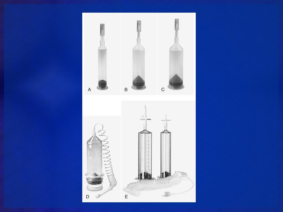
All automatic injectors have certain components in common; each is equipped with a control panel, syringe, heating device, and high-pressure mechanism.

23
Control Panel Each of the automatic injection devices has a control panel that is used to set the parameters of the injection sequence . Depending on the unit and the optional equipment purchased, the control panel will appear more or less intricate. The injector systems are equipped with a touch screen display for ease of programming. Almost all of the units available are digitally controlled and ergonomically designed for ease of use. Some of the systems allow the control panel to be removed from the unit and taken to the control area during the procedure.

24
Syringe In all automatic injectors, the syringe is removable. Disposable syringes are available for use with all automatic injectors; these are presterilized and can be installed easily. The manufacturers also offer prefilled syringes for use with their systems. The syringes are completely disposable, thereby eliminating the possibility of cross-contamination from reusable parts. Syringe capacity varies with brand. The syringes come in a variety of sizes depending on the company and the automatic injector. These range from 60 ml to 200 ml with sizes of 125 ml, 130 ml, and 150 ml also available. The smaller syringes (60 ml) find wide usage in pediatric studies.
 find wide usage in pediatric studies.")
26
Heating Device The heating system is an electronic device that heats and maintains the contrast medium at or near body temperature and reduces the viscosity of certain contrast agents, thereby facilitating the setting of certain flow rates from the injector. It is usually located on the injector head close to the syringe. The syringe temperature is thermostatically controlled and is usually preset at the factory to a nominal 37° C (approximately 98° F). Heating time varies with the injector being used. Most injection device heaters can only maintain the temperature of the contrast agent. Therefore, the contrast agent should be pre-warmed when using injection devices equipped with this type of heater.
. Heating time varies with the injector being used. Most injection device heaters can only maintain the temperature of the contrast agent. Therefore, the contrast agent should be pre-warmed when using injection devices equipped with this type of heater.")
27
TO REDUCE ITS VISCOSITY
CONTRAST WARMER TO REDUCE ITS VISCOSITY

28
Safety Devices Acceleration regulators and pressure-limiting devices can add optional safety features to the automatic injection device. The acceleration regulator allows the drive motor to be accelerated over a specific period of time, which reduces the possibility of catheter whip.

29
Injector Operation When programming the automatic injector, the radiographer must be concerned with one primary factor—the flow rate. Flow rate can be defined simply as the delivery rate (amount delivered per unit of time). It is dependent on the viscosity of the contrast agent, the length and diameter of the catheter, and the injection pressure. The flow rate chosen for a specific procedure is governed by the procedure itself, the vessel entered, the patient, and the nature of the disease. Flow rates can vary from as low as 0.5 ml/s to as high as 10 ml/s, depending on these factors.
. It is dependent on the viscosity of the contrast agent, the length and diameter of the catheter, and the injection pressure. The flow rate chosen for a specific procedure is governed by the procedure itself, the vessel entered, the patient, and the nature of the disease. Flow rates can vary from as low as 0.5 ml/s to as high as 10 ml/s, depending on these factors.")
30
Constant Flow Rate When constant flow injectors are used, the desired flow rate and injection time are set, and the selected volume in milliliters per second will be delivered regardless of the variables involved. For example, if a flow rate of 5 ml/s and a volume of 100 ml are required, they are programmed on the injector as 5 ml/s injection rate for 20 s. This setting will deliver the specific volume at the desired flow rate regardless of the viscosity of the contrast medium or any other parameters involved. Care must be exercised when flow rates are set because too high a flow rate may injure the patient or damage the catheter.

32
Catheter diameter. The catheter with the larger overall internal diameter (ID) (lumen) will provide less resistance against the flow of contrast media and require less pressure to maintain a selected flow rate. 2. Catheter length. The catheter with the overall greater length will provide more resistance to flow than one that is shorter because the resistance is applied over a longer length of catheter tubing

33
CT procedures require a high degree of control over the injection and its parameters to maximize the accuracy of the study. Injection systems used for CT have the same basic components as those used for angiography. The injector head can be either floor mounted or track mounted on an overhead system. In many cases, the console is mounted inside the control booth for maximum operator safety. The Liebel-Flarsheim CT 9000 ADV (Fig. 3-8) allows a variable flow rate, from 10 ml/hr to 10 ml/s. This unit also allows programming injection delays from 0 to 255 s in 0.01-s intervals for use during multiphasic studies.
 allows a variable flow rate, from 10 ml/hr to 10 ml/s. This unit also allows programming injection delays from 0 to 255 s in 0.01-s intervals for use during multiphasic studies..")
36
CT Empower injector has the same safety systems as the previously discussed injectors, including pressure limitation, tilt sensor lockout to prevent air embolism, voice prompts to alert operators to potential problems, and an extravasation detection accessory. The extravasation detector monitors the patient's skin impedance and alerts the technologist to the potential extravasation and also pauses the injection to further protect the patient (Fig. 3-10). The unit is also equipped with a feature that permits a pause in the injection; the injection can then be resumed on command. The parameters of the pause, such as the phase number, pause mode, elapsed time, and volume remaining, are displayed on the console. The display is a high-contrast electroluminescent display that is easily seen under ambient lighting conditions from a variety of different angles The Empower CT is also capable of storing more than 50 anatomically referenced preprogrammed procedures that can be recalled when needed. It also has a touch screen display allowing rapid system control by the technologist
. The unit is also equipped with a feature that permits a pause in the injection; the injection can then be resumed on command. The parameters of the pause, such as the phase number, pause mode, elapsed time, and volume remaining, are displayed on the console. The display is a high-contrast electroluminescent display that is easily seen under ambient lighting conditions from a variety of different angles The Empower CT is also capable of storing more than 50 anatomically referenced preprogrammed procedures that can be recalled when needed. It also has a touch screen display allowing rapid system control by the technologist.")
38
The E-Z-EM Empower injector system is manufactured with a dual-barrel injector head specifically designed for saline chase injections. The routine use of a saline bolus “chaser” for certain CT studies may yield cost savings in terms of the contrast agent and present a decreased risk of contrast nephropathy. The chaser is administered immediately following the injection of the contrast agent. The purpose of the chaser is to provide better vessel enhancement while utilizing a lower dose of contrast medium.

40
Automatic power injectors offer advantage
a. Consistent, reproducible flow rates. b. Precise volume/dosage control. c. Higher injection rates for optimal contrast enhancement. d. Automatic delays for proper enhancement patterns and multiphase imaging.

41
Pharmacology is the study of drugs and their origins, chemical composition, preparations, and use. Pharmacokinetics refers to the mechanisms of bodily absorption, distribution, metabolism, and excretion of drugs. Pharmacodynamics is the action that various drugs have with body tissues.

42
The route of administration will vary not only with the drug used but also the purpose that it has been designed for. The drugs used in vascular and cardiac diagnostic and interventional radiography fall into two major classifications: local and systemic. The local medications are usually administered at a specific site and are injected into the tissues only in that particular area. The route is by direct injection, and the purpose of the drug is the reduction of sensation (pain) in the tissues of the surrounding area. These drugs are used at the beginning of the procedure, and they have an anesthetic and analgesic effect at the puncture site. The anesthetic effect of the “local” medication is almost immediate and is well tolerated by most adult patients. Local medications can also be administered by inhalation and topical application to the skin and other mucous membranes. Inhaled medication is used to produce a rapid response to local respiratory conditions.
 in the tissues of the surrounding area. These drugs are used at the beginning of the procedure, and they have an anesthetic and analgesic effect at the puncture site. The anesthetic effect of the local medication is almost immediate and is well tolerated by most adult patients. Local medications can also be administered by inhalation and topical application to the skin and other mucous membranes. Inhaled medication is used to produce a rapid response to local respiratory conditions.")
43
Systemic medications produce a wide variety of effects to the patient
Systemic medications produce a wide variety of effects to the patient. These drugs are usually used before the procedure begins, at times during the procedure, and often in emergency situations to alleviate a problem. The four major routes of administration for the systemic agents are oral, rectal, sublingual, and parenteral. The first three routes are self-explanatory, and their use will appear obvious. Inhalation can also be considered as a means of administering systemic medications as in the case of general anesthesia.

44
Oral medications are taken by mouth
Oral medications are taken by mouth. The rectal route of administration is used if the patients are unable to take oral medication. This could be due to the inability to retain the drug in the stomach or because the drug would be adversely affected by the gastric contents. Sublingual administration of certain medications is used when quick action of the drug is required. These drugs are manufactured to dissolve quickly when placed under the tongue to provide a rapid effect. It can be seen that these three routes of administration of medication all utilize the digestive system to introduce the medication into the body. The parenteral route pertains to the administration of a medication other than the gastrointestinal tract. This is usually accomplished by injection

45
When any medication is administered it is important to follow some basic principles, known as the “Six Rights.” These basic rules should be followed whenever any medication is dispensed to a patient. Administer the drug to the RIGHT PATIENT 2. Administer the RIGHT DRUG 3. Administer the RIGHT DOSAGE 4. Use the RIGHT ROUTE to administer the drug 5. Provide the drug at the RIGHT TIME 6. Complete the RIGHT DOCUMENTATION

46
Parenteral Route Description Specific Criteria Intravenous Insertion of a needle into a vein Needle held at 15–30-degree angle to patient's arm with needle bevel up. Use the smallest needle size for the product to minimize tissue damage. Most common sizes are 16–18 gauge. Intramuscular Insertion of a needle into a muscle Use a 21–23 gauge needle (1½ inches) to ensure passage into deep muscle tissue. Needle is held at a 90-degree angle. Intrathecal (intraspinal) Insertion of a needle into the subdural, arachnoid, or lumbar areas. Use a 16–25-gauge, 3½ inch long spinal needle. Intradermal Insertion of a needle into the dermis A small syringe or needle combination is used, 26–27 gauge, ¼ to ½ inch needle. The angle of insertion is 5–15 degrees. Subcutaneous Insertion of a needle into the loose connective tissue directly under the dermis Use a 25-gauge, ⅝ inch needle. An angle of 45–90 degrees can be used depending upon the body habitus of the patient. Larger, obese patients will require a 90-degree angle for insertion.
 to ensure passage into deep muscle tissue. Needle is held at a 90-degree angle. Intrathecal (intraspinal) Insertion of a needle into the subdural, arachnoid, or lumbar areas. Use a 16–25-gauge, 3½ inch long spinal needle. Intradermal. Insertion of a needle into the dermis. A small syringe or needle combination is used, 26–27 gauge, ¼ to ½ inch needle. The angle of insertion is 5–15 degrees. Subcutaneous. Insertion of a needle into the loose connective tissue directly under the dermis. Use a 25-gauge, ⅝ inch needle. An angle of 45–90 degrees can be used depending upon the body habitus of the patient. Larger, obese patients will require a 90-degree angle for insertion.")
47
Summary of Drug Types Category Description/Use Route of Administration
Examples Analgesics Reduce pain without loss of consciousness; however, in some individuals a loss of consciousness can occur. Classed as opioids, nonopioids, and other. Parenteral Fentanyl Oral/parenteral Morphine Oral Codeine Meperidine Oxycodone Nonopioid Naproxen Ibuprofen Antiarrhythmics Treat alterations in the rhythm of the heart Beta-blockers Metoprolol Propranolol

48
Anticoagulants Aid in hindering the coagulation of the blood Oral/injection Warfarin Heparin Lepirudin Antiemetics Prevent vomiting Oral/rectal/injection Metoclopramide hydroxyzine Antihypertensives Used in the treatment of high blood pressure (hypertension); usually accompanied by treatment with diuretic. Oral Metoprolol Oral/parenteral Vasotec Antiplatelets Used to destroy blood platelets Aspirin Tirofiban
; usually accompanied by treatment with diuretic. Oral. Metoprolol. Oral/parenteral. Vasotec. Antiplatelets. Used to destroy blood platelets. Aspirin. Tirofiban.")
49
Narcotics Dull the senses, relieve pain, induce sleep; controlled substances that have a wide range of potential for abuse See Analgesics Sedatives Reduce excitement or nervousness, produce a calming effect; also produce sleep or loss of consciousness See Anxiolytics Thrombolytics Used to eliminate or break up a thrombus—blood clots remaining attached to the wall of vessels Oral Warfarin Parenteral Streptokinase Alteplase Duteplase Heparin

50
Vasodilators Cause widening or dilation of blood vessels Parenteral Nitroprusside Oral/parenteral Ethanol Oral/parenteral/ transdermal Nitroglycerine

51
Emergency Medications
The radiographer should also be knowledgeable about those medications that are administered in emergency situations. When an emergency occurs, there is little time to search for the proper medication. The locations, containers, and general dosages of these medications should be known and readily at hand. A number of reactions that occur during and after the examination fall under the allergic reaction category.

52
Levophed Intravenous Causes contraction of smooth muscle resulting in vasoconstriction; used to treat hypotension, increase blood pressure, and maintain circulation Adrenaline Aminophylline Bronchial dilator, used to treat bronchospasm Atropine Intramuscular Increases heart rate Intravenous, oral Preoperative medication Antagonist to cholinergic drugs Benadryl (diphenhydramine) Acute allergic reactions Urticaria Dermatitis Dopamine Continuous intravenous infusion Shock Cardiac stimulant increasing cardiac output and renal blood flow High doses can cause vasoconstriction Diazepam (Valium) Convulsions Antianxiety medication
 Acute allergic reactions. Urticaria. Dermatitis. Dopamine. Continuous intravenous infusion. Shock. Cardiac stimulant increasing cardiac output and renal blood flow. High doses can cause vasoconstriction. Diazepam (Valium) Convulsions. Antianxiety medication.")
53
Types of Contrast Negative Positive Radiopaque Contrast Media (RCM):
intravenous and oral

54
Iodinated ( Radiopaque Contrast Media) RCM are water-soluble compounds that may be administered:
a.Generally into the bloodstream intravenously. b.Directly into a targeted vein or artery for localized enhancement. c.Directly into the intrathecal space during CT myelography. d.Into the joint space during CT arthrography. e.Orally to opacify the gastrointestinal (GI) tract.
 tract.")
55
Suspensions of barium sulfate are commonly employed as positive contrast agents for opacification of the GI tract.

56
Iodinated RCM can be generally divided into the following categories:
•Ionic contrast media are salts consisting of sodium and/or meglumine. Each molecule of ionic contrast agent consists of three iodine atoms. When injected into the bloodstream, each molecule dissociates into two charged particles, or ions. The production of osmotic ions is indicative of high-osmolar contrast media (HOCM). Examples of HOCM are iothalamate meglumine (Conray) and diatrizoate sodium (Hypaque). •Non-ionic contrast media are non-salt chemical compounds that also contain three atoms of iodine per molecule. They do not dissociate in solution. These substances are commonly referred to as low-osmolar contrast media (LOCM). Examples of LOCM are iohexol (Omnipaque), iopamidol (Isovue), and ioversol (Optiray).
. Examples of HOCM are iothalamate meglumine (Conray) and diatrizoate sodium (Hypaque). •Non-ionic contrast media are non-salt chemical compounds that also contain three atoms of iodine per molecule. They do not dissociate in solution. These substances are commonly referred to as low-osmolar contrast media (LOCM). Examples of LOCM are iohexol (Omnipaque), iopamidol (Isovue), and ioversol (Optiray).")
57
Ionic Contrast Media

58
Non-ionic Contrast Media

59
Iso-osmolar contrast media (IOCM) have the same osmolality as blood and therefore may offer improved patient comfort and a reduced potential for untoward side effects. Iodixanol (Visipaque) is an example of a non-ionic iso-osmolar contrast agent. The range of serum iodine concentration for adequate opacification during CT examination is 2 to 8 mg/mL.
 is an example of a non-ionic iso-osmolar contrast agent. The range of serum iodine concentration for adequate opacification during CT examination is 2 to 8 mg/mL.")
62
Iodine Concentration The iodine atoms attached to the basic contrast agent structure are responsible for providing the radiopacity of the substance. As the iodine concentration of the contrast agent increases, the viscosity also increases. Compounds of the meglumine salts have a greater increase in viscosity than those of sodium salts. In considering the use of contrast agents during vascular procedures, viscosity becomes an important factor when long, small-bore catheters are used. More viscous contrast agents require greater injection pressures to deliver the same amount of material, increasing the possibility of patient trauma and catheter damage.

63
Miscibility The miscibility or immiscibility of a contrast agent is an important factor in vascular procedures. Angiographic contrast media must be completely miscible with the blood to prevent any possibility of contrast medium embolization.

64
Persistence A final factor to consider regarding choice of contrast agent is the agent's persistence within the body. Because radiopaque agents instilled either directly or indirectly into body organs are foreign substances, it is important that they be excreted rapidly from the body. However, they should remain and concentrate in the body organs long enough to provide an adequate radiographic study. This is an important criterion in the design of a suitable contrast agent.

67
Contraindications to IV iodinated contrast agents include:
Allergy to iodine. b. Prior severe allergic reaction to an iodinated contrast agent. c. Renal insufficiency/failure.

68
Patients who are in chronic renal failure and undergoing dialysis may be candidates for IV contrast agent administration at the discretion of the radiologist and referring physician. Although iodinated contrast media are known to cross the placental barrier, pregnancy is not a direct contraindication to their administration. However, caution must be used in determining the potential benefit of contrast agent administration in the pregnant patient because the risk of adverse reaction in the fetus is unknown. Nursing mothers may be injected with iodinated contrast agents as clinically warranted. Caution again must be exercised because many such agents are excreted in breast milk. Nursing mothers are typically instructed to pump and discard breast milk for 24 hours after administration of a contrast agent to effectively eliminate risk to the infant.

69
Patients with non–insulin-dependent diabetes who are taking an oral biguanide (metformin) drug may also have a higher risk of CIN-related effects. Patients receiving metformin drug therapy are at increased risk for lactic acidosis, a condition that may be exacerbated by renal insufficiency

71
REACTIONS The onset of reactions is variable; approximately 70% of reactions occur within 5 minutes of the injection, whereas 16% occur more than 5 minutes after the injection and 14% occur 15 minutes after the injection. It is advisable that the radiographer remain with the patient for at least 15 minutes after the injection. When a particular contrast agent is being used, the radiographer should read the supplied product data sheet. Information provided about the contrast agent includes description, indications and contraindications, administration and dosage, warnings or precautions, and adverse reactions or complications. The section on adverse reactions or complications can prepare the radiographer for the possible types of reactions that the agent can produce.

72
The incidence of renal failure after the administration of a contrast medium can be decreased with proper hydration. If the patient is able to take fluids by mouth, a minimum of 500 ml of fluid should be administered prior to the procedure and an additional 2500 ml of fluid should be taken over the 24-hour period following the examination. If intravenous hydration is necessary, the infusion of a 0.45% or 0.9% saline solution should be administered at the rate of 100 ml per hour starting 4 hours before the procedure and continuing for the 24-hour period after the study

78
MOST COMMON SHOCK IN CT ANAPHYLACTIC!

79
SHOCK: PHYSIOLOGIC REACTION TO : ILNESS, TRAUMA OR SEVERE
EMOTIONAL DISTURBANCE

80
SHOCK SYMPTOMS: HYPOTENSION WEAK PULSE RAPID PULSE RAPID BREATHING

81
SHOCK HYPOVOLEMIC SEPTIC CARDIOGENIC NEUROGENIC
ANAPHYLACTIC – MOST COMMON IN CT

82
Overdose Reactions It should not be implied from this classification group that some patients are deliberately overdosed with the contrast agent. However, under certain conditions, some patients may be predisposed to inadvertent contrast agent overdose. Infants, adults with acute renal failure, adults with cardiac failure in the beginning stages, and adults with hepatic failure and ascites are particularly at risk of receiving an excessive osmotic load. A dose that is considered to be within normal limits for the general population may, in fact, be an overdose in patients with these conditions. Obtaining a thorough history from the patient can in most cases identify patients who might be at risk for this type of reaction

83
Cardiovascular Reactions
These reactions tend to cause peripheral vasodilation and a decrease in systemic blood pressure, which can result in reflex tachycardia. Because they are related to the effect of the vagus nerve on the cardiovascular system, they are referred to as vasovagal or vagal-type reactions. Vasovagal reactions can affect myocardial contractility and cardiac electrophysiology and can ultimately be serious. The most severe type of cardiovascular reaction is cardiac arrest. The patient's vital signs should be monitored before, during, and after the procedure.

84
Psychogenic Reactions
Psychogenic factors are important mediators of some major reactions. If patients are overanxious, they may respond to the subjective side effects produced as a result of the introduction of the contrast agent with an autonomic response. The response becomes the basis for the adverse reaction. Substances less likely to cross the blood-brain barrier and enter the central nervous system reduce the likelihood of reactions precipitated by this mechanism.

85
Activation System—Triggering Reactions
The activation of a variety of systems can be triggered as a response to damage to the vascular endothelium at the injection site. These systems do not cause the same type of reaction in every patient because of individual variations in the presence and action of inhibitors in the pathways. Histamine release, tissue damage, thrombus formation, and hemolysis are some manifestations of these types of reactions

86
Enteral Radiopaque Contrast Media
Enteral RCM are administered orally and/or rectally to opacify the GI tract. b. Generally an enteral agent is either a water-soluble iodinated solution or a suspension of barium sulfate. c. Barium sulfate suspensions are readily used as oral and rectal contrast agents for opacification of the GI tract. d. Barium sulfate is an inert compound with excellent attenuation properties. e. Routine transit time for barium sulfate through the GI tract is typically between 30 and 90 minutes.

87
The important considerations in the choice between a water-soluble iodine oral contrast agent and barium sulfate are as follows: •Barium sulfate may be not be utilized in cases of suspected perforation because it may be toxic to the peritoneum. •Barium sulfate is contraindicated in patients who are to undergo surgery or other invasive procedures of the abdomen and/or pelvis. •Barium sulfate can be potentially harmful if aspirated. •Water-soluble oral contrast agents, particularly of the low-osmolar type, are usually more palatable and result in less GI distress. •Water-soluble oral contrast agents may be contraindicated in patients with known iodine allergy.

88
For CT examinations of the entire abdomen and pelvis, substantial opacification of the stomach, small bowel, and proximal large bowel is typically required. An example protocol is: a.450 mL of oral contrast agent minutes before scan. b.300 mL of oral contrast agent 30 minutes before scan. c.150 mL of oral contrast agent immediately before scan

89
f. Water-soluble oral contrast media can also be either high-osmolar or low-osmolar iodinated solutions. g. High-osmolar contrast agents, such as diatrizoate meglumine and diatrizoate sodium, have traditionally been used as oral/rectal CT contrast media. h. Newer low-osmolar contrasts such as iohexol may also be used as oral/rectal contrast media for CT. i. Routine transit time for water-soluble iodinated contrast agents through the GI tract is typically between 30 and 90 minutes.

90
Negative Contrast Agents
Air, gases, and water may be used as negative contrast agents during CT examination. b. Water may be used as an oral contrast agent to fill the GI tract. Advantages include: •Increased palatability and improved patient comfort. •Better demonstration of the enhancing bowel wall. •Does not interfere with three-dimensional (3D) applications. c. Effervescent granules used to treat gas and acid indigestion may also be administered as negative oral contrast agents. When swallowed, these granules add negative contrast in the form of gas to the stomach and proximal small bowel, allowing for better visualization of these structures. d. Water-soluble iodinated solutions may be mixed with carbonated beverages to add negative contrast to the GI tract, improving the demonstration of subtle disease.
 applications. c. Effervescent granules used to treat gas and acid indigestion may also be administered as negative oral contrast agents. When swallowed, these granules add negative contrast in the form of gas to the stomach and proximal small bowel, allowing for better visualization of these structures. d. Water-soluble iodinated solutions may be mixed with carbonated beverages to add negative contrast to the GI tract, improving the demonstration of subtle disease.")
91
e. Air acts as a contrast agent during CT imaging of the chest much like it does on a chest radiograph. Image acquisition at the end of full inspiration improves image quality during a CT examination of the chest. f. Air may also be administered via enema to insufflate the large bowel to improve image quality during CT colonography. g. CT colonography may also involve insufflation of the large intestine with CO2. Distention of the large intestine with room air or CO2 is necessary for optimal visualization of the bowel wall.

92
PREPARATION LIGHT EVENING MEAL BOWEL CLEANSING
NPO AFTER MIDNIGHT (EXCEPT FOR CONTRAST AND MEDS) DETRMINE IF PATIENT HAD BE RECENTLY!
 DETRMINE IF PATIENT HAD BE RECENTLY!")
93
BARIUM SULFATE CONCENTRATION
1-3%

94
WATER SOLUABLE CONTRAST CONCENTRATION
2-5% EX. 100 cc OF WATER AND 20 –50 cc OF GASTROGRAPHIN

Similar presentations


 Oral Administration - substance is ingested through the mouth - digested and absorbed in gastrointestinal tract - passes through.>")


 Pulse (P) Respiration (R) Blood pressure (BP) Pain (often called the fifth vital sign) Oxygen Saturation.>")


 leading to inadequate oxygen delivery to tissues.>")

