Download presentation
Presentation is loading. Please wait.

1
Gastrointestinal Physiology

2
General features of the gastrointestinal tract
Mucosa Epithelium: consists of a single layer of specialized cells; some are involved in secretions and some release hormones Lamina propria: layer of connective tissue which contains glands, hormone- containing cells, lymph nodes, and capillaries Muscularis mucosa: a thin layer of muscle, the contraction of which causes folding and ridges in the mucosal layers

3
Mucosa Epithelium: consists of a single layer of specialized cells; some are involved in secretions and some release hormones Lamina propria: layer of connective tissue which contains glands, hormone- containing cells, lymph nodes, and capillaries Muscularis mucosa: a thin layer of muscle, the contraction of which causes folding and ridges in the mucosal layers Submucosa A layer of connective tissue that contains glands, large blood vessels, and lymphatics. Outermost region has a nerve net called the submucosal (Meissner’s) plexus. Meissner’s plexus is part of the enteric nervous system and is involved in secretory activity.
 plexus. Meissner’s plexus is part of the enteric nervous system and is involved in secretory activity.")
4
Muscularis externa Inner layer of circular muscle Outer layer of longitudinal muscle Myenteric nerve plexus involved in motor activity is between the muscle layers. Serosa Outermost layer of the GI tract Consists of connective tissue and a layer of epithelial cells Within this layer autonomic nerve fibers run and eventually synapse on target cells and the enteric nerve plexes

5
Nervous Control VIP = vasoactive intestinal peptide, an inhibitory parasympathetic transmitter GRP = gastrin-releasing peptide; stimulates the release of gastrin from G cells

6
Neural Control of Gastrointestinal Function— Enteric Nervous System

7
Endocrinal Control CCK = cholecystokinin; GIP = gastric inhibitory peptide (glucose insulinotropic peptide) *Note: In a non–acid-producing stomach (e.g., chronic gastritis), the reduced negative feedback increases circulating gastrin. **All four hormones stimulate insulin release.
, the reduced negative feedback increases circulating gastrin. **All four hormones stimulate insulin release.")
8
MOTILITY

10
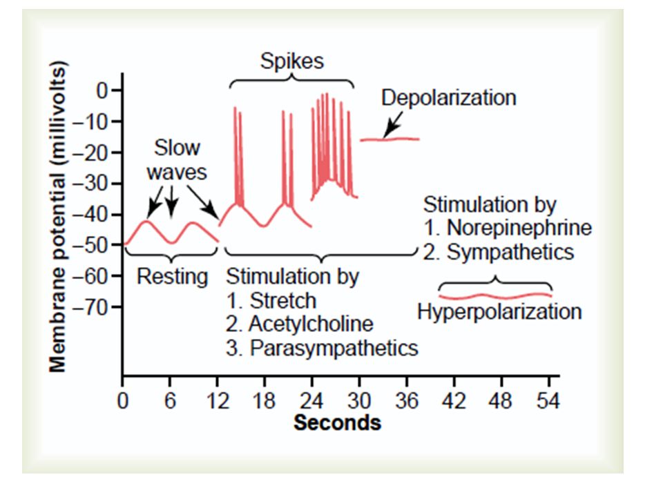
Characteristics of Smooth Muscle
Electrical activity:- Resting membrane potential -40 to -65 mV Oscillation of membrane potential is generated by interstitial cells that act as pacemakers. This is referred to as slow waves or basic electrical rhythm, and if threshold is reached it generates action potentials. Action potentials are generated by the opening of slow channels that allow the entry of both sodium and calcium.

13
Slow waves occur at 3/min in the antrum, as high as
18/min in the duodenum, and 6 to 10/min in the colon

14
Motor activity Stretch produces a contractile response.
Gap junctions create an electrical syncytium within the smooth muscle. Slow waves create low level contractions, and action potentials strengthen the contractions. Pacemaker activity from the interstitial cells creates the intrinsic motor activity. Tonic contraction at sphincters act as valves.

15
Peristaltic propulsion. Peristaltic propulsion involves
formation of a propulsive and a receiving segment, mediated by reflex control of the intestinal musculature

16
Swallowing is a reflex controlled from the brain stem.
anatomic functional Efferent input is via the vagus nerve for all events LES Sensitive to : Nicotine alcohol Caffine and cholacate

17
Events during swallowing:
Relaxation of upper esophageal skeletal muscle sphincter (UES) Primary peristaltic wave Relaxation of lower esophageal smooth muscle sphincter (LES) via VIP acting as an inhibitory transmitter Relaxation of proximal stomach (receptive relaxation)
 Primary peristaltic wave. Relaxation of lower esophageal smooth muscle sphincter (LES) via VIP acting as an inhibitory transmitter. Relaxation of proximal stomach (receptive relaxation)")
18
Achalasia, the LES fails to relax during a swallow, due to abnormalities of the enteric nerves. Primary peristalsis in the esophageal body is poor. Swallowed food is retained in the esophagus. The pressure wave in the esophagus is weak; resting pressure in the LES is abnormally high and does not decrease to allow passage of the swallowed material. Reflux esophagitis, the LES does not maintain adequate tone. The reflux of stomach acid into the esophagus presents serious risk of erosion. Scleroderma may present with reflux due to inadequate LES tone and difficulty swallowing due to poor development of propulsive pressure. Diffuse esophageal spasm is another neuromuscular disorder of the esophagus. It may present with severe chest pain resembling a heart attack. Barium swallowing reveals repeated, spontaneous waves of contraction.

19
Gastric Motility

20
Stimulation Increased parasympathetic activity via acetylcholine and gastrin release Local distension Inhibition Low pH of stomach contents inhibits the release of gastrin Feedback from duodenal overload (neural and hormonal) Stomach Emptying Liquids > CHO > protein > fat (> = faster than) The pyloris of the stomach acts as a sphincter to control the rate of stomach emptying. A wave of contraction closes the sphincter so that only a small volume is moved forward into the duodenum. CCK, GIP, and secretin will increase the degree of pyloric constriction and will slow stomach emptying. ACh = Acetylcholine BER = Basic electrical rhythm GIP = Gastric inhibitory polypeptide GRP = Gastrin-releasing peptide VIP = Vasoactive intestinal

21
Small Intestinal Motility
Rhythmic contractions in adjacent sections create segmentation contractions, which are mixing movements. Waves of contractions preceded by a relaxation of the muscle (peristaltic movements) are propulsive. The ileocecal sphincter, or valve between the small and large intestine, is normally closed. Distension of the ileum creates a muscular wave that relaxes the sphincter. Distension of the colon creates a nervous reflex to constrict the sphincter
 are propulsive. The ileocecal sphincter, or valve between the small and large intestine, is normally closed. Distension of the ileum creates a muscular wave that relaxes the sphincter. Distension of the colon creates a nervous reflex to constrict the sphincter")
22
Colon Motility Segmentation contractions create bulges (haustrations) along the colon. Mass movements, which are propulsive, are more prolonged than the peristaltic movements of the small intestine.
 along the colon. Mass movements, which are propulsive, are more prolonged than the peristaltic movements of the small intestine.")
23
Migrating Myoelectric Complex (MMC)
A propulsive movement initiated during fasting, which begins in the stomach and moves undigested material from the stomach and small intestine into the colon. Repeats every 90–120 minutes during fasting. When one movement reaches the distal ileum, a new one starts in the stomach. Correlated with high circulating levels of motilin, a hormone of the small intestine This movement prevents the backflow of bacteria from the colon into the ileum and its subsequent overgrowth in the distal ileum.

24
Defecation Defecation is a reflex involving the central nervous system. A mass movement in the terminal colon fills the rectum and causes a reflex relaxation of the internal anal sphincter and a reflex contraction of the external anal sphincter. Voluntary relaxation of the external sphincter accompanied with propulsive contraction of the distal colon complete defecation. Lack of a functional innervation of the external sphincter causes involuntary defecation when the rectum fills.

25
Defecation Complex behavior involving voluntary actions and reflexes.
Defecation reflex: sacral spinal cord and efferent cholinergic parasympathetic fibers in pelvic nerves. Distension of rectum and relaxation of internal sphincter. Voluntary actions: relaxation of external sphincter (striated muscle, innervated by somatic fibers via pudendal nerves) and increase of intraabdominal pressure 57
 and increase of intraabdominal pressure. 57.")
26
SECRETIONS

27
Acinar cells → intercalated duct → striated duct
unusual feature - saliva production is stimulated by both parasympathetic and sympathetic nervous systems (although parasympathetic control is dominant). unusually high blood flow
. unusually high blood flow.")
28
When compared with plasma, saliva is hypotonic
has higher K+ and bicarbonate (HCO3-) concentrations, and has lower Na+ and chloride (Cl-) concentrations.
 concentrations, and has lower Na+ and chloride (Cl-) concentrations.")
29
Salivary gland secretion

30
The acinar cells secrete the initial saliva, which is isotonic
The ductal cells modify the initial saliva More NaCl is absorbed than KHCO3 is secreted - there is net absorption of solute.

31
Relation between composition of saliva and the salivary flow rate

32
Regulation of salivary secretion

33
Salivary Secretions Parotid gland secretions are entirely serous (lack mucin). Submandibular and sublingual gland secretions are mixed mucus and serous. They are almost entirely under the control of the parasympathetic system, which promotes secretion. The initial fluid formation in the acinus is via an indirect chloride pump (secondary active transport powered by the Na/K ATPase pump), and the electrolyte composition is isotonic and similar to interstitial fluid. NaCl is reabsorbed in the ducts, and because the water cannot follow the final secretions are hypotonic.
, and the electrolyte composition is isotonic and similar to interstitial fluid. NaCl is reabsorbed in the ducts, and because the water cannot follow the final secretions are hypotonic.")
35
Lipase, amylase

36
Higher vol, thin Lower vol, thick

37
Composition of salivary secretions
Low in Na+, Cl– ; because of reabsorption High in K+, HCO3 because of secretion (pH = 8) α-Amylase (ptyalin): secreted in the active form and begins the digestion of carbohydrates Mucus, glycoprotein Immunoglobulins and lysozymes Low tonicity: Salivary fluid is hypotonic because of reabsorption of NaCl and impermeability of ducts to water
 α-Amylase (ptyalin): secreted in the active form and begins the digestion of carbohydrates. Mucus, glycoprotein. Immunoglobulins and lysozymes. Low tonicity: Salivary fluid is hypotonic because of reabsorption of NaCl and impermeability of ducts to water.")
38
Gastric Secretions The epithelial cells that cover the gastric mucosa secrete a highly viscous alkaline fluid (mucin plus bicarbonate) that protects the stomach lining from the caustic action of HCl. Fluid needs both mucin and bicarbonate to be protective. Nonsteroidal anti-inflammatory drugs such as aspirin decrease the secretion of the mucin and bicarbonate. Surface of the mucosa studded with the openings of the gastric glands Except for the upper cardiac region and lower pyloric region whose glands secrete mainly a mucoid fluid, gastric glands secrete a fluid whose pH can be initially as low as 1.0.
 that protects the stomach lining from the caustic action of HCl. Fluid needs both mucin and bicarbonate to be protective. Nonsteroidal anti-inflammatory drugs such as aspirin decrease the secretion of the mucin and bicarbonate. Surface of the mucosa studded with the openings of the gastric glands. Except for the upper cardiac region and lower pyloric region whose glands secrete mainly a mucoid fluid, gastric glands secrete a fluid whose pH can be initially as low as 1.0.")
40
Secretory products of various gastric cells

41
Secretions of the main cells composing the oxyntic gastric glands
Parietal cells HCl Intrinsic factor combines with vitamin B12 and is reabsorbed in the distal ileum. This is the only substance secreted by the stomach that is required for survival. It is released by the same stimuli that release HCl. Chief Cells Pepsinogen is converted to pepsin by H+ Pepsinogen is initially converted to active pepsin by acid. Active pepsin continues the process. Pepsin is active only in the acid pH medium of the stomach. Pepsin begins the digestion of protein. Mucous Neck Cells . Secrete the protective mucus, HCO3 combination Pepsin is not essential for life

43
Protective and damaging factors in GI Mucosa

44
Control of acid secretion
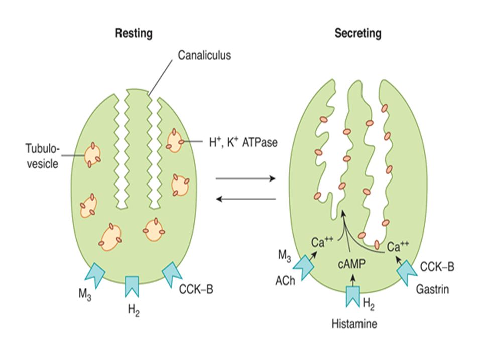
There are 3 natural substances that stimulate parietal cells: Acetylcholine, acting as a transmitter Locally released histamine The hormone gastrin Distension of the stomach following a meal stimulates mechanoreceptors from which the reflex afferents and efferents run in the vagus (vagovagal reflex). The factors that stimulate the parietal cells also stimulate the chief cells. Once the pH of stomach contents decreases to 2.0, negative feedback factors strongly inhibit further acid secretion Somatostatin inhibits parietal cells
. The factors that stimulate the parietal cells also stimulate the chief cells. Once the pH of stomach contents decreases to 2.0, negative feedback factors strongly inhibit further acid secretion. Somatostatin inhibits parietal cells.")
46
Agents that control H secretion

47
Regulation of gastric acid and pepsin secretion by soluble mediators and neural input. Gastrin is released from G cells in the antrum and travels through the circulation to influence the activity of ECL cells and parietal cells. The specific agonists of the chief cell are not well understood. Gastrin release is negatively regulated by luminal acidity via the release of somatostatin from antral D cells.

51
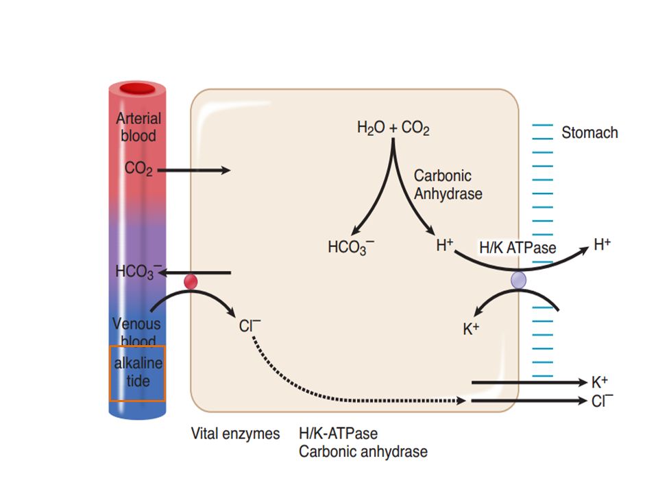
Cellular mechanisms of acid secretion
Within the cell, carbonic anhydrase facilitates the conversion of CO2 into H+ and HCO3–. The demand for CO2 can be so great following a meal that the parietial cells extract CO2 from the arterial blood. Hydrogen ions are secreted by a H/K-ATPase pump similar to that in the distal nephron. The pumping of H+ raises intracellular HCO3– and its gradient across the basal membrane and provides the net force for pumping Cl– into the cell. The chloride diffuses through channels across the apical membrane, creating a negative potential in the stomach lumen. Because of the extraction of CO2 and secretion of HCO3 – , the venous blood leaving the stomach following a meal is alkaline.

52
Ionic composition of gastric secretions
Compared with extracellular fluid, gastric secretions are high in H+, K+, Cl–, but low in Na+. The greater the secretion rate, the higher the H+ and the lower the Na+. Vomiting stomach contents produces a metabolic alkalosis and a loss of body potassium (hypokalemia mainly due to the alkalosis effect on the kidney).
.")
54
Pancreatic Secretions
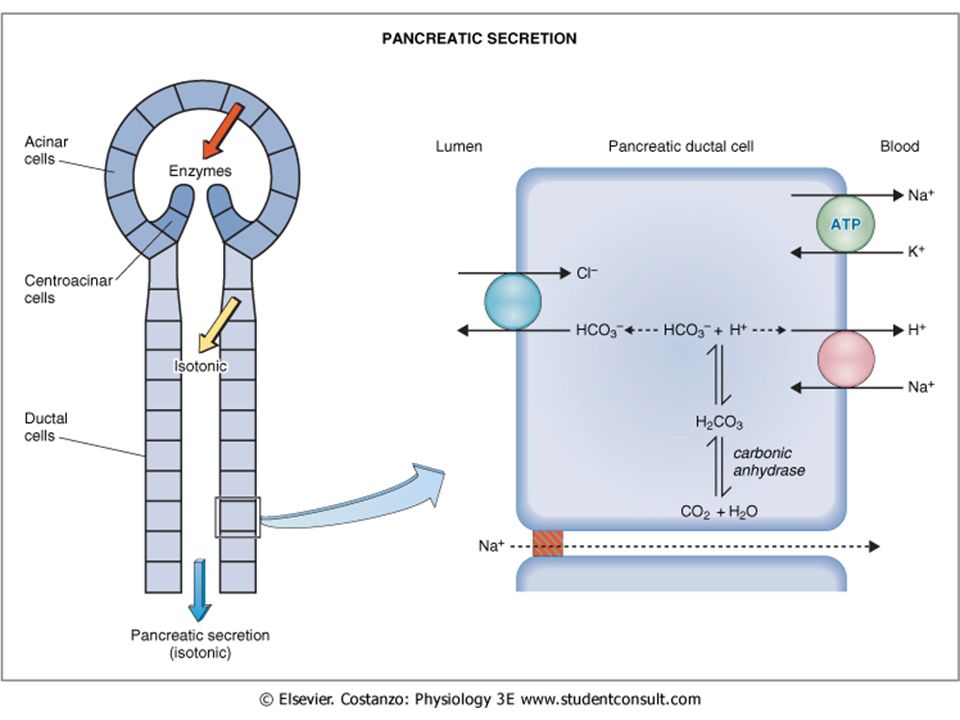
Exocrine tissue is organized into acini and ducts very similar to that of the salivary glands. Cholinergic nerves to the pancreas stimulate the secretion of both the enzyme and aqueous component. Food in the stomach stimulates stretch receptors and, via vagovagal reflexes, stimulates a small secretory volume. Sympathetics inhibit secretion. Most of the control is via secretin and CCK.

55
Fluid and electrolyte components
Aqueous component is secreted by epithelial cells which line the ducts. Fluid is isotonic due to the high permeability of the ducts to water and the concentrations of Na and K are the same as plasma. Bicarbonate and chloride concentrations vary reciprocally. High flow, high bicarbonate, low chloride The high bicarbonate in the lumen is created by a secondary active transport Entry of chloride across the apical membrane into the lumen is via a chloride channel. In cystic fibrosis there is a mutation in the gene that encodes this channel, resulting in less chloride and a reduced fluid component of pancreatic secretions. The smaller volume of highly viscous fluid may also contain few enzymes.

58
Composition of pancreatic juice and flow rate

61
Control of pancreatic secretions
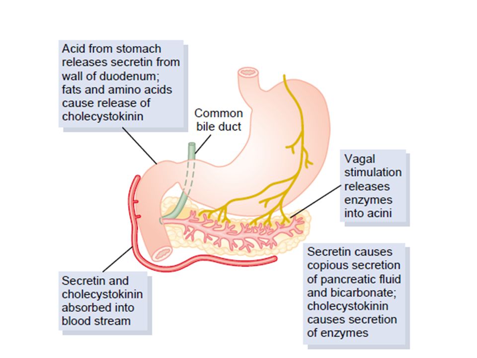
Secretin -- cAMP Released from the duodenum in response to acid entering from the stomach. L Action on the pancreas is the release of fluid high in HCO3–. This released HCO3–-rich fluid is the main mechanism that neutralizes stomach acid entering the duodenum. Cholecystokinin (CCK) -- phospholipase C Released from the duodenum in response to partially digested materials (e.g., fat, petides, and amino acids) lAction on the pancreas is the release of enzymes (amylases, lipases, proteases). Secretion of pancreatic juice is primarily under hormonal control. Secretin acts on the pancreatic ducts to cause copious secretion of a very alkaline pancreatic juice that is rich in HCO3– and poor in enzymes. The effect on duct cells is due to an increase in intracellular cAMP. Secretin also stimulates bile secretion. CCK acts on the acinar cells to cause the release of zymogen granules and production of pancreatic juice rich in enzymes but low in volume. Its effect is mediated by phospholipase C
 -- phospholipase C. Released from the duodenum in response to partially digested materials (e.g., fat, petides, and amino acids) lAction on the pancreas is the release of enzymes (amylases, lipases, proteases). Secretion of pancreatic juice is primarily under hormonal control. Secretin acts on the pancreatic ducts to cause copious secretion of a very alkaline pancreatic juice that is rich in HCO3– and poor in enzymes. The effect on duct cells is due to an increase in intracellular cAMP. Secretin also stimulates bile secretion. CCK acts on the acinar cells to cause the release of zymogen granules and production of pancreatic juice rich in enzymes but low in volume. Its effect is mediated by phospholipase C.")
63
Sodium bicarbonate (NaHCO3), water, and enzyme secretion by
the pancreas, caused by the presence of acid (HCl), fat (soap), or peptone solutions in the duodenum.
, fat (soap), or peptone solutions in the duodenum.")
65
Pancreatic amylases are secreted as active enzymes:
Hydrolyze alpha-1,4-glucoside linkage of complex carbohydrates, forming three smaller compounds: – α-Limit dextrins: still a branched polysaccharide – Maltotriose, a trisaccharide – Maltose, a disaccharide Cannot hydrolyze β linkages of cellulose

66
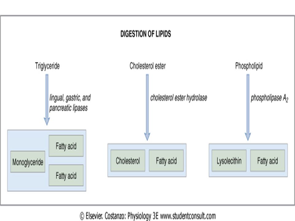
Pancreatic lipases mainly secreted as active enzymes.
Glycerol ester lipase (pancreatic lipase) needs colipase to be effective. Colipase displaces bile salt from the surface of micelles. This allows pancreatic lipase to attach to the droplet and digest it, leading to formation of two free fatty acids and one monoglyceride (a 2-monoglyceride, i.e., an ester on carbon 2)
 needs colipase to be effective. Colipase displaces bile salt from the surface of micelles. This allows pancreatic lipase to attach to the droplet and digest it, leading to formation of two free fatty acids and one monoglyceride (a 2-monoglyceride, i.e., an ester on carbon 2)")
67
Cholesterol esterase (sterol lipase)
Hydrolyzes cholesterol esters to yield cholesterol and fatty acids. Pancreatic proteases are secreted as inactive zymogens. They include trypsinogen, chymotrypsinogen, and procarboxypeptidase

68
SECRETION exocrine glands secrete digestive juices, consisting of
water electrolytes specific organic constituents important for digestive process (enzymes, bile salts, mucus) endocrine glands: hormones for regulation of the GI system
 endocrine glands: hormones for regulation of the GI system.")
69
Small Intestinal Secretions
Most prominent feature of the small intestine is the villi. Surface epithelial cells display microvilli. Water and electrolyte reabsorption greatest at the villus tip. Water and electrolyte secretion greatest at the bottom in the crypts of Lieberkuhn

71
Crypt secretion A Na-K-Cl transporter in the basolateral membrane facilitates the ion uptake by secondary active transport. Na entry drives the entry of K and Cl against their electrochemical gradients. The elevated intracellular Cl and negative intracellular potential drives the diffusion of chloride through channels on the apical membrane. Luminal Cl then pulls water, Na, and other ions into the lumen, creating the isotonic secretion. This is the general scheme of the chloride pump. Neurotransmitter secretagogues include VIP and ACh. Luminal secretagogues include bacterial toxins. A cholera toxin strongly activates the apical Cl channels of the crypts, increasing water and electrolyte secretion.

72
Composition and Formation of Bile

74
Bile secretion = digestive/absorptive function of the liver
Components of bile • bile salts (conjugates of bile acids) • bile pigments (e.g. bilirubin) • cholesterol • phospholipids (lecithins) • proteins • electrolytes (similar to plasma, isotonic with plasma) ml/day
 • bile pigments (e.g. bilirubin) • cholesterol. • phospholipids (lecithins) • proteins. • electrolytes (similar to plasma, isotonic with plasma) ml/day.")
75
Bile acids Primary bile acids known as cholic acid and chenodeoxycholic acid are synthesized by the liver from cholesterol. The lipid-soluble bile acids are then conjugated primarily with glycine. The conjugated forms are water-soluble but contain a lipid soluble segment. Because they are ionized ,at neutral pH, conjugated bile acids exist as salts of cations (Na+) and are, therefore, called bile salts. Bile salts are actively secreted by the liver.
 and are, therefore, called bile salts. Bile salts are actively secreted by the liver.")
76
Biosynthetic pathways of bile salt

77
Micelle Formation When bile salts become concentrated, they form micelles. These are water soluble spheres with a lipid-soluble interior. As such, they provide a vehicle to transport lipid-soluble materials in the aqueous medium of the bile fluid and the small intestine. Micelles are vital in the digestion, transport, and absorption of lipid-soluble substances from the duodenum to the distal ileum.

78
Secondary bile acids are formed by deconjugation and dehydroxylation of the primary bile salts by intestinal bacteria, forming deoxycholic acid (from cholic acid) and lithocholic acid (from chenodeoxycholic acid). Lithocholic acid has hepatotoxic activity and is excreted In the distal ileum, and only in the distal ileum, can the bile salts be actively reabsorbed and recycled (enterohepatic circulation). Lack of active reabsorbing mechanisms (or a distal ileal resection) causes loss in the stool and a general deficiency in bile salts, as the liver has a limited capacity to manufacture them. This deficiency can lead to fat malabsorption a nd cholesterol gallstones.
. Lack of active reabsorbing mechanisms (or a distal ileal resection) causes loss in the stool and a general deficiency in bile salts, as the liver has a limited capacity to manufacture them. This deficiency can lead to fat malabsorption a nd cholesterol gallstones.")
79
2. Bile pigments When old red blood cells are destroyed in the tissue macrophage system, the globin portion of the hemoglobin molecule is split off, & the heme is converted to biliverdin. Most of the biliverdin is converted to bilirubin & excreted in the bile.

80
Bile pigments Bilirubin
A major bile pigment, bilirubin is a lipid-soluble metabolite of hemoglobin. Transported to the liver attached to protein, it is then conjugated and excreted as water-soluble glucuronides. These give a golden yellow color to bile. Stercobilin Produced from metabolism of bilirubin by intestinal bacteria. It gives a brown color to the stool

81
Salts and water The HCO3– component is increased by the action of secretin on the liver. The active pumping of sodium in the gallbladder causes electrolyte and water reabsorption, which concentrates the bile. Bile pigments and bile salts are not reabsorbed from the gallbladder.

82
Phospholipids (mainly lecithin)
Insoluble in water but are solubilized by bile salt micelles. Cholesterol Present in small amounts. It is insoluble in water and must be solubilized by bile salt micelles before it can be secreted in the bile.

83
Control of bile secretion and gallbladder contraction
Secretin causes secretion of HCO3 – and fluid into bile canalicular ducts. Secretion of bile salts by hepatocytes is directly proportional to hepatic portal vein concentration of bile salts. CCK causes gallbladder contraction and sphincter of Oddi relaxation.

84
Functions of bile Digestive function
Emulsification: In the small intestine lipids are emulsified by bile acids (i.e. formation of small droplets of lipids coated with bile acids). Absorptive function – fat & fat soluble vitamins Excretory function – bile pigment , heavy metal Choleretic action – that is bile salts stimulate the liver to secrete bile Prevention of gall stone formation – solution
. Absorptive function – fat & fat soluble vitamins. Excretory function – bile pigment , heavy metal. Choleretic action – that is bile salts stimulate the liver to secrete bile. Prevention of gall stone formation. – solution.")
85
Digestion and Absorption

86
DIGESTION degradation of structurally complex foodstuffs by digestive enzymes 3 categories of energy-rich foodstuffs: carbohydrates, proteins and lipids ABSORPTION absorbable units as a result of the digestive process are transported along with water, vitamins and electrolytes from the lumen of the GI tract into the blood and lymph

87
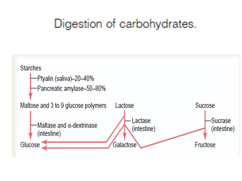
enzymes—α-dextrinase (or α-glucoamylase), isomaltase, and maltase—are all bound to the brush border (apical membrane of enterocytes) Brush border enzymes have their highest activity in the jejunum (upper). These brush border enzymes are required for digestion mainly because disaccharides—e.g., sucrose, lactose—are not absorbed from the gut
. These brush border. enzymes are required for digestion mainly because disaccharides—e.g., sucrose, lactose—are not absorbed from the gut.")
89
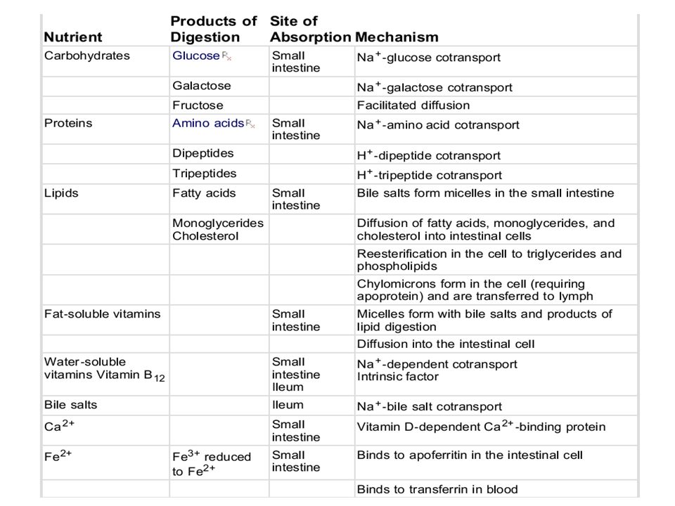
The monosaccharide end products—glucose, galactose, and fructose—
are readily absorbed from the small intestine, also mainly in the jejunum.

90
(secondary active transport linked to sodium
facilitated diffusion. Na-H antiporter. simple diffusion (secondary active transport linked to sodium) facilitated diffusion
 facilitated diffusion.")
92
60% 40%

94
• Emulsification: In the small intestine lipids are emulsified by bile acids (i.e. formation of small droplets of lipids coated with bile acids). Bile salts (bile salts = conjugation of bile acids with taurine or glycine) are polar and water soluble, and function as detergents. Emulsion droplets allow access of the water-soluble lipolytic enzymes by increasing surface area.
 are polar and water soluble, and function as detergents. Emulsion droplets allow access of the water-soluble lipolytic enzymes by increasing surface area.")
95
Big Blobs of Fat Small Blobs of Fat Micelles Fatty Acids and Monoglycerides Enter epithelial cells of duodenum Formation of triglycerides and packed into vesicles Chylomicron Passed into Lacteals

97
Digestive products of fats found in the micelles and absorbed from the intestinal
lumen may include: Fatty acids (long chain) 2-Monoglyceride Cholesterol Lysolecithin Vitamins A, D, E, K Bile salts, which stabilize the micelles Micelles diffuse to the brush border of the intestine. The diffusion through the unstirred layer is the rate-limiting step of fat absorption. The digested lipids then diffuse across the brush border in the lipid matrix. In the mucosal cell, triglyceride is resynthesized and forms lipid droplets (chylomicrons). These leave the intestine via the lymphatic circulation (lacteals). They then enter the bloodstream via the thoracic duct. The more water-soluble short-chain fatty acids can be absorbed by simple diffusion directly into the bloodstream. The bile salts are actively reabsorbed in the distal ileum
 2-Monoglyceride. Cholesterol. Lysolecithin. Vitamins A, D, E, K. Bile salts, which stabilize the micelles. Micelles diffuse to the brush border of the intestine. The diffusion through the. unstirred layer is the rate-limiting step of fat absorption. The digested lipids then diffuse across the brush border in the lipid matrix. In. the mucosal cell, triglyceride is resynthesized and forms lipid droplets (chylomicrons). These leave the intestine via the lymphatic circulation (lacteals). They then. enter the bloodstream via the thoracic duct. The more water-soluble short-chain. fatty acids can be absorbed by simple diffusion directly into the bloodstream. The. bile salts are actively reabsorbed in the distal ileum.")
100
Net Transport Of Electrolytes

101
Duodenum l Hypertonic fluid enters this region, and following the movement of some water into the lumen, the fluid becomes and remains isotonic. l The absorption of most divalent ions and water-soluble vitamins begins here and continues through the small intestine. l Injested iron and calcium tend to form insoluble salts. The acid environment of the stomach redissolves these salts, which facilitates their absorption in the small intestine. Iron and calcium absorption is diminished in individuals with a deficient stomach acid secretion. l Iron, in the Fe++ form only, is absorbed mainly from the duodenum. Jejunum l Overall, there is a net reabsorption of water and electrolytes. l The cellular processes involved are almost identical to those described in the renal physiology section for the cells lining the nephron proximal tubule. Ileum l Net reabsorption of water, sodium, chloride, and potassium continues, but there begins a net secretion of bicarbonate. l It is in the distal ileum, and only in the distal ileum, where the reabsorption of bile salts and intrinsic factor with vitamin B12 takes place. Colon l The colon does not have digestive enzymes or the protein transporters to absorb the products of carbohydrate and protein digestion. l Also, because bile salts are reabsorbed in the distal ileum, very few lipidsoluble substances are absorbed in the colon. l There is a net reabsorption of water and sodium chloride, but there are limitations. l Most of the water and electrolytes must be reabsorbed in the small intestine, or the colon becomes overwhelmed. l Most of the water and electrolytes are absorbed in the ascending and transverse colon; thereafter, the colon has mainly a storage function. l The colon is a target for aldosterone, where it increases sodium and water reabsorption and potassium secretion. l Because there is a net secretion of bicarbonate and potassium, diarrhea usually produces a metabolic acidosis and hypokalemia. It commonly presents as hyperchloremic, nonanion gap metabolic acidosis, as described in the acid-base section

102
DIARRHEA A loss of isotonic fluid that is high in bicarbonate and potassium. Secretory diarrhea: Any oversecretion of fluid and electrolytes can overwhelm the reabsorptive capacity of the gastrointestinal tract. It generally persists during fasting; a good example is that created by cholera toxins. Malabsorptive diarrhea: Created by an improper absorption of nutrients, creating an osmotic effect and the retention of water and electrolytes in the lumen. It generally abates during fasting and typical examples are celiac disease (allergic reaction to gluten, which damages the villi) and lactose intolerance. Executive disease: The mucosal distruction causes the output of a purulent bloody stool that is maintained during fasting. Hyperactivity of the intestine: Accelerated movement of contents from the smallintestine to the colon at a rate faster than they can be reabsorbed, as in inflammatory bowel disease.
 and lactose intolerance. Executive disease: The mucosal distruction causes the output of a purulent bloody stool that is maintained during fasting. Hyperactivity of the intestine: Accelerated movement of contents from the smallintestine to the colon at a rate faster than they can be reabsorbed, as in inflammatory bowel disease.")
103
Which of the following is not normally found in abundance in the portal blood?
A) Amino acids B) Glucose C) Short-chain fatty acids D) Triglycerides
 Amino acids. B) Glucose. C) Short-chain fatty acids. D) Triglycerides.")
104
Compared to plasma, saliva has the highest relative concentration of which of the following ions under basal conditions? A) Bicarbonate B) Chloride C) Potassium D) Sodium under basal conditions, sodium and chloride concentrations in saliva are about 10% to 15% of that of plasma, bicarbonate concentration is about three-fold greater than that of plasma, and potassium concentration is about seven times greater than that of plasma
 Bicarbonate. B) Chloride. C) Potassium. D) Sodium. under basal conditions, sodium and chloride. concentrations in saliva are about 10% to 15% of that of. plasma, bicarbonate concentration is about three-fold. greater than that of plasma, and potassium concentration. is about seven times greater than that of plasma.")
105
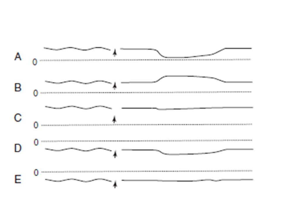
A 33-year-old man comes to the physician because his chest hurts when he eats, especially when he eats meat. He also belches excessively and has heartburn. His wife complains about bad breath. X-ray shows a dilated esophagus. Which one of the pressure tracings was most likely taken at the lower esophageal sphincter of this patient before and after swallowing (indicated by arrow)? The dashed line represents a pressure of 0 mm Hg
. The dashed line represents a pressure of 0 mm Hg .")
107
A 10-year-old boy consumes a cheeseburger, fries, and chocolate shake
A 10-year-old boy consumes a cheeseburger, fries, and chocolate shake. The meal stimulates the release of several gastrointestinal hormones. The presence of fat, carbohydrate, or protein in the duodenum stimulates the release of which of the following hormones from the duodenal mucosa? A) Cholecystokinin (CCK) B) Glucose-dependent insulinotropic peptide (GLIP) C) Gastrin D) Motilin E) Secretin GLIP is the only gastrointestinal hormone released by all three major foodstuffs (fats, proteins, and carbohydrates). The presence of fat and protein in the small intestine stimulates the release of CCK, but carbohydrates do not stimulate its release. The presence of protein in the antrum of the stomach stimulates the release of gastrin, but fat and carbohydrates do not stimulate its release. Fat has a minor effect to stimulate the release of motilin and secretin, but neither hormone is released by the presence of protein or carbohydrate in the gastrointestinal tract.
 Cholecystokinin (CCK) B) Glucose-dependent insulinotropic peptide (GLIP) C) Gastrin D) Motilin E) Secretin GLIP is the only gastrointestinal hormone released. by all three major foodstuffs (fats, proteins, and carbohydrates). The presence of fat and protein in the small. intestine stimulates the release of CCK, but carbohydrates. do not stimulate its release. The presence of. protein in the antrum of the stomach stimulates the. release of gastrin, but fat and carbohydrates do not. stimulate its release. Fat has a minor effect to stimulate. the release of motilin and secretin, but neither hormone. is released by the presence of protein or carbohydrate. in the gastrointestinal tract.")
108
The frequency of slow waves is fixed in various
parts of the gut. The maximum frequency of smooth muscle contractions cannot exceed the slow wave frequency. The slow wave frequency averages about 3 per minute in the stomach, 12 per minute in the duodenum, 10 per minute in the jejunum, and 8 per minute in the ileum. Therefore, the duodenum is most likely to have the highest frequency of smooth muscle contractions.

Similar presentations


 Ass. Prof. Dr. Emre Hamurtekin EMU Faculty of Pharmacy.>")


 Chemical Digestion (mouth, stomach, intestines) Absorption (intestines) Assimilation (at each cell in the.>")


