Download presentation
Presentation is loading. Please wait.

1
THE DIGESTIVE SYSTEM AgriScience 332 Animal Science #8646-E
TEKS: (c)(3)(A)
(3)(A)")
2
Introduction The digestive system works to convert food into simpler molecules that can be absorbed and utilized by the cells of the body.

3
Functions of the digestive system include:
Ingesting food, Grinding food, Digesting food, Absorbing nutrients, and Eliminating solid wastes.

4
The anatomy and physiology of the digestive system differs among species of animals, which have digestive tracts that are adapted to the most efficient use of the feed they consume.

5
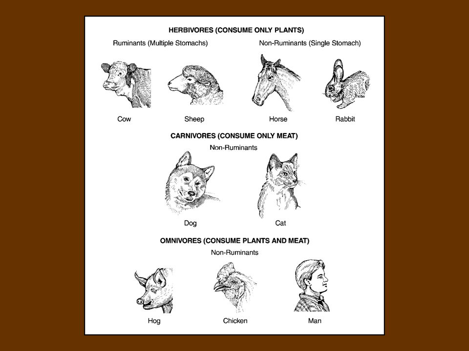
Herbivores are animals that depend entirely on plants for food.
Examples of herbivores are cattle, sheep, horses, and rabbits. Photo by Stephen Ausmus courtesy of USDA Agricultural Research Service.

6
Carnivores are animals that rely almost entirely on meat for food.
Examples of carnivores include cats and dogs.

7
Omnivores are animals that consume both flesh and plants.
Examples of omnivores are swine, chickens, and humans.

8
The length and complexity of the digestive tract depends on the species; carnivores have a relatively short and simple tract, while herbivores’ tracts are much longer and more complex.

9
Among herbivores, there is a difference between species based on the stomach type, monogastric or polygastric.

10
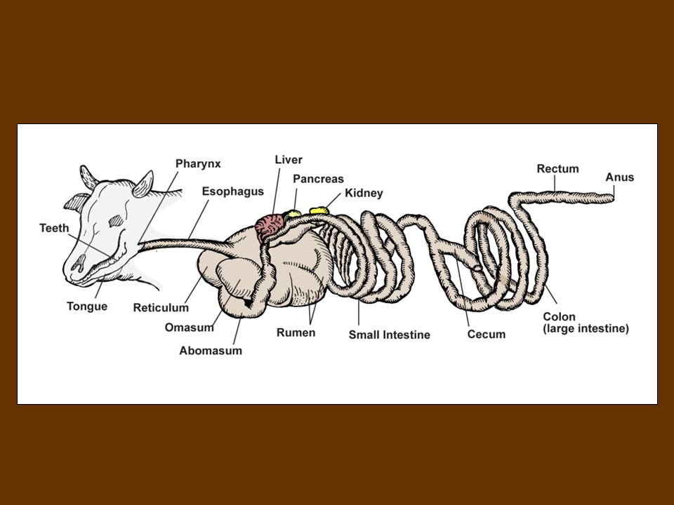
Ruminants are animals that have a four-chambered stomach (polygastric) that includes a large rumen.
Non-ruminants are animals that have a single stomach compartment (monogastric).
.")
12
Capacity of Total Digestive System
The average capacity of the digestive system and the types of feed best suited to the animal varies among the species. Animal: Capacity (in liters) Cattle 356 Sheep & Goat 44 Horse 211 Swine 27 Dog 7 Human 6 (Campbell, Kenealy, and Campbell, 2003)
 Cattle Sheep & Goat. 44. Horse Swine. 27. Dog. 7. Human. 6. (Campbell, Kenealy, and Campbell, 2003)")
13
Swine have larger digestive capacity per pound of body weight than dogs or humans.
Their digestive system is better suited for concentrated feeds with limited amounts of forage.

15
Horses have a much larger digestive system than swine.
Even though they are non-ruminants, horses can utilize large amounts of roughage in their diets because they have an enlarged cecum.

17
Cattle and sheep can utilize large amounts of bulky feeds (roughages) because they are ruminants.
A 1200 pound cow may have a stomach capacity for 300 pounds of feed.

19
Some similarities occur in the composition of the walls of the digestive tract of various species.
The walls of the digestive tract, which extend from the mouth to the anus, have four layers: epithelium, lamina propria, muscles, and visceral peritoneum.

20
Epithelium – mucous membrane that lines the digestive tract from mouth to anus and is continuous with external skin of animals. Lamina Propria – thin layer of connective tissue that supports the epithelium in the intestines.

21
Muscles – muscles of the esophagus are striated, while the remainder of the digestive tract is smooth muscle. Visceral Peritonieum – covers the digestive organs in the abdomen.

22
Anatomy of the Digestive System
The digestive tract extends from the lips to the anus. As previously mentioned, the length and complexity of the digestive tract depends on the species.

23
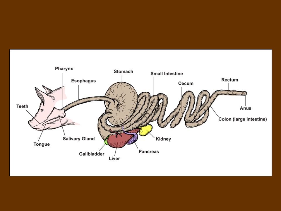
The digestive tract is made up of the following parts:
Mouth, Pharynx, Esophagus, Stomach, Small intestine, and Large intestine.

24
Accessory glands and organs that assist in digesting food include the following:
Salivary glands, Liver, Gallbladder, and Pancreas.

25
Mouth – the primary functions of the mouth are to grasp food, grind food, and mix the food with saliva. The mouth accomplishes these tasks with the use of specialized structures, including lips, tongue, teeth, cheeks, jaw, and salivary glands.

26
Lips of horses are designed to grasp food.
The lips of swine and cattle are used mostly for closing the mouth.

27
The tongue is used by most animals to grasp food.
The tongue helps in the chewing process and in the formation of boluses. Finger-like projections called papillae cover the top surface of the tongue and contain the taste buds.

28
Teeth are responsible for cutting (incisors) and grinding (premolars and molars) food.
After an animal is born, it develops a set of milk or baby teeth. As the animal ages, the milk teeth are replaced by permanent teeth.

29
Animals’ cheeks consist mostly of muscle that is lined with a mucous membrane.
The cheeks line up food with teeth. The movement of the jawbone is controlled by powerful muscles that open and close the jaw and move it from side to side in chewing.

30
Salivary glands secrete saliva that softens food, which aids in swallowing.
Saliva contains mostly water, but does contain some enzymes that begin the chemical breakdown of some starches.

31
Pharynx – a common pathway for food and air.
Food passes from the mouth into the pharynx, where the pharyngeal muscles force food into the esophagus.

32
Esophagus – a muscular tube that connects the pharynx to the stomach.
The esophagus passes through the chest cavity and connects with the stomach just after passing through the diaphragm.

33
The cardia sphincter muscle controls the movement of food into the stomach.
The pyloric sphincter muscle controls movement of food out of the stomach.

34
As previously mentioned, ruminants and non-ruminants differ in the number of compartments that make up the stomach. Non-ruminants, or monogastrics, have a single stomach compartment that is sometimes called the “true” stomach.

35
The stomach of non-ruminants is located just beyond the diaphragm on the left side of the body.
The “true” stomach has folds in the epithelial lining that creates gastric pits. Glands are located throughout the stomach and secrete digestive fluids into the pits, including hydrochloric acid, pepsin, and rennin.

36
Ruminants, or polygastrics, have stomachs with four compartments: rumen, reticulum, omasum, abomasum.

37
The relative sizes of the four stomach compartments of the ruminant vary with age of the animal.
In a calf at birth, the total capacity of the non-glandular compartments (rumen, reticulum, and omasum) is about 30%, but by two months of age, the total capacity of the non-glandular compartments is 70%.
 is about 30%, but by two months of age, the total capacity of the non-glandular compartments is 70%.")
38
The rumen of calves becomes functional at about six to eight weeks of age and, by the time the animal reaches maturity, the rumen makes up 80% of the total stomach capacity.

39
The rumen, reticulum, and omasum contain no glands, but do contain approximately one billion bacteria and one million protozoa (per milliliter). These three compartments soak food and allow microbial digestion to take place.

40
Rumen – the first compartment of the ruminant stomach, which fills most of the left side of the abdomen. The rumen has a very thick muscular wall and consists of two sacs, dorsal sac and ventral sac, that contain many papillae. Food first passes into the rumen, where it can be regurgitated as cud.

41
Reticulum – the forward most portion of the ruminant stomach.
The inner surface of the reticulum has inward folds, resembling a honeycomb shape.

42
The esophageal groove is a groove that extends from the cardia sphincter to the omasum.
It is capable of closing off the rumen and reticulum, allowing food to bypass these two parts and go directly to the omasum.

43
Omasum – the third compartment of the ruminant stomach that contains muscular projections, which are covered by mucous membrane and contain many small papillae. The papillae in the omasum are responsible for grinding roughage.

44
Abomasum – the only glandular stomach of ruminants, the abomasum is located under the omasum.
The epithelial lining and glands of the abomasum are the same as those in the stomach of non-ruminants.

45
Small intestine – a three-part tube that is the site of some digestion and the absorption of nutrients. The small intestine is made up of the duodenum, jejunum, and ileum. The small intestine is lined with many villi, which absorb nutrients.

46
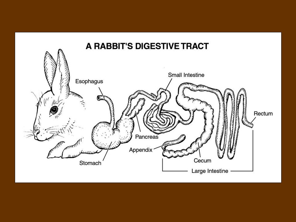
Large intestine – a larger tube of the digestive tract that consists of the cecum, colon, and rectum. The size of the cecum is much greater in horses and rabbits than in other domestic animals.

48
The large intestine is the site of water absorption and some mineral and nutrient absorption, depending on the species. Wastes are eliminated from the rectum through the anus, which is controlled by sphincter muscles.

49
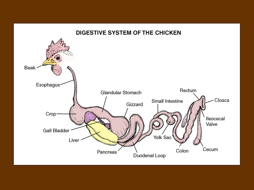
Poultry Digestive System
The anatomy of the poultry digestive system differs from other animals. Poultry do not have teeth and the prehensile structure is the beak.

50
Food passes from the mouth through the esophagus to an enlargement of the gullet called the crop.
The crop temporarily stores food and softens it before it passes to the proventriculus (glandular stomach). From the proventriculus, food quickly passes to the ventriculus, or gizzard.
. From the proventriculus, food quickly passes to the ventriculus, or gizzard.")
51
The gizzard crushes and grinds coarse feed aided by grit and gravel that accumulated in the gizzard during the bird’s life. Food passes from the gizzard into the small intestine where an abundant supply of pancreatic enzymes and bile are used to aid in the bird’s digestion.

53
Anatomy and Physiology of Accessory Digestive Organs
Several organs secrete enzymes into the digestive tract that aid in the digestive process. These organs include the salivary glands, pancreas, liver, and gall bladder.

54
Salivary glands – paired glands, including the parotid, mandibular, and sublingual salivary glands.
The parotid and mandibular salivary glands secrete serous, which is a clear, watery fluid. The sublingual salivary glands secrete serous and mucous, a thick, cloudy protective coating for the mucous membranes of the digestive system.

55
Pancreas – an elongated, lobe-shaped organ located at the beginning of the small intestine, behind the liver. The exocrine functions of the pancreas are to secrete several enzymes into the small intestine to aid in digestion. The endocrine function of the pancreas is to produce insulin, which lowers blood sugar.

56
Liver – a lobe-shaped organ located just behind the diaphragm on the right side of the body.
The liver purifies blood it receives from the stomach, spleen, pancreas, and intestines. The liver also produces bile, which is used in the digestion of fats.

57
Gall Bladder – a small, sac-like organ attached to the liver that collects bile produced by the liver and secretes it into the duodenum. Horses are the only domestic animals that do not have a gall bladder.

58
The Physiology of Digestion
An animal’s appetite is controlled by the hypothalamus and is influenced by the level of glucose in the blood and fill in the stomach. Environmental temperature and the animal’s health also influence appetite.

59
Digestion is the conversion of feedstuffs into nutrients the body can use.
Most feedstuffs are too complex to be used without being broken down into simpler molecules.

60
The digestive process includes mechanical, chemical, and microbial actions.
Mechanical actions include mastication (chewing), deglutition (swallowing), regurgitation, gastric and intestinal motility, and defecation.
, deglutition (swallowing), regurgitation, gastric and intestinal motility, and defecation.")
61
Mastication reduces food particle sizes to create more surface area on which digestive juices can act. Mastication mixes food with saliva. In ruminants, large quantities of ingested food are regurgitated as boluses (cud) so that it can be re-chewed.
 so that it can be re-chewed.")
62
As previously mentioned, poultry have no teeth.
Mechanical digestion for poultry takes place mainly in the gizzard, where grinding reduces the size of food.

63
Microorganisms aid digestion of ruminants in the rumen, reticulum, and omasum and aid digestion of horses and rabbits in the cecum. Microorganisms break down the cellulose of plant cell walls, which provides ruminants with 60% to 80% of their energy.

64
The rumen is the site of approximately 60% to 90% of digestion in ruminants.
In addition to breaking down cellulose, microorganisms also perform important functions in the animal by synthesizing all of the B-complex vitamins and all of the essential amino acids needed by their host.

65
Chemical digestion is mostly caused by enzymes, which speed up the biochemical reactions without being used up in the process. Various body cells make enzymes that are used to break down carbohydrate, protein, and fat compounds into simpler molecules.

66
The following tables show the enzymes that break down compounds into simpler molecules.

67
Chemical Digestion of Carbohydrates
Enzyme: Secreted by: Action: Ptylin Salivary glands of swine & horses. Converts carbohydrates into maltose & dextrin. Amylopsin Pancreas into duodenum. Converts starches & dextrins into simpler dextrins & maltose. Sucrase Small intestine Converts sucrose into glucose and fructose. Maltase Converts maltose into glucose. Lactase Converts lactose into glucose and galactose.

68
Chemical Digestion of Proteins
Enzyme: Secreted by: Action: Hydrochloric Acid (chemical) True stomach Activates enzymes pepsin & rennin. Pepsin Stomach Breaks protein down into proteoses and peptones. Rennin In young nursing animals, coagulates milk to aid digestion. Trypsin Chymotrypsin Carboxypeptidase Pancreas into duodenum. Continue protein digestion by breaking down more complex substances into amino acids.
 True stomach. Activates enzymes pepsin & rennin. Pepsin. Stomach. Breaks protein down into proteoses and peptones. Rennin. In young nursing animals, coagulates milk to aid digestion. Trypsin Chymotrypsin Carboxypeptidase. Pancreas into duodenum. Continue protein digestion by breaking down more complex substances into amino acids.")
69
Chemical Digestion of Fats
Enzyme: Secreted by: Action: Lipase Stomach Converts fats into higher fatty acids & glycerol. Bile (chemical) Produced by liver, stored and secreted by gall bladder into duodenum. Emulsifies fats and breaks them into smaller globules. Steapsin Pancreas into duodenum. Completes conversion of fats into higher fatty acids and glycerol.
 Produced by liver, stored and secreted by gall bladder into duodenum. Emulsifies fats and breaks them into smaller globules. Steapsin. Pancreas into duodenum. Completes conversion of fats into higher fatty acids and glycerol.")
70
Hydrochloric acid in stomach helps dissolve minerals in the diet.
Water and vitamins require no digestion before being utilized by the body.

71
Absorption is the process by which digested nutrients pass from the walls of the digestive tract into the blood. The small intestine is the site of most absorption of nutrients for carnivores and omnivores and is the site of a significant amount of nutrient absorption for herbivores.

72
The small intestine has numerous finger-like projections, called villi, that contain many blood vessels, which are responsible for collecting and absorbing nutrients.

73
Very little, if any, absorption of nutrients occurs in the mouth, esophogus, or stomach.
Some absorption of volatile fatty acids does occur across the rumen wall. Very few nutrients are absorbed in the large intestine, except for a substantial amount of volatile fatty acids in herbivores.

74
The colon of the large intestine is the absorption site of water.

75
The end products of fat digestion are fatty acids and glycerol, which are absorbed by the lymph ducts. The blood absorbs the end products of carbohydrate digestion (monosaccharides and volatile fatty acids), protein digestion (amino acids and peptides), water, and inorganic salts.
, protein digestion (amino acids and peptides), water, and inorganic salts.")
76
Digestion is complete after absorption has made the nutrients available for other parts of body.

77
ALL RIGHTS RESERVED Reproduction or redistribution of all, or
part, of this presentation without written permission is prohibited. Instructional Materials Service Texas A&M University 2588 TAMUS College Station, Texas 2007

Similar presentations


 –>")


