Download presentation
Presentation is loading. Please wait.

1
Introduction to Medical Imaging Instructors: Brian Fleming and Ioana Fleming flembri@pha.jhu.edu, ioana@cs.jhu.edu

2
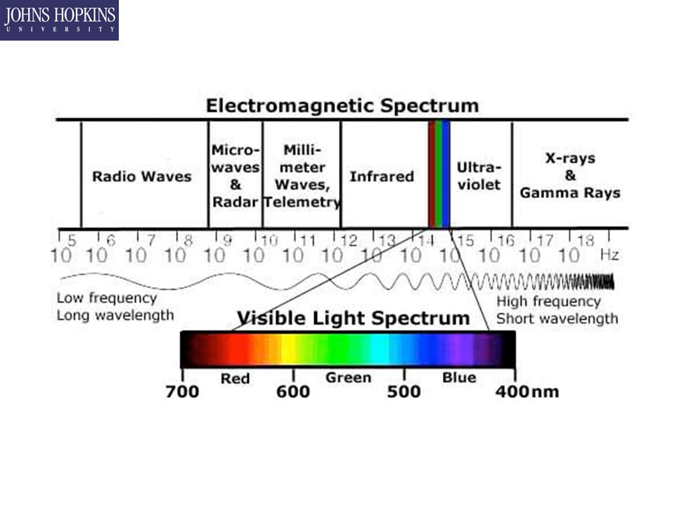
Lecture 1 Meet and greet A brief history of everything Break Intro to Death Rays X-Rays

3
In the Beginning…

4
Where to put the Leeches Hippocrates (460 - 377 BCE) –Muscles, skeleton, kidneys –Observation only Pesky “Oath” prevented human dissection Aristotle and Friends (4 th century BCE) –Animals aren’t people Herophilos and Erasistratus (4 th century BCE) –First human cadavers –Criminals aren’t people either
 –Muscles, skeleton, kidneys –Observation only Pesky Oath prevented human dissection Aristotle and Friends (4 th century BCE) –Animals aren’t people Herophilos and Erasistratus (4 th century BCE) –First human cadavers –Criminals aren’t people either")
5
Where to put the Leeches Dark Ages - Europe –Balance the “humors” –Bleed, burn, drown, or exorcise Dark Ages – Arabia/Persia –Avicenna (1020 AD) Canon of Medicine Premier book of medicine everywhere for 500 years –Ibn Zuhr (1100 AD) Invented Autopsy and discovered parasites Would have killed the idea of humors if logic and fact had been considered a valid argument
 Canon of Medicine Premier book of medicine everywhere for 500 years –Ibn Zuhr (1100 AD) Invented Autopsy and discovered parasites Would have killed the idea of humors if logic and fact had been considered a valid argument")
6
Where to put the Leeches Printing Press – 17 th century –Sharing of ideas brings renaissance (murder?)
")
7
History of Medical Imaging Wilhelm Röntgen (Roentgen) The father of diagnostic radiology German physicist (1845-1923) Discovered x-rays in 1895 X was for "unknown“ First Nobel Prize in physics 1901
 The father of diagnostic radiology German physicist ( ) Discovered x-rays in 1895 X was for unknown First Nobel Prize in physics 1901")
8
Discovery of X-rays

9
How to Irradiate Yourself Step 1 – Force electrons to go where no electron would ever want to go –In air, would cool by giving off light

10
How to Irradiate Yourself Step 2 – Get rid of the air –Air quenches electron escape Unless you really ramp up the voltage… –Try Neon –Or try nothing…

11
How to Irradiate Yourself Step 3 – Run those electrons into a target

13
“Instant” Success Nov 8, 1895: Accidental discovery of x-rays Dec 22, 1895: Bertha’s hand Dec 28, 1895: first publication of results Jan 1, 1896: Roentgen mailed copies to leading scientists Jan 5, 1896: Austrian newspaper story Jan 23, 1896: society presentation Feb 8, 1896: 1st clinical use (in US!)
")
14
Within One Year... 49 serious books on x-rays 1,044 scientific papers Known to: –spot cancer –treat cancer –cause cancer Numerous patents

15
What it REALLY Did…

16
Fluoroscopy

17
Fluorescence

18
Filmed in X-ray!

19
Early Popularity of Fluoroscopy Simple fluoro equipment: –x-ray tube –electrical generator –scintillation screen Convenience of real-time Note: early film required 1- 2 hours of exposure –(where was intensifier screen????)
")
20
Red Goggles Fluoroscopy images were dim 1899: Beclere showed that dark adaptation is a function of the retina 1901: Williams suggested 10-minute dark adaptation 1916: scientific basis of sensitivity of retinal rods in the range of red light 1916-1950’s: red goggles standard gear

21
X-ray Hazards Early 1896: reports of hair falling out Early 1896: skin reddening, inflammation Early 1896: some severe burns (attributed to high voltage in tubes) Early 1896: delayed burns 1902: Edison clear on dangers of x-rays –Clarence Dally’s oozing ulcers, lost fingers, left hand, died in 1904 (Edison never x-rayed again) The use of X-rays for medical purposes (to develop into the field of radiation therapy) was pioneered by Major John Hall-Edwards in Birmingham, England. In 1908, he had to have his left arm amputated owing to the spread of X-ray dermatitis

22
Some More Landmarks 1896: Becquerel discovered radioactivity 1896: stereoradiography developed 1901: contrast agents described 1904: lead glass protection devised 1904: exposure badge invented First angiogram

23
Coolidge X-ray Tube: 1913 Properties of new tube: –high vacuum –hot cathode –tungsten-target Five outstanding properties –accurate adjustment –stable –reproducible –range of x-ray energies –less scattered radiation William Coolidge (expense prevented routine use until 1930’s)
")
24
Potter-Bucky Grid: 1913-1920 Scattered x-rays cause blurring 1913: Gustav Bucky: metal collimator grid –reduce scatter blur 1920: Hollis Potter: movable grids –reduce image of grid

25
Conventional Tomogram: 1929 Overlapping tissues blur tissue of interest Jean Kieffer invented conventional tomogram to image an interior slice –to help diagnose his own TB! Only amateur in >100 years to make a significant discovery in medical imaging

26
Tomography Much as any light/camera, there is a focal plane My moving source and camera in opposite directions, focal plane becomes sharp Basis for almost all modern medical 3-D devices except Ultrasound. –CAT = Computed Axial Tomography –PET = Positron Emission Tomography

27
Tomography (Again!)
")
30
Tomography was hard So it really wasn’t used all that much… Until 1972, when computers and motors led to the development of CAT

31
Impact of X-rays Widespread detection of tuberculosis in 1917 1898: American Roentgen Ray Society 1927: proof of cell damage caused by x-rays 1935: radiologist required to interpret radiograph in court (anybody could previously) Shoe fluoroscopes from 1920’s to 1960’s
 Shoe fluoroscopes from 1920’s to 1960’s")
32
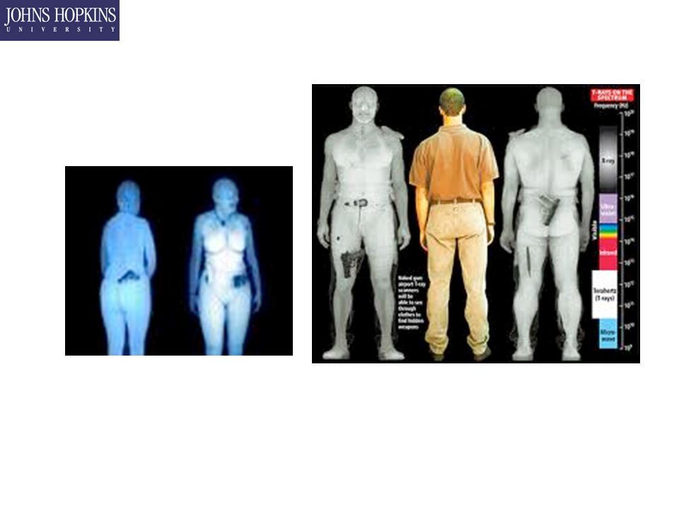
Out with the old, In with the nukes X-rays were (and still are) limited. –Dim, for one (unless subject already dead) –Cannot track temporal events well Blood flow Brain activity Etc Enter nuclear medicine
 –Cannot track temporal events well Blood flow Brain activity Etc Enter nuclear medicine.")
33
Radioactive Decay Antoine Henri Becquerel (1852 – 1908) Shared Nobel Prize of 1903 with Marie and Pierre Curie for discovery of radioactivity –Studying phosphorescence in Uranium salts on one side of his desk and the effect of bright sunlight on fluorescent coated photographic plates on the other.
 Shared Nobel Prize of 1903 with Marie and Pierre Curie for discovery of radioactivity –Studying phosphorescence in Uranium salts on one side of his desk and the effect of bright sunlight on fluorescent coated photographic plates on the other.")
34
Nuclear Physics in a Slide IsotopeMass C 12 12 C 13 13.00335 N 13 13.0057 N 14 14.0031 N 15 15.0001 O 15 15.0031 Neutron1.0087 Proton1.0073 Electron0.00055 Alpha (He)4.0026
4.0026")
35
Spontaneous Decay Every atom in the universe has a chance of spontaneously decaying –p + 0 + e + Happens about once every universe Generally, large isotopes ( > Fe) Alpha decay –Nucleus binding energy is unstable –Mass products have lower mass than parent – 238 U 234 Th + 4 He Beta Decay = emission of electron or positron –p + n 0 + e + + ν –Electronic Transmutation – 15 O 15 N - + e + + ν Gamma Decay – excess from β decay
 Alpha decay –Nucleus binding energy is unstable –Mass products have lower mass than parent – 238 U 234 Th + 4 He Beta Decay = emission of electron or positron –p + n 0 + e + + ν –Electronic Transmutation – 15 O 15 N - + e + + ν Gamma Decay – excess from β decay")
36
Back to Nuclear Imaging Radiopharmaceuticals are injected Biodistribution process causes –absorption, distribution, metabolism, excretion Radioactive decay occurs, producing: –gamma rays (single photons), or Single Photon Emission Computed Tomography (SPECT) –positrons (which yield paired photons) Positron Emission Tomography (PET) Location and counts are recorded as images
, or Single Photon Emission Computed Tomography (SPECT) –positrons (which yield paired photons) Positron Emission Tomography (PET) Location and counts are recorded as images")
37
Positron Annihilation Positrons are Anti-matter (anti-electrons) –When matter and anti-matter collide, they annihilate –Mass energy of electron + positron released as two photons –Total energy = 2*0.511 MeV + extra conserved energy
 –When matter and anti-matter collide, they annihilate –Mass energy of electron + positron released as two photons –Total energy = 2*0.511 MeV + extra conserved energy")
38
Nuclear Medicine Gallium scintigraphy looks for recurrence of malignant melanoma Step 1 – Inject patient with a radioactive substance Alpha, Beta, or Gamma? PET, SPECT Step 2 – Wait for body to distribute Choose radiopharmaceutical depending on target Step 3 – Take photos, make scrapbook

39
Radiopharmaceuticals? IsotopeBeta DecayGamma Decay Half LifeProduct 15 OΒ+Β+ N*122 s 15 N 234 ThΒ-Β- Y24 d 234 Pa 18 FΒ+Β+ N*110 m 18 O 111 InΒ-Β- Y122 s 111 Sn 14 CΒ-Β- N*5730 y 14 N 99m TcNY6 h 99 Tc Tc = Technitium, In = Indium

40
Nuclear Medicine Landmarks Single-photon imaging 1896: Becquerel discovered radioactivity 1930’s: Hevesy mapped internal organs late 1930’s: discovery of technetium 1946: AEC allowed isotopes for medical use 1950’s: Anger invented gamma camera 1968: SPECT introduced by Kuhl 1980’s: Dual/triple headed SPECT systems

41
Gamma Camera For: planar imaging SPECT imaging SPECT = single photon emission computed tomography

42
Commercial Gamma Cameras Toshiba Siemens For planar imaging and SPECT

43
[Normal male, Tc-99m HMPAO, for cerebral blood flow, Brighamrad]
![[Normal male, Tc-99m HMPAO, for cerebral blood flow, Brighamrad]](http://images.slideplayer.com/18/6188643/slides/slide_43.jpg "[Normal male, Tc-99m HMPAO, for cerebral blood flow, Brighamrad]")
44
normal Tc-99m MDP bone scintigram (5 mCi injected dose).
.")
45
Commercial PET Scanners CTI/Siemens

46
PET Images Parkinson's Disease Huntington's disease Myocardial perfusion Dopamine receptors

47
Ultrasound Imaging

48
Medical Ultrasound 1940’s – Ultrasound used to ease pain –Dr. George Ludwig, Naval Medical RI, Bethesda 1949 - Dr. John Wild measures how thick your colon (wall) is –“Father of Medical Ultrasound” 1953 – Inge Edler asks Carl Hertz if he can use radar to see inside the body. –No, but they use ultrasound to measure heart activity, published in 1954
 is – Father of Medical Ultrasound 1953 – Inge Edler asks Carl Hertz if he can use radar to see inside the body. –No, but they use ultrasound to measure heart activity, published in")
49
Medical Ultrasound 1958 – Prof. Ian Donald treats the wife of one of the directors of Babcock and Wilcox –Asks to visit with R&D to see their toys –Asks to play with ultrasound –Uses it on volunteers to measure ultrasonic properties of various people with illnesses –Publishes "Investigation of Abdominal Masses by Pulsed Ultrasound” The most important medical imaging paper… EVER –Goes on to study the growth rate of fetuses, first use of US in obstetrics

50
Medical Ultrasound 1965: First real-time ultrasound scanner 1970: commercial systems widespread mid-1970’s: grayscale and Doppler systems early-1980’s: phased-array systems mid-1990’s: 3-D ultrasound

51
How Does Ultrasound Work? Send a pulse -- receive the pulse Map time-of-arrival to round-trip distance Scan transducer in a plane

52
Ultrasound

53
Sound Propagation Sound travels through different things at different speeds –Speed of sound = c s –Simple Version : MaterialC s (m/s) Air343 Water1482 Steel5960 Muscle1482 Bone?
 Air343 Water1482 Steel5960 Muscle1482 Bone")
54
Ultrasound Images heart Fetal head kidney Corotid artery Fetal spine

55
3D Ultrasound Kretztechnik AG Voluson 530D Gallbladder stone prostate Fetal face

56
Back to X-Ray 1955 – Ronald Bracewell did some maths X ray source and detector move together Pencil thin beam which fans out in 2D Each image is a projection of all tissue in beam Take images at a full 360 degrees Reconstruct using Fourier image analysis 1956 – Allan Cormack gives it a try, succeeds in 1963 1972 – Godfrey Hounsfied demonstrates first CT scanner Hounsfield and Cormack share Nobel prize

57
Computerized Tomography

58
CT Landmarks 1971: Hounsfield scanned first patient (4min) 1972: EMI dominated Chicago RSNA 1974: 26 EMI scanners worldwide 1974: Shepp-Stein reconstruction formula 1975: Commercial 2G, 3G, 4G CT scanners 1979: Hounsfield and Cormack win Nobel Prize 1985: Imatron, 50-100ms per slice Late 80’s: Slip-ring technology 1989: first commercial helical CT scanner 1990s: multislice technology (<30s head-toe)
 1972: EMI dominated Chicago RSNA 1974: 26 EMI scanners worldwide 1974: Shepp-Stein reconstruction formula 1975: Commercial 2G, 3G, 4G CT scanners 1979: Hounsfield and Cormack win Nobel Prize 1985: Imatron, ms per slice Late 80’s: Slip-ring technology 1989: first commercial helical CT scanner 1990s: multislice technology (<30s head-toe)")
59
Early Commercial CT Scanner Siemens Siretom CT Scanner, 1975 Compare to modern CT image

60
Fast and High Resolution CT 2 revs/s 8 slices/s 2.5mm slices 58 s total time Data collected using using Picker Mx8000 TM This image follows image processing and 3D rendering

61
3D Anatomy from CT Facial fractures Lumbar spine CT endoscopy

63
Magnetic Resonance Imaging CT MRI Same patient: acute cerebral infarct

64
How Does MRI Work? Human are “ugly bags of mostly water” H 2 O has protons which have magnetic moments Protons also spin Step 1 – Make protons all spin one way Step 2 – Use radio waves at the resonant frequency to make all the protons suddenly spin the other way Step 3 – Turn off radio and let spins go back to normal Energy difference between spin up and spin down states released as a photon B-field strength dictates resonance frequency Tuned to select individual areas at a time Relaxation time, intensity, all Fourier transform into an image with very high contrast

65
MRI Landmarks 1924: Pauli proposed nuclear magnetism 1937: Rabi measured magnetic moments 1946: Bloch and Purcell described relaxation 1971: Lauterbur invented MRI (published 1973) 1973: Mansfield introduced k-space 1975: Ernst invented NMR Fourier imaging 1977: Damadian’s first whole-body MR scanner 1980: Margulis takes lead at UCSF 1997: Damadian wins patent lawsuit against GE 2003: Lauterbur and Mansfield win Nobel Prize
 1973: Mansfield introduced k-space 1975: Ernst invented NMR Fourier imaging 1977: Damadian’s first whole-body MR scanner 1980: Margulis takes lead at UCSF 1997: Damadian wins patent lawsuit against GE 2003: Lauterbur and Mansfield win Nobel Prize")
66
Some Modern Systems [Siemens, MAGNETOM 42SP] GE 0.5T open magnet (“double donut”)
![Some Modern Systems [Siemens, MAGNETOM 42SP] GE 0.5T open magnet ( double donut )](http://images.slideplayer.com/18/6188643/slides/slide_66.jpg "Some Modern Systems [Siemens, MAGNETOM 42SP] GE 0.5T open magnet ( double donut )")
67
MR Images Tagged MRI Breast implants knee heart

68
What is Next? MEG: magneto-encephalography EEG: electro-encephalography fMRI: functional magnetic resonance micro-PET optical imaging molecular imaging Photo acoustic imaging

69
Why We’re are Afraid to Fly

Similar presentations


 BPKIHS,Dharan.>")


 CT scanning or (CAT scanning) is using X-rays to create a 3D image of the inside of an object. CT stands for computed tomography.>")


; Marie and Pierre Curie (discovery.>")
 – Introduction of medical imaging and MRI – Basic.>")
, was made possible in part by NIH NLM contract# HHSN276201000580P,>")


