Download presentation
Presentation is loading. Please wait.

1
November 2012 American Society for Bone and Mineral Research (ASBMR) Update (+ videos) D Kendler, Vancouver J D Adachi, Hamilton J Brown, Quebec A Cheung, Toronto K. S. Davison, Victoria L Dian, Vancouver A Khan, Oakville D Hanley, Calgary A Hodsman, London R Josse, Toronto A Jovaisis, Ottawa S Kaiser, Halifax L-G Ste. Marie, Montreal C Yuen, Vancouver

2
Credits and Disclosures
Sigma, Canadian Menopause Society Scientific Committee (reviewers) From across Canada, osteoporosis leaders and community practitioners Unrestricted educational grant support: Amgen, Eli Lilly, Merck, Novartis, [Warner Chilcott] Speaker disclosures
 From across Canada, osteoporosis leaders and community practitioners. Unrestricted educational grant support: Amgen, Eli Lilly, Merck, Novartis, [Warner Chilcott] Speaker disclosures.")
3
ASBMR Update: Objectives
To discuss the latest research in the area of metabolic bone disease. To better understand the management of osteoporosis patients, incorporating the most up-to-date information.

4
Navigation Presenter will facilitate presentation with navigation in any order to the abstracts (click on topic: menu and submenu) The respective slides will detail the findings and importance of the chosen investigation Click on “return” slide to return to topic menu. Original abstract is in the “notes” section of the first slide.

5
Topics Fracture Risk Assessment Teriparatide
Atypical fractures/ONJ SERM, Estrogen, Testosterone Bisphosphonates Teriparatide Sclerostin Ab and other Anabolics Denosumab Other and Upcoming therapies Vitamin D / Calcium

6
Fracture Risk Assessment
TBS and fracture risk in DM Cortical porosity in DM Post-fracture care gap OP treatment based on FRAX without BMD (VIDEO) Fall and fracture risk assessment Fracture Liaison Services Models
 Fall and fracture risk assessment. Fracture Liaison Services Models.")
7
TBS (Trabecular Bone Score) is More Sensitive Than BMD to Diabetes-Related Fracture Risk
William Leslie, University of Manitoba, Canada October 14, 03:00 PM - 03:15 PM Room 101C/Minneapolis Convention Center Session:Concurrent Oral Session 22: Osteoporosis Assessment Abstract TBS (Trabecular Bone Score) is More Sensitive Than BMD to Diabetes-Related Fracture Risk Background:Type 2 diabetes (T2D) is associated with increased fracture risk but paradoxically greater BMD. TBS (trabecular bone score), a novel grey-level texture measurement extracted from DXA images, correlates with 3D parameters of bone micro-architecture. We evaluated the ability of lumbar spine (LS) TBS to account for the increased fracture risk in diabetes. Methods:29,407 women ≥50 years at the time of baseline hip and spine DXA were identified from a database containing all clinical BMD results for the Province of Manitoba, Canada. 2,356 of the women satisfied a well-validated definition for diabetes, the vast majority of whom (>90%) would have T2D. LS L14 TBS was derived for each spine DXA examination blinded to clinical parameters and outcomes. Health service records were assessed for incident non-traumatic major osteoporotic fracture codes (mean follow-up 4.7 years). Results:In linear regression adjusted for FRAX risk factors (age,BMI, glucocorticoids, prior major fracture, rheumatoid arthritis, COPD as a smoking proxy, alcohol abuse) and osteoporosis therapy, diabetes was associated with higher BMD for LS, femoral neck and total hip but lower LS TBS (all p<0.001). Similar results were seen after excluding obese subjects withBMI>30. In logistic regression (Figure), the adjusted odds ratio (OR) for a skeletal measurement in the lowest vs highest tertile was less than 1 for all BMD measurements but increased for LS TBS (adjusted OR 2.61, 95%CI ). Major osteoporotic fractures were identified in 175 (7.4%) with and 1,493 (5.5%) without diabetes (p < 0.001). LS TBS predicted fractures in those with diabetes (adjusted HR 1.27, 95%CI ) and without diabetes (HR 1.31, 95%CI ). LS TBS was an independent predictor of fracture (p<0.05) when further adjusted for BMD (LS, femoral neck or total hip). The explanatory effect of diabetes in the fracture prediction model was greatly reduced when LS TBS was added to the model (indicating that TBS captured a large portion of the diabetes-associated risk), but was paradoxically increased from adding any of the BMD measurements. Conclusions:Lumbar spine TBS is sensitive to skeletal deterioration in postmenopausal women with diabetes, whereas BMD is paradoxically greater. LS TBS predicts osteoporotic fractures in those with diabetes, and captures a large portion of the diabetes-associated fracture risk. Combining LS TBS with BMD incrementally improves fracture prediction. William Leslie has nothing to disclose. William Leslie, University of Manitoba, Canada
 is More Sensitive Than BMD to Diabetes-Related Fracture Risk. Background:Type 2 diabetes (T2D) is associated with increased fracture risk but paradoxically greater BMD. TBS (trabecular bone score), a novel grey-level texture measurement extracted from DXA images, correlates with 3D parameters of bone micro-architecture. We evaluated the ability of lumbar spine (LS) TBS to account for the increased fracture risk in diabetes. Methods:29,407 women ≥50 years at the time of baseline hip and spine DXA were identified from a database containing all clinical BMD results for the Province of Manitoba, Canada. 2,356 of the women satisfied a well-validated definition for diabetes, the vast majority of whom (>90%) would have T2D. LS L14 TBS was derived for each spine DXA examination blinded to clinical parameters and outcomes. Health service records were assessed for incident non-traumatic major osteoporotic fracture codes (mean follow-up 4.7 years). Results:In linear regression adjusted for FRAX risk factors (age,BMI, glucocorticoids, prior major fracture, rheumatoid arthritis, COPD as a smoking proxy, alcohol abuse) and osteoporosis therapy, diabetes was associated with higher BMD for LS, femoral neck and total hip but lower LS TBS (all p<0.001). Similar results were seen after excluding obese subjects withBMI>30. In logistic regression (Figure), the adjusted odds ratio (OR) for a skeletal measurement in the lowest vs highest tertile was less than 1 for all BMD measurements but increased for LS TBS (adjusted OR 2.61, 95%CI ). Major osteoporotic fractures were identified in 175 (7.4%) with and 1,493 (5.5%) without diabetes (p < 0.001). LS TBS predicted fractures in those with diabetes (adjusted HR 1.27, 95%CI ) and without diabetes (HR 1.31, 95%CI ). LS TBS was an independent predictor of fracture (p<0.05) when further adjusted for BMD (LS, femoral neck or total hip). The explanatory effect of diabetes in the fracture prediction model was greatly reduced when LS TBS was added to the model (indicating that TBS captured a large portion of the diabetes-associated risk), but was paradoxically increased from adding any of the BMD measurements. Conclusions:Lumbar spine TBS is sensitive to skeletal deterioration in postmenopausal women with diabetes, whereas BMD is paradoxically greater. LS TBS predicts osteoporotic fractures in those with diabetes, and captures a large portion of the diabetes-associated fracture risk. Combining LS TBS with BMD incrementally improves fracture prediction. William Leslie has nothing to disclose. William Leslie, University of Manitoba, Canada.")
8
TBS More Sensitive Than BMD to Diabetes Fracture Risk
Type 2 DM (T2D) associated with increased fracture risk but paradoxically greater BMD. TBS (trabecular bone score), a novel grey-level texture measurement from DXA images. 29,407 women over 50 years from Manitoba database 2,356 had DM (90% assumed T2D) Incidence of major osteoporotic fracture with follow-up 4.7 yr T2D associated with higher BMD LS, femoral neck and total hip, but lower LS TBS (all p<0.001).
 associated with increased fracture risk but paradoxically greater BMD. TBS (trabecular bone score), a novel grey-level texture measurement from DXA images. 29,407 women over 50 years from Manitoba database. 2,356 had DM (90% assumed T2D) Incidence of major osteoporotic fracture with follow-up 4.7 yr. T2D associated with higher BMD LS, femoral neck and total hip, but lower LS TBS (all p<0.001).")
9
TBS More Sensitive Than BMD to Diabetes Fracture Risk
Osteoporotic fractures in 175 (7.4%) with and 1,493 (5.5%) without T2D (p < 0.001). LS TBS predicted fractures in those with T2D (HR 1.27, 95%CI ) and without T2D (HR 1.31, 95%CI ). LS TBS was an independent predictor of fracture (p<0.05) when further adjusted for BMD (LS, FN, or total hip). LS TBS predicts osteoporotic fractures in T2D, and captures a large portion of the diabetes-associated fracture risk. Combining LS TBS with BMD incrementally improves fracture prediction.
 with and 1,493 (5.5%) without T2D (p < 0.001). LS TBS predicted fractures in those with T2D (HR 1.27, 95%CI ) and without T2D (HR 1.31, 95%CI ). LS TBS was an independent predictor of fracture (p<0.05) when further adjusted for BMD (LS, FN, or total hip). LS TBS predicts osteoporotic fractures in T2D, and captures a large portion of the diabetes-associated fracture risk. Combining LS TBS with BMD incrementally improves fracture prediction.")
10
TBS More Sensitive Than BMD to Diabetes Fracture Risk

11
Potential Importance TBS quantitates an aspect of bone architecture
TBS is a “Bone Quality” measurement available by re-analysis of existing DXA capture Particular importance in conditions such as diabetes where DXA BMD does not fully reflect fracture risk

12
return

13
Cortical Porosity as a Distinct Pathomorphology in Postmenopausal, Diabetic Women with Fragility Fractures [1127] Cortical Porosity as a Distinct Pathomorphology in Postmenopausal, Diabetic Women with Fragility Fractures Janina Patsch, University of California, San Francisco, USA October 14, 02:45 PM - 03:00 PM Room 101C/Minneapolis Convention Center Session:Concurrent Oral Session 22: Osteoporosis Assessment Abstract Cortical Porosity as a Distinct Pathomorphology in Postmenopausal, Diabetic Women with Fragility Fractures Cortical porosity (Ct.Po) is increasingly recognized as a skeletal pathomorphology of potential clinical relevance. Type 2 diabetic bone disease is a metabolic bone disease with paradoxically increased fracture rates in spite of relatively high bone mineral density (BMD). The purpose of this study was to assess if Ct.Po was increased in postmenopausal diabetics with fractures when compared with non-fractured diabetics, non-diabetic controls with prevalent fragility fractures, and non-diabetic controls without fractures. All subjects (controls [Co] n=20; controls with fractures [Fx] n=20; diabetics without fracture [DM] n=20; diabetics with fracture [DMFx] n=15) underwent high resolution peripheral quantitative computed tomography (HR-pQCT; distal radius and tibia), DXA (spine, hip, radius), and a blood draw for fasting glucose, HbA1c, and c-peptide. From HR-pQCT scans, volumetric BMD, morphometric standard parameters, and extended measures of cortical bone (including Ct.Po and endocortical bone surface En.BS) [1,2] were obtained. In a 2-way ANCOVA adjusted for age, BMI, and race/ethnicity, Ct.Po was associated with fracture in DM but not controls at the ultradistal tibia (p for interaction=0.038) but not the radius. DMFx displayed the highest cortical porosity of all groups (age-, race & BMI-adjusted means +52.2% vs DM; % vs Fx; +34.2% vs Co). Coherently, DMFx also had a greater En.BS than DM (+11.12%; p=0.035). However, there were no correlations between En.BS or Ct.Po with fasting glucose, HbA1c or c-peptide. In diabetics, En.BS was significantly correlated with tibial Ct.Po (r=0.533). Compartment-specific volumetric BMD and morphometric indices by HR-pQCT (both sites) remained non-significant among the 4 groups. Patients with prevalent fractures had significantly reduced aBMD measured with DXA, but mean aBMD values remained normal to osteopenic in all patients and were particularly high in diabetics stressing the limitations of DXA. In conclusion, we found tibial Ct.Po and En.BS measured by HR-pQCT to be non-invasive surrogate biomarkers to identify type 2 diabetes mellitus postmenopausal women with prevalent fragility fractures. References: [1] Burghardt AJ et al. 2010; Bone 47(3): [2] Burghardt AJ et al. 2010; J Bone Miner Res 25(5): Janina Patsch has nothing to disclose. Janina Patsch, University of California, San Francisco, USA
 is increasingly recognized as a skeletal pathomorphology of potential clinical relevance. Type 2 diabetic bone disease is a metabolic bone disease with paradoxically increased fracture rates in spite of relatively high bone mineral density (BMD). The purpose of this study was to assess if Ct.Po was increased in postmenopausal diabetics with fractures when compared with non-fractured diabetics, non-diabetic controls with prevalent fragility fractures, and non-diabetic controls without fractures. All subjects (controls [Co] n=20; controls with fractures [Fx] n=20; diabetics without fracture [DM] n=20; diabetics with fracture [DMFx] n=15) underwent high resolution peripheral quantitative computed tomography (HR-pQCT; distal radius and tibia), DXA (spine, hip, radius), and a blood draw for fasting glucose, HbA1c, and c-peptide. From HR-pQCT scans, volumetric BMD, morphometric standard parameters, and extended measures of cortical bone (including Ct.Po and endocortical bone surface En.BS) [1,2] were obtained. In a 2-way ANCOVA adjusted for age, BMI, and race/ethnicity, Ct.Po was associated with fracture in DM but not controls at the ultradistal tibia (p for interaction=0.038) but not the radius. DMFx displayed the highest cortical porosity of all groups (age-, race & BMI-adjusted means +52.2% vs DM; % vs Fx; +34.2% vs Co). Coherently, DMFx also had a greater En.BS than DM (+11.12%; p=0.035). However, there were no correlations between En.BS or Ct.Po with fasting glucose, HbA1c or c-peptide. In diabetics, En.BS was significantly correlated with tibial Ct.Po (r=0.533). Compartment-specific volumetric BMD and morphometric indices by HR-pQCT (both sites) remained non-significant among the 4 groups. Patients with prevalent fractures had significantly reduced aBMD measured with DXA, but mean aBMD values remained normal to osteopenic in all patients and were particularly high in diabetics stressing the limitations of DXA. In conclusion, we found tibial Ct.Po and En.BS measured by HR-pQCT to be non-invasive surrogate biomarkers to identify type 2 diabetes mellitus postmenopausal women with prevalent fragility fractures. References: [1] Burghardt AJ et al. 2010; Bone 47(3): [2] Burghardt AJ et al. 2010; J Bone Miner Res 25(5): Janina Patsch has nothing to disclose. Janina Patsch, University of California, San Francisco, USA.")
14
Cortical Porosity in PMP, Diabetic Women with Fractures
DM2 with paradoxically increased fracture rates in spite of high BMD Assess if Ct.Po in PMP DM2 with fractures compared with non-fractured diabetics, non-diabetic controls with prevalent fragility fracture Controls [Co] n=20; controls with fractures [Fx] n=20; diabetics without fracture [DM] n=20; diabetics with fracture [DMFx] n=15)
")
15
Cortical Porosity in PMP, Diabetic Women with Fractures
Ct.Po was significantly associated with fracture in patients with DM2 at the ultradistal tibia (p for interaction=0.038), but not at the radius. No significant association with controls DMFx displayed the highest cortical porosity of all groups (age-, race & BMI-adjusted means +52.2% vs DM; % vs Fx; +34.2% vs Co)
, but not at the radius. No significant association with controls. DMFx displayed the highest cortical porosity of all groups (age-, race & BMI-adjusted means +52.2% vs DM; % vs Fx; +34.2% vs Co)")
16
Cortical Porosity in PMP, Diabetic Women with Fractures
No correlations between Ct.Po and fasting glucose, HbA1c or c-peptide Patients with prevalent fractures had significantly reduced aBMD measured with DXA, but mean aBMD values remained normal to osteopenic in all Tibial Ct.Po measured by HR-pQCT identifies DM2 patients with prevalent fragility fractures.

17
Cortical Porosity in PMP, Diabetic Women with Fractures
Representative HR-pQCT scans of diabetic women with and without fragilityfractures (DMFx vs. DM)
")
18
Potential Importance HR-pQCT may provide additional diagnostic utility when compared to that of DXA alone Differences in cortical porosity may partially explain the increase in appendicular fractures in DM2 as compared to individuals without DM2

19
return

20
Osteoporosis Treatment Following Fragility Fracture Remains Unaddressed Despite Available Therapies and Established Recommendations [SU0333] Osteoporosis Treatment Following Fragility Fracture Remains Unaddressed Despite Available Therapies and Established Recommendations Cynthia O'Malley, Amgen Inc, USA October 14, 11:30 AM - 01:30 PM Discovery Hall-Hall B/Minneapolis Convention Center Session:Poster Session II and Poster Tours Abstract Osteoporosis Treatment Following Fragility Fracture Remains Unaddressed Despite Available Therapies and Established Recommendations Purpose:Clinical practice recommendations state patients with fragility fracture should be evaluated and treated for osteoporosis (OP) as they are at high risk for future fractures. To determine conformity with these recommendations we assessed the use of pharmacologic OP treatment in a large population cohort of patients with fragility fractures. Methods:This retrospective database analysis used data from a large commercially-insured population. Community-dwelling individuals aged ≥50 years with a newly diagnosed fragility fracture and no evidence of OP treatment, malignancy, or Paget’s disease in 12 months pre-fracture were included. OP treatment (including oral and parenteral bisphosphonates, raloxifene, teriparatide and calcitonin) rates in the 12 months post-fracture were evaluated descriptively with Pearson’s chi-squared tests and Cochrane-Armitage tests for significance of trends. Results:The cohort included 88,571 women and 41,984 men with mean ages of 72.3 yrs and 70.5 yrs, respectively. A minority of patients, 18.6% of women and 9.6% of men, initiated OP treatment post-fracture. Treatment rates increased with age, being highest in the age group and then declining in the 'oldest old’ individuals aged ≥85 yrs. Yet even in the age group, only 23% of women and 13% of men were treated. The exception to this peak in the age group was in women with hip fractures, where rates were consistently 20-22% for all ages except the oldest old. Rates declined to 17% for women ≥85, who accounted for 34% of female hip fractures. For both genders, OP treatment initiation was highest following vertebral fracture, intermediate following hip and lowest following wrist and humerus fractures (p<0.0001). OP treatment rates declined during the study period (Figures 1A, 1B). For women, this was observed across all age groups with treatment rates declining steadily by 6-8% between 2001 and 2010 (p<0.0001). The already low treatment rates among men declined steadily over time for those <65 yrs while the decline was less pronounced for older men. Conclusion:OP pharmacotherapy is severely underutilized following fragility fracture, regardless of patient age or fracture site. Treatment rates appear to have declined over the last decade, despite greater emphasis on recommendations for OP treatment following fragility fracture and despite the known increased risk of future fracture conferred by fracture occurrence. Cynthia O'Malley: Amgen Inc #1; Amgen Inc #3 Cynthia O'Malley, Amgen Inc, USA
 as they are at high risk for future fractures. To determine conformity with these recommendations we assessed the use of pharmacologic OP treatment in a large population cohort of patients with fragility fractures. Methods:This retrospective database analysis used data from a large commercially-insured population. Community-dwelling individuals aged ≥50 years with a newly diagnosed fragility fracture and no evidence of OP treatment, malignancy, or Paget’s disease in 12 months pre-fracture were included. OP treatment (including oral and parenteral bisphosphonates, raloxifene, teriparatide and calcitonin) rates in the 12 months post-fracture were evaluated descriptively with Pearson’s chi-squared tests and Cochrane-Armitage tests for significance of trends. Results:The cohort included 88,571 women and 41,984 men with mean ages of 72.3 yrs and 70.5 yrs, respectively. A minority of patients, 18.6% of women and 9.6% of men, initiated OP treatment post-fracture. Treatment rates increased with age, being highest in the age group and then declining in the oldest old’ individuals aged ≥85 yrs. Yet even in the age group, only 23% of women and 13% of men were treated. The exception to this peak in the age group was in women with hip fractures, where rates were consistently 20-22% for all ages except the oldest old. Rates declined to 17% for women ≥85, who accounted for 34% of female hip fractures. For both genders, OP treatment initiation was highest following vertebral fracture, intermediate following hip and lowest following wrist and humerus fractures (p<0.0001). OP treatment rates declined during the study period (Figures 1A, 1B). For women, this was observed across all age groups with treatment rates declining steadily by 6-8% between 2001 and 2010 (p<0.0001). The already low treatment rates among men declined steadily over time for those <65 yrs while the decline was less pronounced for older men. Conclusion:OP pharmacotherapy is severely underutilized following fragility fracture, regardless of patient age or fracture site. Treatment rates appear to have declined over the last decade, despite greater emphasis on recommendations for OP treatment following fragility fracture and despite the known increased risk of future fracture conferred by fracture occurrence. Cynthia O Malley: Amgen Inc #1; Amgen Inc #3. Cynthia O Malley, Amgen Inc, USA.")
21
Treatment Following Fracture Unaddressed Despite Recommendations
Retrospective analysis US data from community-dwelling individuals aged ≥50 years with a newly diagnosed fragility fracture and no OP treatment Initiated OP treatment in the year post-fracture 88,571 women and 41,984 men mean ages of 72.3 and 70.5 yrs. A minority of patients, 18.6% of women and 9.6% of men, initiated OP treatment post-fracture.

22
Treatment Following Fracture Unaddressed Despite Recommendations
Over age 85, only 23% women and 13% men treated. Women ≥85yr accounted for 34% of hip fractures in females For both genders, OP treatment initiation was highest following vertebral fracture, intermediate following hip and lowest following wrist and humerus fractures (p<0.0001). Treatment rates declined over the last decade, despite recommendations for OP treatment following fragility fracture
. Treatment rates declined over the last decade, despite recommendations for OP treatment following fragility fracture.")
23
Treatment Following Fracture Unaddressed Despite Recommendations

24
Potential Importance Secondary prevention of fragility fractures is high priority with osteoporosis experts due to the high risk of future fracture Large care gap indicates the need for innovative programs to identify these high risk patients and target them to evaluation and therapy

25
return

26
Treatment (Rx) of Post-Menopausal Women with High FRAX Scores may be Cost-Effective without First Performing Bone Densitometry [SA0387] Treatment (Rx) of Post-Menopausal Women with High FRAX Scores may be Cost-Effective without First Performing Bone Densitometry John Schousboe, Park Nicollet Clinic University of Minnesota, USA October 13, 11:00 AM - 01:00 PM Discovery Hall-Hall B/Minneapolis Convention Center Session:Poster Session I and Poster Tours Abstract Treatment (Rx) of Post-Menopausal Women with High FRAX Scores may be Cost-Effective without First Performing Bone Densitometry Background: It is uncertain whether drug Rx prevents non-vertebral fractures in postmenopausal women with femoral neck (FN) BMD T-score >-2.5 and no prevalent vertebral fracture. However, among women ≥65 years old, 3 of every 10 meeting FRAX treatment threshold for major osteoporotic fracture and 6 of 10 meeting FRAX treatment threshold for hip fracture have FN T-scores >-2.5. We used a Markov microsimulation model to estimate lifetime costs, health benefits, and cost-effectiveness of Rx of older US White women with high fracture risk by FRAX without BMD compared to first obtaining a BMD test plus vertebral fracture assessment and Rx of only the subset with either FN T-score ≤-2.5 or a prevalent vertebral fracture. Methods:The model had states of hip, morphometric and clinical vertebral, wrist, and humerus fracture, no fracture, and death. Fracture rates, costs, and disutilities were derived from the literature. The prevalence of FN T-score >-2.5 in those with FRAX w/o BMD 10-year hip and major osteoporotic fractures risks ≥ 3% and ≥20% were from the Manitoba Bone Density database. The age-specific prevalences of vertebral fracture were from Rochester Epidemiology Project data. Models were run for women age 65, 75, or 85 until death or age 100, assuming a national health payer perspective and willingness to pay of $75,000 per quality adjusted life year (QALY) gained. For those with FN T-score >-2.5 and no prevalent vertebral fracture, we assumed relative risks of incident clinical vertebral fracture, morphometric vertebral fracture, and non-vertebral fracture on Rx to be, respectively, 0.45, 0.64, and 1.0. (Quandt 2005; Ryder 2008). Sensitivity analyses assumed only 18% reduction of vertebral fractures on Rx in the proportion with T-score > -2.0 (Cummings 1998), or 25% of base case disutility from morphometric vertebral fractures. Results:Table shows estimated costs per QALY gained for Rx of women with high FRAX without BMD score vs. Rx of only those with prevalent vertebral fracture or FN T-score<-2.5; Conclusions:Rx initiation on the basis of high fracture risk estimated by FRAX w/o BMD may be cost-effective, but this is highly sensitive to vertebral fracture disutility and to assumed vertebral fracture reduction on Rx for those with T-score >-2.0 and no prevalent vertebral fracture. A randomized controlled trial of fracture reduction medication in this subset with preference weighted quality of life assessments is needed. John Schousboe has nothing to disclose. John Schousboe, Park Nicollet Clinic University of Minnesota, USA
 of Post-Menopausal Women with High FRAX Scores may be Cost-Effective without First Performing Bone Densitometry. John Schousboe, Park Nicollet Clinic University of Minnesota, USA. October 13, 11:00 AM - 01:00 PM. Discovery Hall-Hall B/Minneapolis Convention Center. Session:Poster Session I and Poster Tours. Abstract. Treatment (Rx) of Post-Menopausal Women with High FRAX Scores may be Cost-Effective without First Performing Bone Densitometry. Background: It is uncertain whether drug Rx prevents non-vertebral fractures in postmenopausal women with femoral neck (FN) BMD T-score >-2.5 and no prevalent vertebral fracture. However, among women ≥65 years old, 3 of every 10 meeting FRAX treatment threshold for major osteoporotic fracture and 6 of 10 meeting FRAX treatment threshold for hip fracture have FN T-scores >-2.5. We used a Markov microsimulation model to estimate lifetime costs, health benefits, and cost-effectiveness of Rx of older US White women with high fracture risk by FRAX without BMD compared to first obtaining a BMD test plus vertebral fracture assessment and Rx of only the subset with either FN T-score ≤-2.5 or a prevalent vertebral fracture. Methods:The model had states of hip, morphometric and clinical vertebral, wrist, and humerus fracture, no fracture, and death. Fracture rates, costs, and disutilities were derived from the literature. The prevalence of FN T-score >-2.5 in those with FRAX w/o BMD 10-year hip and major osteoporotic fractures risks ≥ 3% and ≥20% were from the Manitoba Bone Density database. The age-specific prevalences of vertebral fracture were from Rochester Epidemiology Project data. Models were run for women age 65, 75, or 85 until death or age 100, assuming a national health payer perspective and willingness to pay of $75,000 per quality adjusted life year (QALY) gained. For those with FN T-score >-2.5 and no prevalent vertebral fracture, we assumed relative risks of incident clinical vertebral fracture, morphometric vertebral fracture, and non-vertebral fracture on Rx to be, respectively, 0.45, 0.64, and 1.0. (Quandt 2005; Ryder 2008). Sensitivity analyses assumed only 18% reduction of vertebral fractures on Rx in the proportion with T-score > -2.0 (Cummings 1998), or 25% of base case disutility from morphometric vertebral fractures. Results:Table shows estimated costs per QALY gained for Rx of women with high FRAX without BMD score vs. Rx of only those with prevalent vertebral fracture or FN T-score<-2.5; Conclusions:Rx initiation on the basis of high fracture risk estimated by FRAX w/o BMD may be cost-effective, but this is highly sensitive to vertebral fracture disutility and to assumed vertebral fracture reduction on Rx for those with T-score >-2.0 and no prevalent vertebral fracture. A randomized controlled trial of fracture reduction medication in this subset with preference weighted quality of life assessments is needed. John Schousboe has nothing to disclose. John Schousboe, Park Nicollet Clinic University of Minnesota, USA.")
27
Treatment of PMP Women with High FRAX Scores prior to BMD
In women over 65 yrs, 30% meeting FRAX treatment threshold for major osteoporotic fracture and 60% meeting FRAX treatment threshold for hip fracture have FN T-scores greater than -2.5. Markov model with high fracture risk by FRAX without BMD compared to first obtaining a BMD test plus VFA and Rx of only the subset with either FN T-score ≤-2.5 or a prevalent vertebral fracture. Prevalence of FN T-score >-2.5 in those with FRAX w/o BMD 10-year hip and major osteoporotic fractures risks ≥ 3% and ≥20% from Manitoba database.

28
Treatment of PMP Women with High FRAX Scores prior to BMD
Age-specific prevalence of vertebral fracture from Rochester Epidemiology Project. Costs per QALY gained for Rx of women with high FRAX without BMD score vs. Rx of only those with prevalent vertebral fracture or FN T-score<-2.5; Treatment initiation on the basis of high fracture risk estimated by FRAX w/o BMD may be cost-effective, but this is highly sensitive to vertebral fracture disutility and to assumed vertebral fracture reduction on Rx for those with T-score >-2.0 and no prevalent vertebral fracture. A randomized controlled trial of medication in this population is needed.

29
Treatment of PMP Women with High FRAX Scores prior to BMD

30
Potential Importance Introduces debate as to whether clinical risk factors are sufficient, without a BMD, to identify a patient population at risk of fracture (and likely to benefit from therapy) Counter to this is Clinical trials rely on BMD to identify treatment candidates FRAX without BMD correlates imprecisely to FRAX with BMD BMD may be desirable for follow-up
 Counter to this is. Clinical trials rely on BMD to identify treatment candidates. FRAX without BMD correlates imprecisely to FRAX with BMD. BMD may be desirable for follow-up.")
31
FRAX alone may be cost-effective in identifying patients for pharmacotherapy: but specific intervention trials in this population are not available

32
return

33
System-Level Approaches to the Secondary Prevention of Osteoporotic Fractures: A Systematic Review and Meta-analysis [SA0356] System-level Approaches to the Secondary Prevention of Osteoporotic Fractures: A Systematic Review and Meta-analysis. Kirtan Ganda1, Markus Seibel*2, Michele Puech3, Jian Sheng Chen4. 1Concord Hospital, Australia, 2Bone Research Program, ANZAC Research Institute, University of Sydney, Australia, 3Public Health Unit, Hornsby Ku-ringai Hospital, Australia, 4Institute of Bone & Joint Research, The University of Sydney, Australia Introduction: Despite the availability of effective treatments that reduce refracture and mortality rates in patients presenting with incident osteoporotic fractures, the majority of these patients are not being assessed or treated for osteoporosis. In the past, numerous clinical care pathways have been trialed to close this treatment gap. We evaluated the effectiveness of published system-level care approaches for the secondary prevention of fractures in patients with osteoporosis. Methods: We searched eight medical literature databases using relevant key words to identify reports published between 1996 and 2011, describing clinical or system level care models for secondary fracture prevention. The following information was extracted from each publication by two independent researchers: Study design, patient characteristics, identification strategies, assessment and treatment initiation strategies and outcome measures (rates of BMD testing, anti-resorptive treatment initiation, refractures, and cost-effectiveness). The outcomes within each model of care were analysed in two ways: Firstly, we conducted meta-analyses of studies with valid control groups for two outcome measures, BMD testing and anti-resorptive treatment initiation. Secondly, we described the outcomes of studies not included in the metaanalysis, stratified by the model of care. Results: The literature search identified 43 studies and re-grouped them into four general models of care: Type A, where patients are identified, assessed and treated as part of a fully integrated service; Type B, as in A, but the service only makes treatment recommendations (no initiation of therapy); Type C, which consists of patient education plus alerting the primary care physicians to the need for further assessment and treatment; Type D, consists of patient education only. The meta-analyses showed a trend towards better rates of BMD testing and treatment initiation with increasing intensity of intervention (table). Only five studies examined re-fracture rates but only one Type A had a control group and enough power to show a significant decrease in re-fractures at 4 years follow-up. Type A & B services have been shown to be cost-effective. Conclusions: Fully co-ordinated system-level approaches to secondary prevention of osteoporotic fractures appear more effective in improving patient outcomes than approaches involving alerts and/or education only, whilst still being cost-effective. Andrea Kirtan Ganda, Markus Seibel, Michele Puech, Jian Sheng Chen, University of Sydney, NSW
. The outcomes within each model of care were analysed in two ways: Firstly, we conducted meta-analyses of studies with valid control groups for two outcome measures, BMD testing and anti-resorptive treatment initiation. Secondly, we described the outcomes of studies not included in the metaanalysis, stratified by the model of care. Results: The literature search identified 43 studies and re-grouped them into four general models of care: Type A, where patients are identified, assessed and treated as part of a fully integrated service; Type B, as in A, but the service only makes treatment recommendations (no initiation of therapy); Type C, which consists of patient education plus alerting the primary care physicians to the need for further assessment and treatment; Type D, consists of patient education only. The meta-analyses showed a trend towards better rates of BMD testing and treatment initiation with increasing intensity of intervention (table). Only five studies examined re-fracture rates but only one Type A had a control group and enough power to show a significant decrease in re-fractures at 4 years follow-up. Type A & B services have been shown to be cost-effective. Conclusions: Fully co-ordinated system-level approaches to secondary prevention of osteoporotic fractures appear more effective in improving patient outcomes than approaches involving alerts and/or education only, whilst still being cost-effective. Andrea Kirtan Ganda, Markus Seibel, Michele Puech, Jian Sheng Chen, University of Sydney, NSW.")
34
Systems-Level Secondary Fracture Prevention
Identified 43 studies of fracture prevention programs and grouped them in four general models of care: Type A: patients identified, assessed & treated - a fully integrated service Type B, as in A, but the service only makes treatment recommendations Type C, provides patient education & alerts PCP re. need for assessment and treatment Type D,provides patient education only.

35
Systems-Level Secondary Fracture Prevention
Summary Of Meta-analyses: Treatment Initiation Rates by Treatment Type Intervention Type Risk Difference 95% Confidence Interval Model of Care ‘A’ (n=8) 0.29 Model of Care ‘B’ (n=5) 0.21 Model of Care ‘C’ (n=7) 0.16 Model of Care ‘D’ (n=81) 0.03
 Model of Care ‘B’ (n=5) Model of Care ‘C’ (n=7) Model of Care ‘D’ (n=81)")
36
Systems-Level Secondary Fracture Prevention
Better rates of BMD testing and treatment initiation with increasing intensity of intervention. Only five studies examined re-fracture rates but only one Type A had a control group and enough power to show a significant decrease in re-fractures at 4 years follow-up. Type A & B services have been shown to be cost effective

37
Potential Importance Fracture Liaison Service (FLS) with initiation of assessment and therapy = most effective approach. Role for the Primary Care Physician? Simple education programs are not effective. Perhaps the FLS could turn over the work to primary care practitioners after long experience with success is appreciated by the medical community.

38
return

39
Effect of a Multifactorial Fall-and-Fracture Risk Assessment and Management Program on Gait and Balance and Disability in Hospitalized Older Adults: a Controlled Study [FR0020] Effect of a Multifactorial Fall-and-Fracture Risk Assessment and Management Program on Gait and Balance and Disability in Hospitalized Older Adults: a Controlled Study Andrea Trombetti, Division of Bone Diseases, University Hospitals and Faculty of Medicine of Geneva, Switzerland October 12, 05:45 PM - 07:00 PM Discovery Hall-Hall B/Minneapolis Convention Center Session:Welcome Reception and Plenary Poster Session Abstract Effect of a Multifactorial Fall-and-Fracture Risk Assessment and Management Program on Gait and Balance and Disability in Hospitalized Older Adults: a Controlled Study Purpose: Falls and fragility fractures among older adults are common, devastating and costly. Hospitalization affords a major opportunity for identifying high-risk individuals and for interdisciplinary cooperation to manage fall and fracture risk factors. We assessed the effects on physical performances and the level of independence in activities of daily living (ADL), of a multifactorial fall-and-fracture risk assessment and management program including targeted exercise applied in a specific geriatric hospital setting. Methods: We conducted a controlled study among 122 geriatric in-patients (mean±SD age, 84±7 years) admitted with a fall-related diagnosis to an acute rehabilitation ward. Among them, 92 were admitted to a dedicated unit and enrolled into a multidisciplinary, multifactorial program addressing risk factors for fall and fracture, including intensive targeted exercise, who constituted the intervention group. Thirty age-matched patients who received standard usual care in a general geriatric unit formed the control group. Primary outcomes included gait and balance performances, as assessed by instrumental gait analysis and functional tests, and the level of independence in ADL, measured on two separate occasions during stay (11.6±6 days apart). Secondary outcomes included length of hospital stay, incidence of in-hospital falls, hospital readmission and mortality rates. Results: Compared to the usual care group, the intervention group improved their Timed Up & Go (P=0.017), Tinetti (P<0.001), and Functional Independence Measure (P=0.027) tests performances, as well as several gait parameters (comfortable gait velocity: adjusted mean difference, 8.6 cm/s; 95% CI, ; P=0.003). Furthermore, this program favourably impacted three-month hospital readmission rate (HR, 0.30; 95% CI, ; P=0.02). Three patients in both groups (10% and 3% of control and intervention patients, respectively) were readmitted for fall-related causes (P=0.14), two control patients (7%) with a fall-related fracture. Conclusions: A multidisciplinary, multifactorial fall-and-fracture risk-based intervention program incorporating targeted exercise, delivered during hospitalization in a dedicated unit, was effective and more beneficial than usual care in improving physical parameters related to the risk of fall and reducing disability among high-risk oldest old patients. Andrea Trombetti has nothing to disclose. Andrea Trombetti, Division of Bone Diseases, University Hospitals and Faculty of Medicine of Geneva, Switzerland
, of a multifactorial fall-and-fracture risk assessment and management program including targeted exercise applied in a specific geriatric hospital setting. Methods: We conducted a controlled study among 122 geriatric in-patients (mean±SD age, 84±7 years) admitted with a fall-related diagnosis to an acute rehabilitation ward. Among them, 92 were admitted to a dedicated unit and enrolled into a multidisciplinary, multifactorial program addressing risk factors for fall and fracture, including intensive targeted exercise, who constituted the intervention group. Thirty age-matched patients who received standard usual care in a general geriatric unit formed the control group. Primary outcomes included gait and balance performances, as assessed by instrumental gait analysis and functional tests, and the level of independence in ADL, measured on two separate occasions during stay (11.6±6 days apart). Secondary outcomes included length of hospital stay, incidence of in-hospital falls, hospital readmission and mortality rates. Results: Compared to the usual care group, the intervention group improved their Timed Up & Go (P=0.017), Tinetti (P<0.001), and Functional Independence Measure (P=0.027) tests performances, as well as several gait parameters (comfortable gait velocity: adjusted mean difference, 8.6 cm/s; 95% CI, ; P=0.003). Furthermore, this program favourably impacted three-month hospital readmission rate (HR, 0.30; 95% CI, ; P=0.02). Three patients in both groups (10% and 3% of control and intervention patients, respectively) were readmitted for fall-related causes (P=0.14), two control patients (7%) with a fall-related fracture. Conclusions: A multidisciplinary, multifactorial fall-and-fracture risk-based intervention program incorporating targeted exercise, delivered during hospitalization in a dedicated unit, was effective and more beneficial than usual care in improving physical parameters related to the risk of fall and reducing disability among high-risk oldest old patients. Andrea Trombetti has nothing to disclose. Andrea Trombetti, Division of Bone Diseases, University Hospitals and Faculty of Medicine of Geneva, Switzerland.")
40
Fall-and-Fracture Risk Assessment and Management
Effects on physical performance and ADL of fall-and-fracture risk assessment and management program 122 in-patients (mean age, 84±7 years) with fall 92 multidisciplinary program re: fall and fracture 30 age-matched controls. Intervention group improved Timed Up & Go (P=0.017), Tinetti (P<0.001), and Functional Independence Measure (P=0.027) tests Decreased hospital readmission (HR, 0.30; 95%, P=0.02). More beneficial than usual care in improving physical parameters related to the risk of fall
 with fall. 92 multidisciplinary program re: fall and fracture. 30 age-matched controls. Intervention group improved Timed Up & Go (P=0.017), Tinetti (P<0.001), and Functional Independence Measure (P=0.027) tests. Decreased hospital readmission (HR, 0.30; 95%, P=0.02). More beneficial than usual care in improving physical parameters related to the risk of fall.")
41
Potential Importance Integrated fall and fracture risk assessment and intervention may reduce future hospitalization

42
return

43
Atypical Femoral Fractures/ONJ
TPTD and healing of AFF (VIDEO) Subtroch fractures and AFF criteria AFF histomorphometry before and after TPTD BP delivery to mandible
 Subtroch fractures and AFF criteria. AFF histomorphometry before and after TPTD. BP delivery to mandible.")
44
Effect of Teriparatide on Fracture Healing in Patients with Non-Displaced Incomplete Atypical Femur Fractures [SU0362] Effect of Teriparatide on Fracture Healing in Patients with Non-Displaced Incomplete Atypical Femur Fractures Angela Cheung, University Health Network, Canada October 14, 11:30 AM - 01:30 PM Discovery Hall-Hall B/Minneapolis Convention Center Session:Poster Session II and Poster Tours Abstract Effect of Teriparatide on Fracture Healing in Patients with Non-Displaced Incomplete Atypical Femur Fractures Purpose: With the increased awareness of atypical femur fractures (AFFs), incomplete non-displaced fractures associated with bisphosphonate therapy are diagnosed more frequently, but optimal therapy for these fractures is unclear. We describe a case series of 13 patients with incomplete AFFs who have been treated with teriparatide therapy. Methods: Thirteen patients who sustained an incomplete non-displaced AFF according to the criteria set forth by ASBMR were included. We assessed radiographic fracture healing using CT scans and plain radiographs, measuring the depth of the lucency line through the cortex and the degree that it extends around the circumference every 6 months. We also measured areal BMD at the total hip, femoral neck, lumbar spine and forearm by DXA (Hologic Discovery A, Hologic Inc.) every 6 months. Outcomes, such as pain, mobility, and progression or regression of incomplete fracture as well as need for surgical intervention, were also noted. Results: All 13 patients were postmenopausal women (69.2% Caucasians, 15.4% Southeast Asians, 15.4% South Asians) with mean age of 68.6 (range ) years, mean BMI of 27.5kg/cm2 and mean serum 25-hydroxyvitamin D of nmol/L at the time of diagnosis of incomplete AFF. All had normal ionized calcium and intact PTH. Eight out of thirteen had a previous complete AFF. Average duration of bisphosphonate use was 12.6 years (range years). Mean BMD T-scores at the lumbar spine, total hip and femoral neck at the time of diagnosis of incomplete AFF were -1.87, and -1.85, respectively. As of March 5, 2012, average duration of teriparatide therapy was 13.4 months (range: 1.4 to 20.2 months). Out of the 13 patients, 3 had to have prophylactic surgical repair for their incomplete AFFs (2 for debilitating pain and progression of incomplete fracture and 1 because of patient / physician preference). For the other 10 patients, 5 had radiographic improvement, 4 had no change and 1 progressed despite teriparatide. Improvement in pain corresponded to radiographic healing, but often preceded it. Of the 13 patients at last follow-up, only 2 did not require the use of an assistive device for mobility. Conclusions: Based on limited observational data without a control group for comparison, it is unclear whether teriparatide improves fracture healing in patients with incomplete non-displaced AFFs. A randomized controlled trial examining the use of teriparatide in this population is urgently needed. Angela Cheung: Eli Lilly #5 Angela Cheung, University Health Network, Canada
, incomplete non-displaced fractures associated with bisphosphonate therapy are diagnosed more frequently, but optimal therapy for these fractures is unclear. We describe a case series of 13 patients with incomplete AFFs who have been treated with teriparatide therapy. Methods: Thirteen patients who sustained an incomplete non-displaced AFF according to the criteria set forth by ASBMR were included. We assessed radiographic fracture healing using CT scans and plain radiographs, measuring the depth of the lucency line through the cortex and the degree that it extends around the circumference every 6 months. We also measured areal BMD at the total hip, femoral neck, lumbar spine and forearm by DXA (Hologic Discovery A, Hologic Inc.) every 6 months. Outcomes, such as pain, mobility, and progression or regression of incomplete fracture as well as need for surgical intervention, were also noted. Results: All 13 patients were postmenopausal women (69.2% Caucasians, 15.4% Southeast Asians, 15.4% South Asians) with mean age of 68.6 (range ) years, mean BMI of 27.5kg/cm2 and mean serum 25-hydroxyvitamin D of nmol/L at the time of diagnosis of incomplete AFF. All had normal ionized calcium and intact PTH. Eight out of thirteen had a previous complete AFF. Average duration of bisphosphonate use was 12.6 years (range years). Mean BMD T-scores at the lumbar spine, total hip and femoral neck at the time of diagnosis of incomplete AFF were -1.87, and -1.85, respectively. As of March 5, 2012, average duration of teriparatide therapy was 13.4 months (range: 1.4 to 20.2 months). Out of the 13 patients, 3 had to have prophylactic surgical repair for their incomplete AFFs (2 for debilitating pain and progression of incomplete fracture and 1 because of patient / physician preference). For the other 10 patients, 5 had radiographic improvement, 4 had no change and 1 progressed despite teriparatide. Improvement in pain corresponded to radiographic healing, but often preceded it. Of the 13 patients at last follow-up, only 2 did not require the use of an assistive device for mobility. Conclusions: Based on limited observational data without a control group for comparison, it is unclear whether teriparatide improves fracture healing in patients with incomplete non-displaced AFFs. A randomized controlled trial examining the use of teriparatide in this population is urgently needed. Angela Cheung: Eli Lilly #5. Angela Cheung, University Health Network, Canada.")
45
TPTD and Fracture Healing in Patients with AFF
13 patients with incomplete AFF treated with TPTD. All were PMP women (69.2% Caucasians, 15.4% Southeast Asians, 15.4% South Asians) mean age 68.6 (range ) years 8/13 had previous complete AFF. Average duration of BP was 12.6 years (range ). Mean BMD T-scores at diagnosis of AFF were LS -1.87, TH and FN
 mean age 68.6 (range ) years. 8/13 had previous complete AFF. Average duration of BP was 12.6 years (range ). Mean BMD T-scores at diagnosis of AFF were LS -1.87, TH and FN")
46
TPTD and Fracture Healing in Patients with AFF
TPTD therapy mean 13.4 months (range: 1.4 to 20.2 months). 3 pts prophylactic surgical repair (2 for progression of fracture and 1 for preference). Other 10 patients: 5 had radiographic improvement, 4 had no change and 1 progressed despite TPTD. Unclear whether TPTD improves fracture healing in patients with incomplete non-displaced AFFs.
. 3 pts prophylactic surgical repair (2 for progression of fracture and 1 for preference). Other 10 patients: 5 had radiographic improvement, 4 had no change and 1 progressed despite TPTD. Unclear whether TPTD improves fracture healing in patients with incomplete non-displaced AFFs.")
47
Potential Importance ASBMR Task Force on AFF indeterminate on role of TPTD in incomplete AFF Potential role in fracture healing, reversal of bone turnover suppression after BP therapy Indeterminate response due to low numbers, short duration of TPTD in some patients, heterogeneity of patients studied

48
Incomplete Atypical Femoral Fractures may respond to teriparatide therapy but a definitive clinical trial needs to be performed

49
return

50
Evaluation of 42 Cases of Subtrochanteric Fractures using the ASBMR Taskforce Criteria for Atypical Femoral Fractures [SU0370] Evaluation of 42 Cases of Subtrochanteric Fractures using the ASBMR Taskforce Criteria for Atypical Femoral Fractures Angela Juby, University of Alberta, Canada October 14, 11:30 AM - 01:30 PM Discovery Hall-Hall B/Minneapolis Convention Center Session:Poster Session II and Poster Tours Abstract Evaluation of 42 Cases of Subtrochanteric Fractures using the ASBMR Taskforce Criteria for Atypical Femoral Fractures Purpose The purpose of this study was to evaluate the radiographs of cases of subtrochanteric fracture identified in a database search, and see whether they fulfilled the ASBMR taskforce criteria for atypical femoral fracture. Methods Data was obtained from a retrospective chart review of all cases with an ICD 10 code for subtrochanteric fracture or unspecified hip or femur fracture, referred to two tertiary care hospitals over a seven year period ( ), in Edmonton, Alberta, Canada. 50 cases of isolated subtrochanteric fracture or femoral shaft fracture were identified after chart review, out of 232 probable cases suggested by coding alone. A radiologist independently reviewed all 50 cases using the ASBMR Taskforce criteria to assess for atypical femoral fracture. Ethics approval was obtained from the regional ethics review board. Results Radiographic films were available for review in 42 cases. In 8 cases (4 each from 2002 and 2003) films were not available. Of those 42, 19 fulfilled the criteria for atypical fracture, that is, they all had the five major features: location; appropriate history; transverse or short oblique; non-comminuted; and a possible medial spike. 7 of these cases also had radiological minor features (cortical thickening, delayed healing or bilaterality). Clinical data review identified a further 11 cases with positive minor features, bringing 18/19 cases having all five major and some minor features. The clinical minor features included prodromal symptoms (5 cases), comorbidities (4), bisphosphonate use (13), glucocorticoid use (4), anticonvulsant use(1). Of interest, the 5 previously reported cases based on general X ray reports and prodromal symptoms, 3 did not meet the new criteria for atypical femoral fracture. Conclusions Atypical femoral fractures are rare fractures. Over a seven year period, from two major referral hospitals servicing Northern Alberta ( a catchment area of approximately 2 million people), only 50 possible cases were identified from general radiographic reports. However, only 19/42 (45%) of these were true atypical femoral fractures based on the ASBMR criteria. During the study period, approximately typical osteoporotic hip fractures would have occurred, highlighting the much greater risk of typical versus atypical fractures in osteoporotic patients. Angela Juby has nothing to disclose. Angela Juby, University of Alberta, Canada
, in Edmonton, Alberta, Canada. 50 cases of isolated subtrochanteric fracture or femoral shaft fracture were identified after chart review, out of 232 probable cases suggested by coding alone. A radiologist independently reviewed all 50 cases using the ASBMR Taskforce criteria to assess for atypical femoral fracture. Ethics approval was obtained from the regional ethics review board. Results. Radiographic films were available for review in 42 cases. In 8 cases (4 each from 2002 and 2003) films were not available. Of those 42, 19 fulfilled the criteria for atypical fracture, that is, they all had the five major features: location; appropriate history; transverse or short oblique; non-comminuted; and a possible medial spike. 7 of these cases also had radiological minor features (cortical thickening, delayed healing or bilaterality). Clinical data review identified a further 11 cases with positive minor features, bringing 18/19 cases having all five major and some minor features. The clinical minor features included prodromal symptoms (5 cases), comorbidities (4), bisphosphonate use (13), glucocorticoid use (4), anticonvulsant use(1). Of interest, the 5 previously reported cases based on general X ray reports and prodromal symptoms, 3 did not meet the new criteria for atypical femoral fracture. Conclusions. Atypical femoral fractures are rare fractures. Over a seven year period, from two major referral hospitals servicing Northern Alberta ( a catchment area of approximately 2 million people), only 50 possible cases were identified from general radiographic reports. However, only 19/42 (45%) of these were true atypical femoral fractures based on the ASBMR criteria. During the study period, approximately typical osteoporotic hip fractures would have occurred, highlighting the much greater risk of typical versus atypical fractures in osteoporotic patients. Angela Juby has nothing to disclose. Angela Juby, University of Alberta, Canada.")
51
42 Cases of Subtrochanteric Fractures for AFF
Evaluate subtrochanteric fracture for ASBMR taskforce criteria for AFF. Chart review all cases coded for subtrochanteric fracture or unspecified hip or femur fracture, referred to two tertiary care Edmonton hospitals over 7 years ( ) 50 isolated subtrochanteric fracture or femoral shaft fracture identified out of 232 probable cases suggested by coding Radiologist review all 50 cases using ASBMR Taskforce criteria to assess for AFF
 50 isolated subtrochanteric fracture or femoral shaft fracture identified out of 232 probable cases suggested by coding. Radiologist review all 50 cases using ASBMR Taskforce criteria to assess for AFF.")
52
42 Cases of Subtrochanteric Fractures for AFF
Films reviewed in 42 cases; 19 had five major features: location; appropriate history; transverse or short oblique; non-comminuted; medial spike. 7 of these cases also had radiological minor features (cortical thickening, delayed healing or bilaterality). Clinical data review identified a further 11 cases with positive minor features, bringing 18/19 cases having all five major and some minor features.
. Clinical data review identified a further 11 cases with positive minor features, bringing 18/19 cases having all five major and some minor features.")
53
42 Cases of Subtrochanteric Fractures for AFF
Clinical minor features included prodromal symptoms (5 cases), comorbidities (4), bisphosphonate use (13), glucocorticoid use (4), anticonvulsant use(1). AFF rare fractures. Only 19/42 (45%) of these were true atypical femoral fractures based on the ASBMR criteria. During the study period, approximately typical osteoporotic hip fractures would have occurred Much greater risk of typical v. AFF
, comorbidities (4), bisphosphonate use (13), glucocorticoid use (4), anticonvulsant use(1). AFF rare fractures. Only 19/42 (45%) of these were true atypical femoral fractures based on the ASBMR criteria. During the study period, approximately typical osteoporotic hip fractures would have occurred. Much greater risk of typical v. AFF.")
54
Potential Importance Diagnostic coding may identify many subtrochanteric fractures, but few meet criteria for AFF Overwhelmingly greater incidence of typical hip fractures Importance of evaluating thoroughly for AFF using all criteria

55
return

56
Quantitative Bone Histomorphometry in Patients with Bisphosphonate-Associated Atypical Subtrochanteric Femur Fractures Before and After 12 Months of Teriparatide [1030] Quantitative Bone Histomorphometry in Patients with Bisphosphonate-Associated Atypical Subtrochanteric Femur Fractures Before and after 12 months of Teriparatide Paul Miller, Colorado Center for Bone Research, USA October 13, 10:45 AM - 11:00 AM Auditorium-Main/Minneapolis Convention Center Session:Concurrent Oral Session 05: John H. Carsten's Memorial Session for Osteoporosis Treatment Abstract Quantitative Bone Histomorphometry in Patients with Bisphosphonate-Associated Atypical Subtrochanteric Femur Fractures Before and after 12 months of Teriparatide Purpose: Atypical subtrochanteric femur fractures (ASFF) have been associated with long-term bisphosphonate (BP) administration. The mechanism whereby BP’s may be linked to the greater risk of ASFF is unknown, but may be associated with a reduction in bone remodeling. Recombinant human 1-34 parathyroid hormone (rh-1-34 PTH) increases bone remodeling. It is unknown what effect administration of rh 1-34 PTH may have on remodeling in these patients. We report the results of paired bone biopsies done before and 12 months after treatment with rh 1-34 PTH in 15 patients with bisphosphonate associated ASFF. Methods: Fifteen post-menopausal women on long-term alendronate who presented with ASFF underwent double tetracycline labeled transiliac bone biopsies between 6 weeks to 7 months following their ASFF. All patients already had intramedullary rods placed. The patients then received teriparatide (rh 1-34 PTH), 20ug/day for 12 months at which time a second biopsy was obtained from the opposite iliac crest. Quantitative histomorphmetric analysis was performed and parameters were compared to published normative reference standards as well as to each patient’s baseline biopsy. Results: The baseline mineral apposition rate (MAR) in the group averaged um/day (normal: um/day) and was zero in 7 patients. After teriparatide administration the average MAR increased to um/day. All 7 patients who had immeasurable MAR at baseline increased their MAR following teriparatide (average: um/day). Likewise the bone formation rate (BFR) at baseline averaged %/yr (normal: %/yr) and was also zero in the same 7 patients that had immeasurable MAR. Following teriparatide the BFR increased in all patients, including the 7 with immeasurable MAR to an average of 4.03 %/yr. Conclusions: Bone remodeling in patients with bisphosphonate associated ASFF is, on average, lower than published normal reference values but increase after discontinuation of bisphosphonates and 12 months of teriparatide administration. The baseline biopsies were heterogeneous in their quantitative parameters, though 7/15 patients had immeasurable MAR and BFR. The observations in this uncontrolled analysis do not confirm any pathophysiological link between bisphosphonate exposure and the development of ASFF but do suggest that (along with BP discontinuation) teriparatide may increase MAR and BFR in these patients who present with these ASFF. It remains unknown if teriparatide would prevent early fractures from progressing. Paul Miller has nothing to disclose. Paul Miller, Colorado Center for Bone Research, USA
 have been associated with long-term bisphosphonate (BP) administration. The mechanism whereby BP’s may be linked to the greater risk of ASFF is unknown, but may be associated with a reduction in bone remodeling. Recombinant human 1-34 parathyroid hormone (rh-1-34 PTH) increases bone remodeling. It is unknown what effect administration of rh 1-34 PTH may have on remodeling in these patients. We report the results of paired bone biopsies done before and 12 months after treatment with rh 1-34 PTH in 15 patients with bisphosphonate associated ASFF. Methods: Fifteen post-menopausal women on long-term alendronate who presented with ASFF underwent double tetracycline labeled transiliac bone biopsies between 6 weeks to 7 months following their ASFF. All patients already had intramedullary rods placed. The patients then received teriparatide (rh 1-34 PTH), 20ug/day for 12 months at which time a second biopsy was obtained from the opposite iliac crest. Quantitative histomorphmetric analysis was performed and parameters were compared to published normative reference standards as well as to each patient’s baseline biopsy. Results: The baseline mineral apposition rate (MAR) in the group averaged um/day (normal: um/day) and was zero in 7 patients. After teriparatide administration the average MAR increased to um/day. All 7 patients who had immeasurable MAR at baseline increased their MAR following teriparatide (average: um/day). Likewise the bone formation rate (BFR) at baseline averaged %/yr (normal: %/yr) and was also zero in the same 7 patients that had immeasurable MAR. Following teriparatide the BFR increased in all patients, including the 7 with immeasurable MAR to an average of 4.03 %/yr. Conclusions: Bone remodeling in patients with bisphosphonate associated ASFF is, on average, lower than published normal reference values but increase after discontinuation of bisphosphonates and 12 months of teriparatide administration. The baseline biopsies were heterogeneous in their quantitative parameters, though 7/15 patients had immeasurable MAR and BFR. The observations in this uncontrolled analysis do not confirm any pathophysiological link between bisphosphonate exposure and the development of ASFF but do suggest that (along with BP discontinuation) teriparatide may increase MAR and BFR in these patients who present with these ASFF. It remains unknown if teriparatide would prevent early fractures from progressing. Paul Miller has nothing to disclose. Paul Miller, Colorado Center for Bone Research, USA.")
57
Bone Histomorphometry with BP-AFF after 12 months of TPTD
15 AFF patients had intramedullary rods inserted, then treated with TPTD 20ug/day for 12 mo Baseline and 12 mo iliac crest biopsy Baseline mineral apposition rate (MAR) in the group averaged um/day (normal: um/day) and was zero in 7 patients. After TPTD administration the average MAR increased to um/day. All 7 patients who had immeasurable MAR at baseline increased their MAR following TPTD (average: um/day).
 in the group averaged um/day (normal: um/day) and was zero in 7 patients. After TPTD administration the average MAR increased to um/day. All 7 patients who had immeasurable MAR at baseline increased their MAR following TPTD (average: um/day).")
58
Bone Histomorphometry with BP-AFF after 12 months of TPTD
Following TPTD, BFR increased in all patients, including the 7 with immeasurable MAR to an average of 4.03 %/yr. Along with BP discontinuation, TPTD may increase MAR and BFR in patients with AFF. Unknown if TPTD would prevent early fractures from progressing.

59
Potential Importance TPTD (and stopping BP) can increase bone turnover evidenced by bone biopsy in most patients with AFF post BP Uncertain whether TPTD should be used after AFF

60
return

61
Fluorescence Imaging Reveals High Bisphosphonate Delivery to the Mandible Regardless of Bone Turnover Status [SA0411] Fluorescence Imaging Reveals High Bisphosphonate Delivery to the Mandible Regardless of Bone Turnover Status Joseph E. Perosky, University of Michigan Department of Orthopaedic Surgery, USA October 13, 11:00 AM - 01:00 PM Discovery Hall-Hall B/Minneapolis Convention Center Session:Poster Session I and Poster Tours Abstract Fluorescence Imaging Reveals High Bisphosphonate Delivery to the Mandible Regardless of Bone Turnover Status Osteonecrosis of the jaw (ONJ) is a detrimental condition of exposed bone in the oral cavity. While associated with bisphosphonate (BP) use, it is unknown what role BPs play in the etiology of ONJ. If local BP concentration is responsible for the occurrence of ONJ, then understanding factors that regulate BP localization is of high importance. The purpose of this study was to investigate the role of bone turnover in the local accumulation of BPs at skeletal sites (mandible, femur, and tibia). High and low bone turnover states were induced by parathyroid hormone (PTH) and BP treatment, and compared to a control group (PBS) using fluorescent-labeled BPs to assess local drug concentration. Fourteen week male Balb/c mice were treated with rhPTH[1-34] (80µg/kg daily), pamidronate (PAM, 1.07 mg/kg daily), or PBS injection for 1 week (n=14-16/group), after which mice were injected with a single dose of a far-red fluorescent pamidronate (FRFP, 100 nmol/kg). Twenty-four hours after injection, mice were euthanized and femora, tibiae, and mandibles were excised and imaged under fluorescence illumination to quantify average FRFP signal at anatomic sites of interest. Statistical changes were at the level p<0.05 unless otherwise noted. Serum collected at euthanasia from a subset of mice revealed the bone turnover markers, osteocalcin and TRACP5b, increased and decreased after 1 week of PTH or PAM respectively. However significant changes in bone mass or surface area by µCT were not observed at any site. FRFP signal was normalized by bone surface area (FRFP/BS) measured by µCT to compare BP delivery at multiple sites. For all treatment groups, FRFP/BS in the mandible was higher than both femur (PTH=26%, PBS=69%, PAM=39%) and tibia (PTH=99%, PBS=187%, PAM=120%). For femora and tibiae, FRFP delivery in the context of PTH-induced high bone turnover increased compared to PBS (Femur=37%, Tibia=43%) and increased marginally, but not significantly vs. PAM low turnover groups (Femur=26%, Tibia=29%). Interestingly, mandibles were less sensitive to treatment effects, as FRFP/BS was equivalent across treatment groups. Dynamic histomorphometry revealed FRFP spatially followed calcein and alizarin labels administered 6 and 1 day prior to FRFP. These findings suggest that, at the femur and tibia, local bone turnover regulates local BP delivery, however the mandible may be particularly susceptible to high levels of BP delivery regardless of bone turnover condition. Joseph E. Perosky has nothing to disclose. Joseph E. Perosky, University of Michigan Department of Orthopaedic Surgery, USA
 is a detrimental condition of exposed bone in the oral cavity. While associated with bisphosphonate (BP) use, it is unknown what role BPs play in the etiology of ONJ. If local BP concentration is responsible for the occurrence of ONJ, then understanding factors that regulate BP localization is of high importance. The purpose of this study was to investigate the role of bone turnover in the local accumulation of BPs at skeletal sites (mandible, femur, and tibia). High and low bone turnover states were induced by parathyroid hormone (PTH) and BP treatment, and compared to a control group (PBS) using fluorescent-labeled BPs to assess local drug concentration. Fourteen week male Balb/c mice were treated with rhPTH[1-34] (80µg/kg daily), pamidronate (PAM, 1.07 mg/kg daily), or PBS injection for 1 week (n=14-16/group), after which mice were injected with a single dose of a far-red fluorescent pamidronate (FRFP, 100 nmol/kg). Twenty-four hours after injection, mice were euthanized and femora, tibiae, and mandibles were excised and imaged under fluorescence illumination to quantify average FRFP signal at anatomic sites of interest. Statistical changes were at the level p<0.05 unless otherwise noted. Serum collected at euthanasia from a subset of mice revealed the bone turnover markers, osteocalcin and TRACP5b, increased and decreased after 1 week of PTH or PAM respectively. However significant changes in bone mass or surface area by µCT were not observed at any site. FRFP signal was normalized by bone surface area (FRFP/BS) measured by µCT to compare BP delivery at multiple sites. For all treatment groups, FRFP/BS in the mandible was higher than both femur (PTH=26%, PBS=69%, PAM=39%) and tibia (PTH=99%, PBS=187%, PAM=120%). For femora and tibiae, FRFP delivery in the context of PTH-induced high bone turnover increased compared to PBS (Femur=37%, Tibia=43%) and increased marginally, but not significantly vs. PAM low turnover groups (Femur=26%, Tibia=29%). Interestingly, mandibles were less sensitive to treatment effects, as FRFP/BS was equivalent across treatment groups. Dynamic histomorphometry revealed FRFP spatially followed calcein and alizarin labels administered 6 and 1 day prior to FRFP. These findings suggest that, at the femur and tibia, local bone turnover regulates local BP delivery, however the mandible may be particularly susceptible to high levels of BP delivery regardless of bone turnover condition. Joseph E. Perosky has nothing to disclose. Joseph E. Perosky, University of Michigan Department of Orthopaedic Surgery, USA.")
62
Fluorescence Imaging Bisphosphonate Delivery to the Mandible
Role of bone turnover in the local accumulation of BPs at mandible, femur, and tibia in mice. High and low bone turnover induced by PTH and BP treatment fluorescent-labeled BPs to assess local drug concentration. TPTD (80µg/kg daily), pamidronate (PAM, 1.07 mg/kg daily), or PBS for 1 week (n=14-16/group), after which injected with single dose far-red fluorescent pamidronate (FRFP, 100 nmol/kg).
, pamidronate (PAM, 1.07 mg/kg daily), or PBS for 1 week (n=14-16/group), after which injected with single dose far-red fluorescent pamidronate (FRFP, 100 nmol/kg).")
63
Fluorescence Imaging Bisphosphonate Delivery to the Mandible
For all treatment groups, FRFP/BS in the mandible was higher than both femur (PTH=26%, PBS=69%, PAM=39%) and tibia (PTH=99%, PBS=187%, PAM=120%). Mandibles less sensitive to treatment effects, as FRFP/BS was equivalent across treatment groups. At femur and tibia, local bone turnover regulates local BP delivery, however the mandible may be particularly susceptible to high levels of BP delivery regardless of bone turnover condition.
 and tibia (PTH=99%, PBS=187%, PAM=120%). Mandibles less sensitive to treatment effects, as FRFP/BS was equivalent across treatment groups. At femur and tibia, local bone turnover regulates local BP delivery, however the mandible may be particularly susceptible to high levels of BP delivery regardless of bone turnover condition.")
64
Fluorescence Imaging Bisphosphonate Delivery to the Mandible

65
Potential Importance ONJ of uncertain pathogenesis
Selective concentration of BP in mandible may contribute to pathology Blood supply to mandible may contribute to BP accumulation at sites of bone trauma

66
return

67
SERM/Estrogen/Testosterone
Sex steroid and Fx in WHI Sex steroid and Fx in MrOs HRpQCT in early menopause: KEEPS

68
Sex Steroid Hormones and Fracture in a Multi-ethnic Cohort of Women: The Women’s Health Initiative (WHI) [SA0335] Sex Steroid Hormones and Fracture in a Multi-ethnic Cohort of Women: The Women’s Health Initiative (WHI) Jane Cauley, University of Pittsburgh Graduate School of Public Health, USA October 13, 11:00 AM - 01:00 PM Discovery Hall-Hall B/Minneapolis Convention Center Session:Poster Session I and Poster Tours Abstract Sex Steroid Hormones and Fracture in a Multi-ethnic Cohort of Women: The Women’s Health Initiative (WHI) Sex steroid hormones have been linked to the risk of hip fracture. To our knowledge, however, all of these studies have been carried out in White women. The objective of the current analysis was to test the hypothesis that sex steroid hormones influence the risk of fracture across race/ethnicity. We performed a nested case-control study within the prospective WHI Observational Study. Fractures were self-reported except for hip fractures. We excluded spine, facial and digit fractures. Incident fractures were identified in 381 Black, 192 Hispanic, 112 Asian, and 46 Native American women over an average of 8.6 years. A random sample of 400 White women who fractured was chosen. One control was selected per case and matched on age, race/ethnicity, and blood draw date. Serum bioavailable estradiol (BioE2), testosterone (BioT) and sex hormone-binding globulin (SHBG) were measured using baseline fasting serum. Conditional logistic models were used to calculate the odds ratio (OR) and 95% confidence interval (CI). Tertile cutoffs were determined in the total sample of controls. The mean age of the women across ethnicity ranged from yrs. Comparison of median sex hormones among controls showed that BioE2 (pg/ml) levels were lowest in Asian women (5.3) and highest in Black and Native American women (8.1). Median BioT (ng/dL) levels were also lowest in Asian women (9.5) and highest in Black women (12.1). Median SHBG (nmol/L) levels were lowest in Black women (37.9) and highest in White women (48.1). In multivariable adjusted models, higher BioE2 was associated with a decreased risk of fracture among White and among Black women. Higher BioT was associated with a lower risk of fracture in Black women but it was not significant in White women. Increasing SHBG was associated with a lower risk of fracture in Whites but a higher risk of fracture in Blacks (p race x SHBG interaction =0.004). There was no association between sex steroid hormones and fracture in the other ethnic groups except higher BioT was associated with a lower risk of fractures in Native American women; OR per 1 standard deviation increase in BioT=0.20(0.05,0.76). In conclusion, serum E2 is an important biomarker for fracture risk in both White and Black women. Testosterone levels appear to have greater influence on fracture risk in Black and Native American women. The differential association between SHBG and fracture in Whites and Blacks needs replication. Jane Cauley has nothing to disclose. Jane Cauley, University of Pittsburgh Graduate School of Public Health, USA
 Jane Cauley, University of Pittsburgh Graduate School of Public Health, USA. October 13, 11:00 AM - 01:00 PM. Discovery Hall-Hall B/Minneapolis Convention Center. Session:Poster Session I and Poster Tours. Abstract. Sex Steroid Hormones and Fracture in a Multi-ethnic Cohort of Women: The Women’s Health Initiative (WHI) Sex steroid hormones have been linked to the risk of hip fracture. To our knowledge, however, all of these studies have been carried out in White women. The objective of the current analysis was to test the hypothesis that sex steroid hormones influence the risk of fracture across race/ethnicity. We performed a nested case-control study within the prospective WHI Observational Study. Fractures were self-reported except for hip fractures. We excluded spine, facial and digit fractures. Incident fractures were identified in 381 Black, 192 Hispanic, 112 Asian, and 46 Native American women over an average of 8.6 years. A random sample of 400 White women who fractured was chosen. One control was selected per case and matched on age, race/ethnicity, and blood draw date. Serum bioavailable estradiol (BioE2), testosterone (BioT) and sex hormone-binding globulin (SHBG) were measured using baseline fasting serum. Conditional logistic models were used to calculate the odds ratio (OR) and 95% confidence interval (CI). Tertile cutoffs were determined in the total sample of controls. The mean age of the women across ethnicity ranged from yrs. Comparison of median sex hormones among controls showed that BioE2 (pg/ml) levels were lowest in Asian women (5.3) and highest in Black and Native American women (8.1). Median BioT (ng/dL) levels were also lowest in Asian women (9.5) and highest in Black women (12.1). Median SHBG (nmol/L) levels were lowest in Black women (37.9) and highest in White women (48.1). In multivariable adjusted models, higher BioE2 was associated with a decreased risk of fracture among White and among Black women. Higher BioT was associated with a lower risk of fracture in Black women but it was not significant in White women. Increasing SHBG was associated with a lower risk of fracture in Whites but a higher risk of fracture in Blacks (p race x SHBG interaction =0.004). There was no association between sex steroid hormones and fracture in the other ethnic groups except higher BioT was associated with a lower risk of fractures in Native American women; OR per 1 standard deviation increase in BioT=0.20(0.05,0.76). In conclusion, serum E2 is an important biomarker for fracture risk in both White and Black women. Testosterone levels appear to have greater influence on fracture risk in Black and Native American women. The differential association between SHBG and fracture in Whites and Blacks needs replication. Jane Cauley has nothing to disclose. Jane Cauley, University of Pittsburgh Graduate School of Public Health, USA.")
69
Sex Steroid Hormones and Fracture in WHI
Nested case-control study within WHI Incident fractures in 381 Black, 192 Hispanic, 112 Asian, and 46 Native American women over 8.6 years. Mean age yrs. BioE2 (pg/ml) lowest in Asian (5.3) and highest in Black and Native American (8.1). BioT (ng/dL) lowest in Asian women (9.5) and highest in Black (12.1). SHBG (nmol/L) lowest in Black (37.9) and highest in White (48.1).
 lowest in Asian (5.3) and highest in Black and Native American (8.1). BioT (ng/dL) lowest in Asian women (9.5) and highest in Black (12.1). SHBG (nmol/L) lowest in Black (37.9) and highest in White (48.1).")
70
Sex Steroid Hormones and Fracture in WHI
Serum E2 important biomarker for fracture risk in both White and Black women. Testosterone has greater influence on fracture risk in Black and Native American women.

71
Sex Steroid Hormones and Fracture in WHI

72
Potential Importance Sex steroids in postmenopausal women may be dependent on ethnicity (reference ranges not ethnicity dependent) Different fracture risk prediction may be seen in different ethnic groups with various sex steroid assays.

73
return

74
Lack of Value of Serum Sex Steroid Measures in the Prediction of Osteoporosis and Fracture Risk in Community-Dwelling, Ambulatory Older Men [1019] Lack of value of serum sex steroid measures in the prediction of osteoporosis and fracture risk in community-dwelling, ambulatory older men Eric Orwoll, Oregon Health and Science University, USA October 13, 09:30 AM - 09:45 AM Room 200DE/Minneapolis Convention Center Session:Concurrent Oral Session 04: Osteoporosis – Assessment Abstract Lack of Value of Serum Sex Steroid Measures in the Prediction of Osteoporosis and Fracture Risk in Community-dwelling, Ambulatory Older Men Serum estradiol (E), sex hormone binding globulin (SHBG) and testosterone (T) have all been associated with BMD and fracture risk in populations of older men. Measures of T are commonly used in clinical evaluations of osteoporosis in men, but the value of these measures for risk assessment in individuals has not been formally assessed. In the MrOS Study, 1563 community-dwelling, ambulatory men>65 yrs had baseline assays of E and T by mass spec, and SHBG by RIA. We assessed baseline BMD and subsequent BMD change by DXA, and ascertained incident fractures over 10 yrs. The value of T, E and SHBG levels for predicting the rate of BMD change was assessed via fit statistics from linear regression models. The ability of T, E and SHBG to predict BMD status (T score<-2.5 vs >-2.5) and fracture risk was assessed via logistic regression models. We initially considered models that were unadjusted, thus simulating the clinical situation, but also considered models with age and BMI adjustments. Finally, we examined clinical thresholds of E, T and SHBG to assess predictive utility. For each model we computed the area under the receiver operating characteristic curve (AUROC) to evaluate discriminative ability, and used net reclassification improvement (NRI) and integrated discrimination improvement (IDI) to assess improvement in risk prediction over a baseline model containing age, BMI and BMD. Results: T, E and SHBG provided virtually no benefit for the prediction of BMD status, BMD loss or fracture risk. For instance, the AUROCs for hip fracture prediction using continuous T, E or SHBG alone were between whereas the AUROC using baseline age+BMI+ BMD was Moreover, when added to age+BMI+BMD, measures of sex steroids and SHBG (examined as continuous variables) did not improve the predictive value for any endpoint (p values for NRI and IDI insignificant for all). When conventional cutpoints for low sex steroids were examined there was no improvement. Results were unchanged when bioavailable T and E were examined. When only men with low BMD (T score -1.0 to -2.5) were analyzed the results were essentially the same. Summary: In individual older men there appears to be low predictive value for sex steroid and SHBG measurements in assessing BMD status, rate of BMD change or fracture risk. There is little indication for including T, E or SHBG measures for estimating the risk of developing osteoporosis or fracture in community-dwelling older men. Eric Orwoll has nothing to disclose. Eric Orwoll, Oregon Health and Science University, USA
, sex hormone binding globulin (SHBG) and testosterone (T) have all been associated with BMD and fracture risk in populations of older men. Measures of T are commonly used in clinical evaluations of osteoporosis in men, but the value of these measures for risk assessment in individuals has not been formally assessed. In the MrOS Study, 1563 community-dwelling, ambulatory men>65 yrs had baseline assays of E and T by mass spec, and SHBG by RIA. We assessed baseline BMD and subsequent BMD change by DXA, and ascertained incident fractures over 10 yrs. The value of T, E and SHBG levels for predicting the rate of BMD change was assessed via fit statistics from linear regression models. The ability of T, E and SHBG to predict BMD status (T score<-2.5 vs >-2.5) and fracture risk was assessed via logistic regression models. We initially considered models that were unadjusted, thus simulating the clinical situation, but also considered models with age and BMI adjustments. Finally, we examined clinical thresholds of E, T and SHBG to assess predictive utility. For each model we computed the area under the receiver operating characteristic curve (AUROC) to evaluate discriminative ability, and used net reclassification improvement (NRI) and integrated discrimination improvement (IDI) to assess improvement in risk prediction over a baseline model containing age, BMI and BMD. Results: T, E and SHBG provided virtually no benefit for the prediction of BMD status, BMD loss or fracture risk. For instance, the AUROCs for hip fracture prediction using continuous T, E or SHBG alone were between whereas the AUROC using baseline age+BMI+ BMD was Moreover, when added to age+BMI+BMD, measures of sex steroids and SHBG (examined as continuous variables) did not improve the predictive value for any endpoint (p values for NRI and IDI insignificant for all). When conventional cutpoints for low sex steroids were examined there was no improvement. Results were unchanged when bioavailable T and E were examined. When only men with low BMD (T score -1.0 to -2.5) were analyzed the results were essentially the same. Summary: In individual older men there appears to be low predictive value for sex steroid and SHBG measurements in assessing BMD status, rate of BMD change or fracture risk. There is little indication for including T, E or SHBG measures for estimating the risk of developing osteoporosis or fracture in community-dwelling older men. Eric Orwoll has nothing to disclose. Eric Orwoll, Oregon Health and Science University, USA.")
75
Sex steroid measures in osteoporosis in men
Estradiol (E), SHBG and testosterone (T) associated with BMD and fracture risk in older men. MrOS: 1563 men > 65 yrs, baseline assays of E, T and SHBG. Ability of T, E and SHBG to predict BMD status (T score<-2.5 vs >-2.5) and fracture risk was assessed.
, SHBG and testosterone (T) associated with BMD and fracture risk in older men. MrOS: 1563 men > 65 yrs, baseline assays of E, T and SHBG. Ability of T, E and SHBG to predict BMD status (T score<-2.5 vs >-2.5) and fracture risk was assessed.")
76
Sex steroid measures in osteoporosis in men
T, E and SHBG provided virtually no benefit for the prediction of BMD status, BMD loss or fracture risk. Results were unchanged when bioavailable T and E were examined. In older men there is low predictive value for sex steroid and SHBG measurements in assessing BMD status, rate of BMD change or fracture risk.

77
Potential Importance Male sex steroid determinations may not be predictive of bone density, bone loss or fracture risk OP therapies (ALN, RIS, ZOL, TPTD, DMAB) generally work well in either the presence or absence of sex steroid Non-sex steroid therapies should be the standard of care for men with idiopathic male osteoporosis
 generally work well in either the presence or absence of sex steroid. Non-sex steroid therapies should be the standard of care for men with idiopathic male osteoporosis.")
78
return

79
HRpQCT Reveals That Four Years of Estrogen Therapy in Early Postmenopausal Women Prevents Cortical, but Not Trabecular, Bone Loss [1138] HRpQCT Reveals That Four Years of Estrogen Therapy in Early Postmenopausal Women Prevents Cortical, but Not Trabecular, Bone Loss Joshua Farr, Mayo Clinic, USA October 14, 04:00 PM - 04:15 PM Auditorium-Main/Minneapolis Convention Center Session:Concurrent Oral Session 23: Osteoporosis - Treatment (Clinical) Abstract HRpQCT Reveals That Four Years of Estrogen Therapy in Early Postmenopausal Women Prevents Cortical, but Not Trabecular, Bone Loss Cross-sectional and longitudinal studies in women have shown that cortical bone mass remains relatively stable until the menopause and then decreases significantly. By contrast, trabecular bone loss begins early in life (in the third decade) and continues throughout life, albeit with an acceleration following menopause. These observational data suggest the hypothesis that loss of cortical bone is closely tied to estrogen (E) deficiency, whereas trabecular bone loss is largely due to intrinsic age-related processes independent of E. Although a number of clinical trials of E therapy have been conducted, none could directly address this hypothesis as virtually all previous studies used DXA, which cannot separate trabecular from cortical bone or assess bone microstructure. Thus, we used HRpQCT to assess cortical and trabecular microstructure at the distal radius at baseline and at 4 years in a subset of early postmenopausal women (mean age, 53 yrs) enrolled in the Kronos Early Estrogen Prevention Study (KEEPS) in which the women were randomized to placebo, oral (0.45 mg/d conjugated equine estrogens) or transdermal (50 µg/d 17β-estradiol) E (both in combination with progesterone, 200 mg/d for 12 d each month). For this mechanistic analysis, we included women who had completed 4 years of treatment. Since the changes in bone microstructure were similar in the oral and transdermal E groups, they were combined and we report data on 31 placebo (PL) and 45 E-treated (ET) subjects. As shown in the Figure (Panel A), ET prevented the decreases in cortical vBMD and increases in cortical porosity observed in the PL group. By contrast, ET was unable to prevent decreases in trabecular bone volume fraction or trabecular thickness (Panel B). Despite these differences in cortical versus trabecular bone responses to ET observed using HRpQCT, ET consistently prevented decreases in hip, spine, and distal radius areal BMD assessed by DXA, suggesting that changes in cortical bone at these sites may have masked ongoing trabecular bone loss. These direct interventional data thus support the observational findings showing fundamentally different regulation of cortical versus trabecular bone by E. Further animal and human studies are needed to define the mechanisms for the differential responses of trabecular and cortical bone to E as well as the underlying, E-independent processes leading to trabecular bone loss even early in life. Joshua Farr has nothing to disclose. Joshua Farr, Mayo Clinic, USA
 Abstract. HRpQCT Reveals That Four Years of Estrogen Therapy in Early Postmenopausal Women Prevents Cortical, but Not Trabecular, Bone Loss. Cross-sectional and longitudinal studies in women have shown that cortical bone mass remains relatively stable until the menopause and then decreases significantly. By contrast, trabecular bone loss begins early in life (in the third decade) and continues throughout life, albeit with an acceleration following menopause. These observational data suggest the hypothesis that loss of cortical bone is closely tied to estrogen (E) deficiency, whereas trabecular bone loss is largely due to intrinsic age-related processes independent of E. Although a number of clinical trials of E therapy have been conducted, none could directly address this hypothesis as virtually all previous studies used DXA, which cannot separate trabecular from cortical bone or assess bone microstructure. Thus, we used HRpQCT to assess cortical and trabecular microstructure at the distal radius at baseline and at 4 years in a subset of early postmenopausal women (mean age, 53 yrs) enrolled in the Kronos Early Estrogen Prevention Study (KEEPS) in which the women were randomized to placebo, oral (0.45 mg/d conjugated equine estrogens) or transdermal (50 µg/d 17β-estradiol) E (both in combination with progesterone, 200 mg/d for 12 d each month). For this mechanistic analysis, we included women who had completed 4 years of treatment. Since the changes in bone microstructure were similar in the oral and transdermal E groups, they were combined and we report data on 31 placebo (PL) and 45 E-treated (ET) subjects. As shown in the Figure (Panel A), ET prevented the decreases in cortical vBMD and increases in cortical porosity observed in the PL group. By contrast, ET was unable to prevent decreases in trabecular bone volume fraction or trabecular thickness (Panel B). Despite these differences in cortical versus trabecular bone responses to ET observed using HRpQCT, ET consistently prevented decreases in hip, spine, and distal radius areal BMD assessed by DXA, suggesting that changes in cortical bone at these sites may have masked ongoing trabecular bone loss. These direct interventional data thus support the observational findings showing fundamentally different regulation of cortical versus trabecular bone by E. Further animal and human studies are needed to define the mechanisms for the differential responses of trabecular and cortical bone to E as well as the underlying, E-independent processes leading to trabecular bone loss even early in life. Joshua Farr has nothing to disclose. Joshua Farr, Mayo Clinic, USA.")
80
HRpQCT in KEEPS Trial: Estrogen in Early Postmenopause Prevents Cortical, But Not Trabecular, Bone Loss HRpQCT cortical and trabecular bone at radius in early PMP women (mean age, 53 yrs) in Kronos Early Estrogen Prevention Study (KEEPS) Randomized to 4yr PBO, oral (0.45 mg/d CEE) or transdermal (50 µg/d 17β-estradiol) E (with progesterone, 200 mg/d for 12 d each month). 31 PBO and 45 ET ET prevented decreases in cortical vBMD and increases in cortical porosity observed in PBO group. ET unable to prevent decreases in trabecular bone volume fraction or trabecular thickness
 in Kronos Early Estrogen Prevention Study (KEEPS) Randomized to 4yr PBO, oral (0.45 mg/d CEE) or transdermal (50 µg/d 17β-estradiol) E (with progesterone, 200 mg/d for 12 d each month). 31 PBO and 45 ET. ET prevented decreases in cortical vBMD and increases in cortical porosity observed in PBO group. ET unable to prevent decreases in trabecular bone volume fraction or trabecular thickness.")
81
HRpQCT in KEEPS Trial: Estrogen in Early Postmenopause Prevents Cortical, but Not Trabecular, Bone Loss ET prevented decreases in hip, spine, and distal radius areal BMD, suggesting that changes in cortical bone at these sites may have masked ongoing trabecular bone loss. Different regulation of cortical versus trabecular bone by E. Further studies needed to define mechanisms for the differential responses of trabecular and cortical bone to E

82
HRpQCT in KEEPS Trial: Estrogen in Early Postmenopause Prevents Cortical, but Not Trabecular, Bone Loss

83
Potential Importance Different cortical and trabecular bone effects of estrogen therapy Preservation of cortical envelope with decreases in trabecular bone Possibly related to lower than usual dose of estrogen

84
return

85
Bisphosphonate BMD changes and fracture risk: ZOL extn
Drug access to bone compartments (VIDEO)
")
86
Richard Eastell, University of Sheffield, UNITED KINGDOM
Relationship Between Change in Total Hip BMD in Response to Zoledronic Acid 5 mg and Post-treatment Change in Total Hip BMD: the HORIZON-PFT Extension Study [FR0376] Relationship Between Change in Total Hip BMD in Response to Zoledronic Acid 5 mg and Post-treatment Change in Total Hip BMD: the HORIZON-PFT Extension Study Richard Eastell, University of Sheffield, UNITED KINGDOM October 12, 05:45 PM - 07:00 PM Discovery Hall-Hall B/Minneapolis Convention Center Session:Welcome Reception and Plenary Poster Session Abstract Relationship Between Change in Total Hip BMD in Response to Zoledronic Acid 5 mg and Post-treatment Change in Total Hip BMD: the HORIZON-PFT Extension Study There is a small mean decrease in hip bone mineral density (BMD) after stopping zoledronic acid 5 mg annually. It would be helpful to better understand the determinants of this bone loss. The purpose of this study was to determine whether greater rates of bone loss after stopping therapy are associated with greater BMD gain in response to initiation of anti-resorptive therapy or to baseline bone turnover and change in bone turnover. In the Health Outcomes and Reduced Incidence with Zoledronic acid Once Yearly–Pivotal Fracture Trial (HORIZON-PFT) Extension study, one group of women who were randomized to receive zoledronic acid 5 mg annually for 3 years (years 0, 1 and 2) were then randomized to receive placebo annually for three infusions (at years 3, 4 and 5, Z3P3 treatment group, n=617). We measured total hip BMD by DXA at baseline, 1, 2, and 3 years after annual zoledronic acid 5 mg and at 4.5 and 6 years after annual placebo. Total PINP was measured by automated immunoassay analyser at 0, 3, 4.5, and 6 years, but results were excluded if patients had a fracture in the past year (PINP increases for up to a year following a fracture). The change in total hip BMD in years 0-3 was related to the change in total hip BMD on placebo in years 3-6 (r= -0.15, P=0.0015). Thus, those with greater gains during years 0-3 had greater loss during years 3-6. The PINP at baseline was correlated with the change in total hip BMD in years 0-3 on zoledronic acid 5 mg annually (r= 0.32, P=0.0002) and to the change in total hip BMD in years 3-6 after stopping treatment (r= -0.26, P=0.007). The change in PINP (years 0-3) was correlated with the change in total hip BMD in years 0-3 on zoledronic acid 5 mg annually (r= -0.35, P=0.0003) and the change in PINP (years 3-6) was correlated with the change in total hip BMD in years 3-6 on zoledronic acid 5 mg annually (r=-0.18, P=0.0005). We conclude that the BMD loss after stopping zoledronic acid is greater in those with greater gains in BMD on zoledronic acid treatment. One possible mechanism for this association is that higher bone turnover before initiating treatment is associated with both greater BMD response to zoledronic acid and greater rates of loss when stopping therapy. Richard Eastell: Novartis #99 Richard Eastell, University of Sheffield, UNITED KINGDOM
 after stopping zoledronic acid 5 mg annually. It would be helpful to better understand the determinants of this bone loss. The purpose of this study was to determine whether greater rates of bone loss after stopping therapy are associated with greater BMD gain in response to initiation of anti-resorptive therapy or to baseline bone turnover and change in bone turnover. In the Health Outcomes and Reduced Incidence with Zoledronic acid Once Yearly–Pivotal Fracture Trial (HORIZON-PFT) Extension study, one group of women who were randomized to receive zoledronic acid 5 mg annually for 3 years (years 0, 1 and 2) were then randomized to receive placebo annually for three infusions (at years 3, 4 and 5, Z3P3 treatment group, n=617). We measured total hip BMD by DXA at baseline, 1, 2, and 3 years after annual zoledronic acid 5 mg and at 4.5 and 6 years after annual placebo. Total PINP was measured by automated immunoassay analyser at 0, 3, 4.5, and 6 years, but results were excluded if patients had a fracture in the past year (PINP increases for up to a year following a fracture). The change in total hip BMD in years 0-3 was related to the change in total hip BMD on placebo in years 3-6 (r= -0.15, P=0.0015). Thus, those with greater gains during years 0-3 had greater loss during years 3-6. The PINP at baseline was correlated with the change in total hip BMD in years 0-3 on zoledronic acid 5 mg annually (r= 0.32, P=0.0002) and to the change in total hip BMD in years 3-6 after stopping treatment (r= -0.26, P=0.007). The change in PINP (years 0-3) was correlated with the change in total hip BMD in years 0-3 on zoledronic acid 5 mg annually (r= -0.35, P=0.0003) and the change in PINP (years 3-6) was correlated with the change in total hip BMD in years 3-6 on zoledronic acid 5 mg annually (r=-0.18, P=0.0005). We conclude that the BMD loss after stopping zoledronic acid is greater in those with greater gains in BMD on zoledronic acid treatment. One possible mechanism for this association is that higher bone turnover before initiating treatment is associated with both greater BMD response to zoledronic acid and greater rates of loss when stopping therapy. Richard Eastell: Novartis #99. Richard Eastell, University of Sheffield, UNITED KINGDOM.")
87
Change TH BMD and Post-ZOL Change in TH BMD
Bone loss after stopping ZOL associated with greater BMD gain in response to initiation of ZOL or to baseline bone turnover and change in bone turnover. HORIZON-PFT Extension, Z3P3 group, n=617 Change in TH BMD in years 0-3 related to change in TH BMD on PBO in years 3-6 (r= -0.15, P=0.0015). Those with greater gains during years 0-3 had greater loss during years 3-6.
. Those with greater gains during years 0-3 had greater loss during years 3-6.")
88
Change TH BMD and Post-ZOL Change in TH BMD
Baseline P1NP correlated with change in THBMD in years 0-3 on ZOL (r= 0.32, P=0.0002) and to change in TH BMD in years 3-6 after stopping (r= -0.26, P=0.007). Change in P1NP (years 0-3) correlated with change in THBMD in years 0-3 on ZOL (r= -0.35, P=0.0003) and the change in PINP (years 3-6) was correlated with change in THBMD in years 3-6 on zoledronic acid 5 mg annually (r=-0.18, P=0.0005). BMD loss after stopping ZOL greater in those with greater gains in BMD on ZOL
 and to change in TH BMD in years 3-6 after stopping (r= -0.26, P=0.007). Change in P1NP (years 0-3) correlated with change in THBMD in years 0-3 on ZOL (r= -0.35, P=0.0003) and the change in PINP (years 3-6) was correlated with change in THBMD in years 3-6 on zoledronic acid 5 mg annually (r=-0.18, P=0.0005). BMD loss after stopping ZOL greater in those with greater gains in BMD on ZOL.")
89
Potential Importance Bone turnover markers corresponded to changes in BMD, especially P1NP Potential utility of BTM as an early predictor of BMD response Possible “regression to the mean” with greatest gainers in first 3 years the greatest losers in next 3 years

90
return

91
Antiresorptive Action is Dependent on Access to Remodeling Upon Cortical and Trabecular Surfaces: Comparison of Denosumab and Alendronate FR0389 Antiresorptive Action is Dependent on Access to Remodeling Upon Cortical and Trabecular Surfaces: Comparison of Denosumab and Alendronate. Roger Zebaze*1, Cesar Libanati2, Matthew Austin2, John Bilezikian3, Ego Seeman1. 1Austin Health, University of Melbourne, Australia, 2Amgen Inc., USA, 3Columbia University College of Physicians & Surgeons, USA Purpose: Antiresorptives differ in the extent to which they suppress remodeling. Since remodeling is surface dependent, the degree to which antiresorptives suppress remodeling could be due, in part, to differences in their accessibility to bone surfaces. Cortical bone has a low surface/bone volume configuration so accessibility to remodeling on intracortical surfaces, the main site of bone loss during aging, may be more difficult than to trabecular surfaces. Denosumab rapidly and markedly reduces remodeling and does so more than alendronate. We hypothesized that differences in distribution and mechanism of action between denosumab and alendronate will result in a greater reduction in intracortical porosity with denosumab. Methods: Postmenopausal women aged 61¡5 years were randomized doubleblind to denosumab 60 mg Q6M (N=83), alendronate 70 mg QW (N=82), or placebo (N=82) for 12 months. Trabecular bone volume fraction (BV/TV) and porosity in both the compact and the trabecularized cortex (outer and inner transitional zones) were measured from HRpQCT distal radius images obtained at baseline, 6 months, and 12 months using Strax 1.0 software. Reported results are from repeated measures models fit separately for each compartment allowing for heterogeneity in variance between treatment groups. Results: Both denosumab and alendronate improved trabecular BV/TV compared with placebo at 6 and 12 months. Alendronate reduced porosity at month 6 in the compact and trabecularized cortex compared with placebo; however, at month 12 there was no evidence of a difference with alendronate relative to placebo in any of these cortical compartments (Table). Denosumab reduced porosity at both 6 and 12 months in all cortical compartments compared with baseline and placebo. These reductions were larger than those with alendronate at month 12 in all cortical compartments (Figure). Furthermore, denosumab resulted in a more homogeneous response in the compact cortex and outer transitional zone. Conclusions: Cortical bone comprises most mineralized volume and increasingly determines bone strength as trabeculae disappear with aging. Access to cortical bone by therapeutic agents is a challenge because of its surface/bone volume configuration. The broader distribution of denosumab compared with alendronate is associated with greater and more uniform reductions in porosity throughout the cortex, effects likely to increase bending and compressive strength. Roger Zebaze, Austin Health, University of Melbourne, Australia
, alendronate 70 mg QW (N=82), or placebo. (N=82) for 12 months. Trabecular bone volume fraction (BV/TV) and porosity in. both the compact and the trabecularized cortex (outer and inner transitional zones) were measured from HRpQCT distal radius images obtained at baseline, 6 months, and 12 months using Strax 1.0 software. Reported results are from repeated measures. models fit separately for each compartment allowing for heterogeneity in variance. between treatment groups. Results: Both denosumab and alendronate improved trabecular BV/TV compared. with placebo at 6 and 12 months. Alendronate reduced porosity at month 6 in the. compact and trabecularized cortex compared with placebo; however, at month 12. there was no evidence of a difference with alendronate relative to placebo in any of. these cortical compartments (Table). Denosumab reduced porosity at both 6 and 12. months in all cortical compartments compared with baseline and placebo. These. reductions were larger than those with alendronate at month 12 in all cortical. compartments (Figure). Furthermore, denosumab resulted in a more homogeneous. response in the compact cortex and outer transitional zone. Conclusions: Cortical bone comprises most mineralized volume and increasingly. determines bone strength as trabeculae disappear with aging. Access to cortical bone. by therapeutic agents is a challenge because of its surface/bone volume configuration. The broader distribution of denosumab compared with alendronate is associated with. greater and more uniform reductions in porosity throughout the cortex, effects likely. to increase bending and compressive strength. Roger Zebaze, Austin Health, University of Melbourne, Australia.")
92
ALN v. Dmab Porosity The degree to which antiresorptives suppress remodeling could be due, in part, to differences in their access to bone compartments. Cortical bone access may be more difficult than trabecular bone PMP women randomized to Dmab (N=83) or ALN (N=82), or PBO (N=82) for 12 months. HRpQCT distal radius at 0, 6, and 12 months Both Dmab and ALN improved trabecular BV/TV v. PBO.
 or ALN (N=82), or PBO (N=82) for 12 months. HRpQCT distal radius at 0, 6, and 12 months. Both Dmab and ALN improved trabecular BV/TV v. PBO.")
93
ALN v. Dmab Porosity ALN reduced porosity at month 6 in the cortex; at month 12 no difference ALN v. PBO. Dmab reduced porosity in all cortical compartments with reductions larger than ALN Access to cortical bone by therapeutic agents may be a challenge Broader distribution of Dmab v. ALN associated with greater and more uniform reductions in porosity throughout the cortex, effects likely to increase bone strength.

94
Porosity effects of ALN v. Dmab

96
Access of medications to the cortical compartment of bone may be crucial to their efficacy. Bisphosphonates (v. Dmab) may be limited in their ability to access cortical bone.
 may be limited in their ability to access cortical bone..")
97
return

98
Denosumab BMD changes and Fx risk: FREEDOM ext
BTM profile: FREEDOM extn Dmab v. RIS in ALN non-adherent pts Dmab in men: Baseline BTM effects Histomorphometry after 5 yr Dmab (VIDEO) Dmab and TBS (Trabecular Bone Score) Dmab in men v. Women v. men with prostate ca Hypophosphatasia: Enzyme Replacement Therapy for Children Using Bone- Targeted, Tissue-Nonspecific Alkaline Phosphatase.
 Dmab and TBS (Trabecular Bone Score) Dmab in men v. Women v. men with prostate ca. Hypophosphatasia: Enzyme Replacement Therapy for Children Using Bone- Targeted, Tissue-Nonspecific Alkaline Phosphatase.")
99
Relationship Between Changes in Bone Mineral Density and Incidence of Fracture With 6 Years of Denosumab Treatment [1099] Relationship Between Changes in Bone Mineral Density and Incidence of Fracture With 6 Years of Denosumab Treatment Paul D. Miller, University of Colorado Health Sciences Center and Colorado Center for Bone Research, USA October 14, 10:30 AM - 10:45 AM Auditorium-Main/Minneapolis Convention Center Session:Concurrent Oral Session 17: Osteoporosis - Treatment (Clinical) Abstract Relationship Between Changes in Bone Mineral Density and Incidence of Fracture With 6 Years of Denosumab Treatment Purpose: During the first 3 years of denosumab (DMAb) treatment in FREEDOM, there were continued increases in bone mineral density (BMD) and a robust reduction in fracture risk (Cummings et al., NEJM 2009). The changes in total hip BMD explained a considerable proportion of the reduction in new or worsening vertebral and nonvertebral fracture risk (Austin et al., JBMR 2011). Here, we conducted a BMD responder analysis and explored if the progressive BMD gains with 6 years of DMAb therapy continued to relate to the observed fracture incidence. Methods: The long-term efficacy and safety of DMAb for up to 10 years is being investigated in the open-label extension of the 3-year FREEDOM trial. During the extension, all participants receive 60 mg DMAb every 6 months. For the analyses presented here, women from the FREEDOM DMAb group received 3 more years of DMAb for a total of 6 years. The percentages of women treated with DMAb who achieved BMD increases from FREEDOM baseline at the lumbar spine, total hip, and femoral neck were determined. A logistic regression model was used to examine the relationship between change in total hip BMD and new or worsening vertebral fracture. A comparable approach was employed for nonvertebral fracture using the Cox proportional hazards model. Results: For women who received 3 additional years of DMAb treatment (N=2343 enrolled), further significant increases in BMD occurred for cumulative 6-year mean gains of 15.2% (lumbar spine), 7.5% (total hip), and 6.7% (femoral neck). At year 6, almost all women treated with DMAb had gains in BMD at the lumbar spine (98%), total hip (96%), and femoral neck (91%). Additionally, 99% of women had gains in BMD at any of these sites, and of these, the gains were >3% in 98% of women and >6% in 95% of women. Fracture incidence remained low during the extension. The relationships between total hip BMD gains and new or worsening vertebral and nonvertebral fractures with 6 years of DMAb treatment are shown in Figures 1 and 2, respectively. Conclusion: Almost all women who received 6 years of DMAb treatment had gains in BMD at the lumbar spine, total hip, or femoral neck; and those gains were >6% in 95% of them. While on denosumab treatment, the risk of new or worsening vertebral fracture and nonvertebral fracture decreased with increasing percentage change in total hip BMD over 6 years. This association provides clinical relevance to the progressive and continued BMD gains reported with DMAb over time. Paul D. Miller: Procter & Gamble, Sanofi/Aventis, Roche, Eli Lilly, Merck, Novartis, Amgen, Takeda, Radius, GE #2; Warner Chilcott, Merck, Eli Lilly, Amgen, Novartis, Roche, GlaxoSmithKline, Baxter, Wright #5; Warner Chilcott, Amgen, Novartis, Roche, #8 Paul D. Miller, University of Colorado Health Sciences Center and Colorado Center for Bone Research, USA
 Abstract. Relationship Between Changes in Bone Mineral Density and Incidence of Fracture With 6 Years of Denosumab Treatment. Purpose: During the first 3 years of denosumab (DMAb) treatment in FREEDOM, there were continued increases in bone mineral density (BMD) and a robust reduction in fracture risk (Cummings et al., NEJM 2009). The changes in total hip BMD explained a considerable proportion of the reduction in new or worsening vertebral and nonvertebral fracture risk (Austin et al., JBMR 2011). Here, we conducted a BMD responder analysis and explored if the progressive BMD gains with 6 years of DMAb therapy continued to relate to the observed fracture incidence. Methods: The long-term efficacy and safety of DMAb for up to 10 years is being investigated in the open-label extension of the 3-year FREEDOM trial. During the extension, all participants receive 60 mg DMAb every 6 months. For the analyses presented here, women from the FREEDOM DMAb group received 3 more years of DMAb for a total of 6 years. The percentages of women treated with DMAb who achieved BMD increases from FREEDOM baseline at the lumbar spine, total hip, and femoral neck were determined. A logistic regression model was used to examine the relationship between change in total hip BMD and new or worsening vertebral fracture. A comparable approach was employed for nonvertebral fracture using the Cox proportional hazards model. Results: For women who received 3 additional years of DMAb treatment (N=2343 enrolled), further significant increases in BMD occurred for cumulative 6-year mean gains of 15.2% (lumbar spine), 7.5% (total hip), and 6.7% (femoral neck). At year 6, almost all women treated with DMAb had gains in BMD at the lumbar spine (98%), total hip (96%), and femoral neck (91%). Additionally, 99% of women had gains in BMD at any of these sites, and of these, the gains were >3% in 98% of women and >6% in 95% of women. Fracture incidence remained low during the extension. The relationships between total hip BMD gains and new or worsening vertebral and nonvertebral fractures with 6 years of DMAb treatment are shown in Figures 1 and 2, respectively. Conclusion: Almost all women who received 6 years of DMAb treatment had gains in BMD at the lumbar spine, total hip, or femoral neck; and those gains were >6% in 95% of them. While on denosumab treatment, the risk of new or worsening vertebral fracture and nonvertebral fracture decreased with increasing percentage change in total hip BMD over 6 years. This association provides clinical relevance to the progressive and continued BMD gains reported with DMAb over time. Paul D. Miller: Procter & Gamble, Sanofi/Aventis, Roche, Eli Lilly, Merck, Novartis, Amgen, Takeda, Radius, GE #2; Warner Chilcott, Merck, Eli Lilly, Amgen, Novartis, Roche, GlaxoSmithKline, Baxter, Wright #5; Warner Chilcott, Amgen, Novartis, Roche, #8. Paul D. Miller, University of Colorado Health Sciences Center and Colorado Center for Bone Research, USA.")
100
BMD and Fracture With 6 Years of Denosumab Treatment
BMD gains with 6 yrs (3 yr core 3 yr extension) DMAb and fracture 3 additional years of DMAb treatment (N=2343 enrolled), further increases BMD for 6-year gains of 15.2% (LS), 7.5% (TH), and 6.7% (FN) At year 6, responders (gains in BMD) with DMAb were 98% at LS BMD, 96% at TH, and 91% at FN
 DMAb and fracture. 3 additional years of DMAb treatment (N=2343 enrolled), further increases BMD for 6-year gains of 15.2% (LS), 7.5% (TH), and 6.7% (FN) At year 6, responders (gains in BMD) with DMAb were 98% at LS BMD, 96% at TH, and 91% at FN.")
101
BMD and Fracture With 6 Years of Denosumab Treatment
99% of women had gains in BMD at any of these sites >3% in 98% of women >6% in 95% of women Gains in BMD LS TH or FN >6% in 95% On denosumab, the risk of new or worsening vertebral fracture and nonvertebral fracture decreased with increasing percentage change in total hip BMD over 6 years.

102
Miller et al. ASBMR; Minneapolis, MN; October 14, 2012

103
Miller et al. ASBMR; Minneapolis, MN; October 14, 2012
Amgen Corporate Template

104
Miller et al. ASBMR; Minneapolis, MN; October 14, 2012
Amgen Corporate Template

105
Potential Importance The percent of fracture risk reduction accounted for by increases in BMD has led to confusion as to the role of BMD follow-up testing Significant and progressive increases in BMD on DMAB BMD improvement with Dmab may provide assurance of fracture risk reduction

106
return

107
The Dynamic Profile of CTX Observed With Denosumab Is Maintained Over 6 Years of Treatment: Results From the First 3 Years of the Pivotal Phase 3 Fracture Trial (FREEDOM) Extension [SU0393] The Dynamic Profile of CTX Observed With Denosumab Is Maintained Over 6 Years of Treatment: Results From the First 3 Years of the Pivotal Phase 3 Fracture Trial (FREEDOM) Extension Christian Roux, Paris Descartes University, France October 14, 11:30 AM - 01:30 PM Discovery Hall-Hall B/Minneapolis Convention Center Session:Poster Session II and Poster Tours Abstract The Dynamic Profile of CTX Observed With Denosumab Is Maintained Over 6 Years of Treatment: Results From the First 3 Years of the Pivotal Phase 3 Fracture Trial (FREEDOM) Extension Denosumab (DMAb) is associated with a unique profile of bone resorption inhibition: a rapid decrease in CTX by 3 days and a release of inhibition at the end of the 6-month dosing interval when DMAb serum levels decrease (McClung NEJM 2006). This dynamic profile of CTX inhibition is sustained with continued treatment. In FREEDOM, CTX values at 6 months were influenced by baseline CTX values and days since the 1st injection (Eastell JBMR 2011). The 3-year FREEDOM study has been extended, and 3 additional years of data are available. Here we explore whether the rapid and dynamic inhibition of CTX continues to be observed, and whether CTX values after 6 years of continued DMAb treatment remained highly predicted by pre-treatment CTX values as well as by the days since last injection. During the FREEDOM extension, subjects received 60 mg DMAb every 6 months and daily supplemental calcium/vitamin D. CTX was measured in fasting serum samples by ELISA (Nordic Bioscience). We evaluated the dynamic profile of CTX in 50 subjects from the long-term group (3 years DMAb in FREEDOM, 3 years in extension) who had CTX measurements at 10 days and 6 months following the 1st DMAb dose in the extension. To determine if pre-treatment CTX values and days since last injection continued to predict CTX values over time, data were assessed from 79 subjects who had also received 6 years of DMAb and had CTX measurements available at FREEDOM and extension baseline and year 6. A Tobit-style model was used to handle the censoring of the CTX values below the quantification limit at year 6 and to evaluate its relationship with days since the last injection, and the FREEDOM and extension baseline CTX values. Ten days after the 1st DMAb dose in the extension, CTX values were decreased with a median reduction of 91%, and at 6 months after DMAb administration, CTX values were reduced by 77% (n=50). At the end of the dosing interval at year 6, the median reduction in CTX was 57% (n=79). CTX values at year 6 were significantly correlated with CTX values at FREEDOM baseline (p<0.01) and time since the last DMAb dose at year 5.5 (p<0.0001) (Fig.). CTX values at year 6 also were significantly correlated with CTX values at the extension study baseline (after 3 years of DMAb in FREEDOM, p<0.0001). Long-term DMAb treatment is associated with a dynamic profile of CTX reduction. Pre-treatment CTX values and time since the last DMAb injection continue to be significant predictors of the CTX values at year 6. Christian Roux: Amgen Inc., MSD, Bongrain #2; Amgen Inc., Lilly, MSD, Novartis, Roche #5 Christian Roux, Paris Descartes University, France
 Extension. Christian Roux, Paris Descartes University, France. October 14, 11:30 AM - 01:30 PM. Discovery Hall-Hall B/Minneapolis Convention Center. Session:Poster Session II and Poster Tours. Abstract. The Dynamic Profile of CTX Observed With Denosumab Is Maintained Over 6 Years of Treatment: Results From the First 3 Years of the Pivotal Phase 3 Fracture Trial (FREEDOM) Extension. Denosumab (DMAb) is associated with a unique profile of bone resorption inhibition: a rapid decrease in CTX by 3 days and a release of inhibition at the end of the 6-month dosing interval when DMAb serum levels decrease (McClung NEJM 2006). This dynamic profile of CTX inhibition is sustained with continued treatment. In FREEDOM, CTX values at 6 months were influenced by baseline CTX values and days since the 1st injection (Eastell JBMR 2011). The 3-year FREEDOM study has been extended, and 3 additional years of data are available. Here we explore whether the rapid and dynamic inhibition of CTX continues to be observed, and whether CTX values after 6 years of continued DMAb treatment remained highly predicted by pre-treatment CTX values as well as by the days since last injection. During the FREEDOM extension, subjects received 60 mg DMAb every 6 months and daily supplemental calcium/vitamin D. CTX was measured in fasting serum samples by ELISA (Nordic Bioscience). We evaluated the dynamic profile of CTX in 50 subjects from the long-term group (3 years DMAb in FREEDOM, 3 years in extension) who had CTX measurements at 10 days and 6 months following the 1st DMAb dose in the extension. To determine if pre-treatment CTX values and days since last injection continued to predict CTX values over time, data were assessed from 79 subjects who had also received 6 years of DMAb and had CTX measurements available at FREEDOM and extension baseline and year 6. A Tobit-style model was used to handle the censoring of the CTX values below the quantification limit at year 6 and to evaluate its relationship with days since the last injection, and the FREEDOM and extension baseline CTX values. Ten days after the 1st DMAb dose in the extension, CTX values were decreased with a median reduction of 91%, and at 6 months after DMAb administration, CTX values were reduced by 77% (n=50). At the end of the dosing interval at year 6, the median reduction in CTX was 57% (n=79). CTX values at year 6 were significantly correlated with CTX values at FREEDOM baseline (p<0.01) and time since the last DMAb dose at year 5.5 (p<0.0001) (Fig.). CTX values at year 6 also were significantly correlated with CTX values at the extension study baseline (after 3 years of DMAb in FREEDOM, p<0.0001). Long-term DMAb treatment is associated with a dynamic profile of CTX reduction. Pre-treatment CTX values and time since the last DMAb injection continue to be significant predictors of the CTX values at year 6. Christian Roux: Amgen Inc., MSD, Bongrain #2; Amgen Inc., Lilly, MSD, Novartis, Roche #5. Christian Roux, Paris Descartes University, France.")
108
CTX with Dmab Over 6 Years: FREEDOM Extension
FREEDOM extension, CTX in 50 subjects every 6 months for 6 years on DMAb 79 subjects who received 6 years of DMAb had CTX at FREEDOM extension baseline and year 6. 10 days after 1st DMAb dose in extension, CTX decreased 91%, and 6 mo after DMAb, CTX reduced 77% (n=50). At year 6, pre-dose CTX was decreased 57% (n=79). CTX at year 6 correlated with CTX at FREEDOM baseline (p<0.01) and time since the last DMAb dose at year 5.5 (p<0.0001)
. At year 6, pre-dose CTX was decreased 57% (n=79). CTX at year 6 correlated with CTX at FREEDOM baseline (p<0.01) and time since the last DMAb dose at year 5.5 (p<0.0001)")
109
CTX with Dmab Over 6 Years: FREEDOM Extension
CTX at year 6 correlated with CTX at the extension study baseline (after 3 years of DMAb in FREEDOM, p<0.0001). Long-term DMAb treatment is associated with a dynamic profile of CTX reduction. Pre-treatment CTX values and time since the last DMAb injection continue to be significant predictors of CTX at year 6.
. Long-term DMAb treatment is associated with a dynamic profile of CTX reduction. Pre-treatment CTX values and time since the last DMAb injection continue to be significant predictors of CTX at year 6.")
110
Potential Importance No tachyphylaxis seen with DMAB to 6 years
Dynamic BMT changes persist with recovery prior to next dose Reversibility of Dmab effect does not diminish over time

111
return

112
Denosumab Compared With Risedronate in Postmenopausal Women Suboptimally Adherent With Alendronate Therapy: Efficacy and Safety Results From a Randomized Open-label Study [1228] Denosumab Compared With Risedronate in Postmenopausal Women Suboptimally Adherent With Alendronate Therapy: Efficacy and Safety Results From a Randomized Open-label Study JP Brown, CHUQ-CHUL Research Centre, Canada October 15, 04:00 PM - 04:15 PM Auditorium-Main/Minneapolis Convention Center Session:Late-Breaking Abstract Presentations - Clinical I Abstract Denosumab Compared With Risedronate in Postmenopausal Women Suboptimally Adherent With Alendronate Therapy: Efficacy and Safety Results From a Randomized Open-label Study Denosumab (DMAb), a fully human monoclonal antibody that specifically targets RANKL to inhibit osteoclast formation, function, and survival, reduces the risk for new vertebral, nonvertebral, and hip fractures (Cummings NEJM 2009). In subjects who were treatment naïve or previously treated with alendronate, DMAb was associated with greater gains in bone mineral density (BMD) and decreases in bone turnover markers when compared with alendronate-treated subjects (Brown JBMR 2009; Kendler JBMR 2010). The purpose of this open-label trial was to compare the efficacy and safety of DMAb with risedronate over 12 months in postmenopausal women who transitioned from daily or weekly alendronate treatment and were considered to be suboptimally adherent with therapy. This was a multicenter, international, randomized, open-label, parallel-group study in which postmenopausal women aged 55 and older were randomized 1:1 to receive open-label DMAb 60 mg subcutaneously every 6 months or risedronate 150 mg orally every month (one 75 mg tablet on each of two consecutive days) for 12 months. The primary endpoint was percent change from baseline in total hip BMD at month 12. Secondary endpoints included percent change from baseline in femoral neck and lumbar spine BMD at month 12, and percent change from baseline in serum CTX at months 1 and 6 (exploratory). Safety endpoints were also assessed. A total of 870 subjects were randomized (435, DMAb; 435, risedronate) who had a mean (SD) age of 68 (7) years, mean (SD) BMD T-score of –1.6 (0.9), –1.9 (0.7), and –2.2 (1.2) at the total hip, femoral neck, and lumbar spine, respectively, and median CTX of 0.3 ng/mL. DMAb significantly increased BMD at the total hip compared with risedronate at month 12 (2.0% vs 0.5%, respectively; p < ; Figure). DMAb also significantly increased BMD at the femoral neck (1.4% vs 0%) and lumbar spine (3.4% vs 1.1%) compared with risedronate (p < at both sites). DMAb significantly decreased CTX compared with risedronate at month 1 (median change from baseline of –78% vs –17%; p < ) and month 6 (–61% vs –23%; p < ). In this open-label study, overall adverse events (AEs) and serious AEs were similar between groups. In postmenopausal women who were suboptimally adherent with alendronate, switching to DMAb is more effective than risedronate based on significantly greater increases in BMD at all measured sites and greater reductions in CTX with DMAb. JP Brown has nothing to disclose. JP Brown, CHUQ-CHUL Research Centre, Canada
, a fully human monoclonal antibody that specifically targets RANKL to inhibit osteoclast formation, function, and survival, reduces the risk for new vertebral, nonvertebral, and hip fractures (Cummings NEJM 2009). In subjects who were treatment naïve or previously treated with alendronate, DMAb was associated with greater gains in bone mineral density (BMD) and decreases in bone turnover markers when compared with alendronate-treated subjects (Brown JBMR 2009; Kendler JBMR 2010). The purpose of this open-label trial was to compare the efficacy and safety of DMAb with risedronate over 12 months in postmenopausal women who transitioned from daily or weekly alendronate treatment and were considered to be suboptimally adherent with therapy. This was a multicenter, international, randomized, open-label, parallel-group study in which postmenopausal women aged 55 and older were randomized 1:1 to receive open-label DMAb 60 mg subcutaneously every 6 months or risedronate 150 mg orally every month (one 75 mg tablet on each of two consecutive days) for 12 months. The primary endpoint was percent change from baseline in total hip BMD at month 12. Secondary endpoints included percent change from baseline in femoral neck and lumbar spine BMD at month 12, and percent change from baseline in serum CTX at months 1 and 6 (exploratory). Safety endpoints were also assessed. A total of 870 subjects were randomized (435, DMAb; 435, risedronate) who had a mean (SD) age of 68 (7) years, mean (SD) BMD T-score of –1.6 (0.9), –1.9 (0.7), and –2.2 (1.2) at the total hip, femoral neck, and lumbar spine, respectively, and median CTX of 0.3 ng/mL. DMAb significantly increased BMD at the total hip compared with risedronate at month 12 (2.0% vs 0.5%, respectively; p < ; Figure). DMAb also significantly increased BMD at the femoral neck (1.4% vs 0%) and lumbar spine (3.4% vs 1.1%) compared with risedronate (p < at both sites). DMAb significantly decreased CTX compared with risedronate at month 1 (median change from baseline of –78% vs –17%; p < ) and month 6 (–61% vs –23%; p < ). In this open-label study, overall adverse events (AEs) and serious AEs were similar between groups. In postmenopausal women who were suboptimally adherent with alendronate, switching to DMAb is more effective than risedronate based on significantly greater increases in BMD at all measured sites and greater reductions in CTX with DMAb. JP Brown has nothing to disclose. JP Brown, CHUQ-CHUL Research Centre, Canada.")
113
Dmab v. RIS In PMO pts not adherent to ALN
Compare efficacy and safety of DMAb with RIS over 12 months in PMO transitioned from ALN and were non-adherent Randomized to DMAb or mthly RIS for 12 mo. 870 randomized (435, DMAb; 435, RIS), age 68 years, BMD T-score –1.6, –1.9, and –2.2 at the total hip, FN, and LS, respectively
, age 68 years, BMD T-score –1.6, –1.9, and –2.2 at the total hip, FN, and LS, respectively.")
114
Dmab v. RIS In PMO pts not adherent to ALN
DMAb increased BMD total hip v. RIS (2.0% v. 0.5%; p < ) Overall adverse events similar between groups. In postmenopausal women who were suboptimally adherent with ALN, switching to DMAb is more effective than RIS Greater increases in BMD Greater reductions in CTX with DMAb.
 Overall adverse events similar between groups. In postmenopausal women who were suboptimally adherent with ALN, switching to DMAb is more effective than RIS. Greater increases in BMD. Greater reductions in CTX with DMAb.")
115
Dmab v. RIS In PMO pts not adherent to ALN

116
Potential Importance BMD and BTM benefits in ALN-nonadherent patients switched to DMAB v. RIS monthly Fracture endpoints not available in this small group May reinforce data suggesting improved adherence to DMAB over oral BP

117
return

118
The Effect of Denosumab on Bone Mineral Density (BMD) Assessed by Baseline Bone Turnover in Men With Low BMD [FR0393] The Effect of Denosumab on Bone Mineral Density (BMD) Assessed by Baseline Bone Turnover in Men With Low BMD Paul Miller, Colorado Center for Bone Research, USA October 12, 05:45 PM - 07:00 PM Discovery Hall-Hall B/Minneapolis Convention Center Session:Welcome Reception and Plenary Poster Session Abstract The Effect of Denosumab on Bone Mineral Density (BMD) Assessed by Baseline Bone Turnover in Men With Low BMD Purpose:Denosumab (DMAb), a fully human monoclonal RANKL antibody, has been shown to increase BMD in postmenopausal women with high and low bone turnover,1 and reduce the risk for new vertebral, non-vertebral, and hip fractures.2 ADAMO evaluated DMAb in men with low BMD and demonstrated increases in BMD at all skeletal sites measured and reductions in serum CTX (sCTX).3 We assessed the efficacy of DMAb in men across a range of baseline bone turnover in ADAMO. Methods:ADAMO was a multicenter, randomized, double-blind, placebo-controlled study. Subjects were randomized 1:1 to receive either 60mg DMAb or placebo administered subcutaneously once every 6 months over a 12-month period. Subjects were included if they were aged ≥30 and ≤85yrs; had a BMD T-score ≤-2.0 and ≥-3.5 at the lumbar spine (LS) or femoral neck (FN), or had a prior major osteoporotic fracture and a T-score ≤-1.0 and ≥-3.5 at the LS or FN. Subjects received ≥1000mg calcium and ≥800IU vitamin D supplementation daily. Percent change in sCTX was assessed at day 15. Percent change in BMD from baseline to month 12 at the LS, total hip (TH), FN, trochanter (TR), and 1/3 radius (1/3R) BMD was assessed by tertile baseline sCTX levels. Results:A total of 242 subjects (121, placebo; 121, DMAb) were enrolled. After 12 months of treatment with DMAb, BMD increased from baseline by 5.7%, 2.4%, 2.1%, 3.1%, and 0.6% at the LS, TH, FN, TR, and 1/3R, respectively (all p<0.02 compared with placebo and adjusted for multiplicity). sCTX was reduced by 81% (DMAb) vs 7% (placebo) from baseline at day 15. Subjects treated with DMAb, compared with placebo, demonstrated greater gains in LS and TH BMD at month 12 at each tertile of baseline sCTX (Figure). Subjects in the highest tertile of baseline resorption had numerically greatest gains in BMD when compared with the lowest tertile, although differences were not statistically significant. Associations between baseline sCTX and 12-month BMD improvements were weaker at the FN and 1/3R, sites with greater variability in measurements.4 Conclusions:Men with low BMD treated for one year with DMAb, compared with placebo, demonstrated greater gains in BMD independent of baseline sCTX at key skeletal sites routinely used to diagnose and manage patients with osteoporosis. The data also suggest that men at all levels of bone turnover may benefit from DMAb therapy. 1. Bone, JCEM 2008 2. Cummings, NEJM 2009 3. Gruntmanis, ENDO 2012 4. Brown, ASBMR 2009 Paul Miller: Procter & Gamble, sanofi-aventis, Roche, Eli-Lilly, Merck, Novartis, Amgen, Takeda, Radius, GE #2; Warner Chilcott, Merck, Eli Lilly, Amgen, Novartis, Roche, GSK, Baskter, Wright #5; Warner Chilcott, Amgen, Novartis, Roche #8 Paul Miller, Colorado Center for Bone Research, USA
 Assessed by Baseline Bone Turnover in Men With Low BMD. Paul Miller, Colorado Center for Bone Research, USA. October 12, 05:45 PM - 07:00 PM. Discovery Hall-Hall B/Minneapolis Convention Center. Session:Welcome Reception and Plenary Poster Session. Abstract. The Effect of Denosumab on Bone Mineral Density (BMD) Assessed by Baseline Bone Turnover in Men With Low BMD. Purpose:Denosumab (DMAb), a fully human monoclonal RANKL antibody, has been shown to increase BMD in postmenopausal women with high and low bone turnover,1 and reduce the risk for new vertebral, non-vertebral, and hip fractures.2 ADAMO evaluated DMAb in men with low BMD and demonstrated increases in BMD at all skeletal sites measured and reductions in serum CTX (sCTX).3 We assessed the efficacy of DMAb in men across a range of baseline bone turnover in ADAMO. Methods:ADAMO was a multicenter, randomized, double-blind, placebo-controlled study. Subjects were randomized 1:1 to receive either 60mg DMAb or placebo administered subcutaneously once every 6 months over a 12-month period. Subjects were included if they were aged ≥30 and ≤85yrs; had a BMD T-score ≤-2.0 and ≥-3.5 at the lumbar spine (LS) or femoral neck (FN), or had a prior major osteoporotic fracture and a T-score ≤-1.0 and ≥-3.5 at the LS or FN. Subjects received ≥1000mg calcium and ≥800IU vitamin D supplementation daily. Percent change in sCTX was assessed at day 15. Percent change in BMD from baseline to month 12 at the LS, total hip (TH), FN, trochanter (TR), and 1/3 radius (1/3R) BMD was assessed by tertile baseline sCTX levels. Results:A total of 242 subjects (121, placebo; 121, DMAb) were enrolled. After 12 months of treatment with DMAb, BMD increased from baseline by 5.7%, 2.4%, 2.1%, 3.1%, and 0.6% at the LS, TH, FN, TR, and 1/3R, respectively (all p<0.02 compared with placebo and adjusted for multiplicity). sCTX was reduced by 81% (DMAb) vs 7% (placebo) from baseline at day 15. Subjects treated with DMAb, compared with placebo, demonstrated greater gains in LS and TH BMD at month 12 at each tertile of baseline sCTX (Figure). Subjects in the highest tertile of baseline resorption had numerically greatest gains in BMD when compared with the lowest tertile, although differences were not statistically significant. Associations between baseline sCTX and 12-month BMD improvements were weaker at the FN and 1/3R, sites with greater variability in measurements.4. Conclusions:Men with low BMD treated for one year with DMAb, compared with placebo, demonstrated greater gains in BMD independent of baseline sCTX at key skeletal sites routinely used to diagnose and manage patients with osteoporosis. The data also suggest that men at all levels of bone turnover may benefit from DMAb therapy. 1. Bone, JCEM Cummings, NEJM Gruntmanis, ENDO Brown, ASBMR Paul Miller: Procter & Gamble, sanofi-aventis, Roche, Eli-Lilly, Merck, Novartis, Amgen, Takeda, Radius, GE #2; Warner Chilcott, Merck, Eli Lilly, Amgen, Novartis, Roche, GSK, Baskter, Wright #5; Warner Chilcott, Amgen, Novartis, Roche #8. Paul Miller, Colorado Center for Bone Research, USA.")
119
Dmab Effect on BMD by Baseline Bone Turnover in Men
Efficacy of DMAb in men across a range of baseline bone turnover in ADAMO. Men randomized to DMAb or placebo for 1 year 30 to 85yrs; BMD T-score -2.0 to -3.5 LS or FN, or prior osteoporotic fracture and a T-score -1.0 to -3.5 at the LS or FN. 242 subjects (121, placebo; 121, DMAb) With DMAb, BMD increased 5.7%, 2.4%, 2.1%, 3.1%, and 0.6% at the LS, TH, FN, TR, and 1/3R, respectively (all p<0.02 v. placebo). sCTX reduced by 81% (DMAb) vs 7% (placebo) from baseline at day 15.
 With DMAb, BMD increased 5.7%, 2.4%, 2.1%, 3.1%, and 0.6% at the LS, TH, FN, TR, and 1/3R, respectively (all p<0.02 v. placebo). sCTX reduced by 81% (DMAb) vs 7% (placebo) from baseline at day 15.")
120
Dmab Effect on BMD by Baseline Bone Turnover in Men
DMAb –associated greater gains in LS and TH BMD at month 12 at each tertile of baseline sCTX Highest tertile of baseline resorption had numerically greatest gains in BMD when compared with the lowest tertile (p=NS) Men with low BMD treated 1 year with DMAb, v. PBO, demonstrated greater gains in BMD independent of baseline sCTX Men at all levels of bone turnover may benefit from DMAb therapy.
 Men with low BMD treated 1 year with DMAb, v. PBO, demonstrated greater gains in BMD independent of baseline sCTX. Men at all levels of bone turnover may benefit from DMAb therapy.")
121
Dmab Effect on BMD by Baseline Bone Turnover in Men

122
Potential Importance DMAB efficacy not dependent on baseline bone turnover BTM not necessary to determine treatment choice in men with osteoporosis

123
return

124
Effects of 5 Years of Denosumab on Bone Histology and Histomorphometry: the FREEDOM Study Extension
Jacques Brown, CHUQ Research Centre Laval University, CANADA October 14, 03:00 PM - 03:15 PM Auditorium-Main/Minneapolis Convention Center Session:Concurrent Oral Session 23: Osteoporosis - Treatment (Clinical) Abstract Effects of 5 Years of Denosumab on Bone Histology and Histomorphometry: the FREEDOM Study Extension Purpose:Denosumab (DMAb), a fully human monoclonal antibody to RANKL, reduces bone resorption, increases bone mineral density, and decreases risk for vertebral, nonvertebral, and hip fractures in postmenopausal women with osteoporosis.1 Consistent with DMAb’s mechanism of action, transiliac crest bone biopsies from subjects treated with DMAb for 1 to 3 years demonstrated reduced bone turnover, which was reversible with treatment cessation.2,3 Since long-term treatment with DMAb has shown sustained reduction in bone turnover and low incidence of vertebral and non-vertebral fractures through 6 years,4 we evaluated the effects of DMAb on remodeling at the tissue level. Methods:Transiliac crest bone biopsy assessment was conducted as a substudy of the phase 3 pivotal fracture trial (FREEDOM1) extension Results:A total of 41 subjects participated in the bone biopsy substudy at month 24, which corresponded to year 5 of study participation (13 cross-over and 28 long-term subjects). Demographics for this subset were comparable with that of the overall FREEDOM study extension population. Mean (SD) number of months from the last dose of DMAb to the first dose of tetracycline was 5.7 (0.5) months. Qualitative bone histology, assessed in all samples was unremarkable, showing normally mineralized lamellar bone. Five subjects in the long-term group did not have osteoid that could be visualized; samples in 4 of 5 were intact and were reported to show normal mineralization. Structural indices, including cancellous bone volume, trabecular number and surface were similar between the cross-over and long-term groups. Consistent with DMAb’s mechanism of action, resorption was decreased as reflected by eroded surface in both cross-over and long-term subjects compared with placebo-treated subjects in FREEDOM (Table). A total of 10/13 (77%) cross-over subjects and 14/28 (50%) long-term subjects had specimens with double-tetracycline label in trabecular and/or cortical compartments; 5 cross-over subjects and 10 long-term subjects were evaluable for dynamic trabecular bone parameters at month 24. Dynamic remodeling indices were low for the cross-over and long-term groups, and consistent with reduced bone turnover with DMAb therapy (Table). Conclusions:DMAb treatment through 5 years results in normal bone quality with reduced bone turnover, consistent with its mechanism of action. 1. Cummings, NEJM 2009 2. Reid, JBMR 2010 3. Brown, JBMR 2011 4. Papapoulos, JBMR 2012 Jacques Brown: Abbott, Amgen, Bristol-Myers Squibb, Eli Lilly, Merck, Novartis, Pfzer, Roche, sanofi-aventis, Servier, Warner Chilcott #2; Amgen, Eli Lilly, Merck, Novartis, sanofi-aventis, Warner Chilcott #5; Amgen, Eli Lilly, Novartis #8 Jacques Brown, CHUQ Research Centre Laval University, CANADA
 Abstract. Effects of 5 Years of Denosumab on Bone Histology and Histomorphometry: the FREEDOM Study Extension. Purpose:Denosumab (DMAb), a fully human monoclonal antibody to RANKL, reduces bone resorption, increases bone mineral density, and decreases risk for vertebral, nonvertebral, and hip fractures in postmenopausal women with osteoporosis.1 Consistent with DMAb’s mechanism of action, transiliac crest bone biopsies from subjects treated with DMAb for 1 to 3 years demonstrated reduced bone turnover, which was reversible with treatment cessation.2,3 Since long-term treatment with DMAb has shown sustained reduction in bone turnover and low incidence of vertebral and non-vertebral fractures through 6 years,4 we evaluated the effects of DMAb on remodeling at the tissue level. Methods:Transiliac crest bone biopsy assessment was conducted as a substudy of the phase 3 pivotal fracture trial (FREEDOM1) extension. Results:A total of 41 subjects participated in the bone biopsy substudy at month 24, which corresponded to year 5 of study participation (13 cross-over and 28 long-term subjects). Demographics for this subset were comparable with that of the overall FREEDOM study extension population. Mean (SD) number of months from the last dose of DMAb to the first dose of tetracycline was 5.7 (0.5) months. Qualitative bone histology, assessed in all samples was unremarkable, showing normally mineralized lamellar bone. Five subjects in the long-term group did not have osteoid that could be visualized; samples in 4 of 5 were intact and were reported to show normal mineralization. Structural indices, including cancellous bone volume, trabecular number and surface were similar between the cross-over and long-term groups. Consistent with DMAb’s mechanism of action, resorption was decreased as reflected by eroded surface in both cross-over and long-term subjects compared with placebo-treated subjects in FREEDOM (Table). A total of 10/13 (77%) cross-over subjects and 14/28 (50%) long-term subjects had specimens with double-tetracycline label in trabecular and/or cortical compartments; 5 cross-over subjects and 10 long-term subjects were evaluable for dynamic trabecular bone parameters at month 24. Dynamic remodeling indices were low for the cross-over and long-term groups, and consistent with reduced bone turnover with DMAb therapy (Table). Conclusions:DMAb treatment through 5 years results in normal bone quality with reduced bone turnover, consistent with its mechanism of action. 1. Cummings, NEJM Reid, JBMR Brown, JBMR Papapoulos, JBMR Jacques Brown: Abbott, Amgen, Bristol-Myers Squibb, Eli Lilly, Merck, Novartis, Pfzer, Roche, sanofi-aventis, Servier, Warner Chilcott #2; Amgen, Eli Lilly, Merck, Novartis, sanofi-aventis, Warner Chilcott #5; Amgen, Eli Lilly, Novartis #8. Jacques Brown, CHUQ Research Centre Laval University, CANADA.")
125
5 Yr Dmab on Bone Histomorphometry: FREEDOM
41 subjects at month 24, (year 5 of study 13 cross-over and 28 long-term subjects) Normally-mineralized lamellar bone. 5 pts in long-term group had no observable osteoid Structural indices, including cancellous bone volume, trabecular number and surface were similar between the cross-over and long-term groups.
 Normally-mineralized lamellar bone. 5 pts in long-term group had no observable osteoid. Structural indices, including cancellous bone volume, trabecular number and surface were similar between the cross-over and long-term groups.")
126
5 Yr Dmab on Bone Histomorphometry: FREEDOM
Resorption decreased in both cross-over and long-term subjects compared with placebo-treated subjects 10/13 (77%) cross-over subjects and 14/28 (50%) long-term subjects had double-tetracycline label in trabecular and/or cortical compartments DMAb treatment through 5 years results in normal bone quality with reduced bone turnover, consistent with its mechanism of action.
 cross-over subjects and 14/28 (50%) long-term subjects had double-tetracycline label in trabecular and/or cortical compartments. DMAb treatment through 5 years results in normal bone quality with reduced bone turnover, consistent with its mechanism of action.")
127
5 Yr Dmab on Bone Histomorphometry: FREEDOM

128
Potential Importance Histomorphometry essential for demonstration of long-term safety of OP medications At 5 years, no safety issues identified with DMAB. Suppression of bone turnover is marker of DMAB efficacy

129
Histomorphometry evidence of bone turnover suppression in patients five years on Denosumab is expected given the mechanism of action

130
return

131
Denosumab Significantly Improved Trabecular Bone Score (TBS), an Index of Trabecular Microarchitecture, in Postmenopausal Women With Osteoporosis [1174] Denosumab Significantly Improved Trabecular Bone Score (TBS), an Index of Trabecular Microarchitecture, in Postmenopausal Women With Osteoporosis Michael R. McClung, Oregon Osteoporosis Center, USA October 15, 11:15 AM - 11:30 AM Auditorium-Main/Minneapolis Convention Center Session:Concurrent Oral Session 29: Osteoporosis - Treatment (Clinical) Abstract Denosumab Significantly Improved Trabecular Bone Score (TBS), an Index of Trabecular Microarchitecture, in Postmenopausal Women With Osteoporosis Purpose: The trabecular bone score (TBS), a novel gray-level texture index determined from lumbar spine DXA scans, correlates with 3D parameters of trabecular bone microarchitecture known to predict fracture. TBS may enhance the identification of patients at increased risk for vertebral fracture independently of bone mineral density (BMD) (Hans JBMR 2011; Boutroy JBMR 2010). Denosumab (DMAb) decreased bone turnover, increased BMD, and reduced new vertebral fractures in postmenopausal women with osteoporosis. We explored the effect of DMAb on TBS over 36 months and evaluated the association between TBS and lumbar spine BMD in women who had DXA scans obtained from eligible scanners for TBS evaluation in FREEDOM. Methods: FREEDOM was a 3-year, randomized, double-blind trial that enrolled postmenopausal women with a lumbar spine or total hip DXA T-score <–2.5, but not <–4.0 at both sites. Women received placebo or 60 mg DMAb every 6 months. A subset of women in FREEDOM participated in a DXA substudy where lumbar spine DXA scans were obtained at baseline and months 1, 6, 12, 24, and 36. We retrospectively applied, in a blinded-to-treatment manner, a novel software program (TBS iNsight® v1.9, Med-Imaps, Pessac, France) to the standard lumbar spine DXA scans obtained in these women to determine their TBS indices at baseline and months 12, 24, and 36. From previous studies, a TBS >1.35 is considered as normal microarchitecture, a TBS between 1.35 and >1.20 as partially deteriorated, and ≤1.20 reflects degraded microarchitecture. Results: There were 285 women (128 placebo, 157 DMAb) with a TBS value at baseline and ≥1 post-baseline visit. Their mean age was 73, their mean lumbar spine BMD T-score was –2.79, and their mean lumbar spine TBS was In addition to the robust gains in DXA lumbar spine BMD observed with DMAb (9.8% at month 36), there were consistent, progressive, and significant increases in TBS compared with placebo and baseline (Table & Figure). BMD explained a very small fraction of the variance in TBS at baseline (r2<0.07). In addition, the variance in the TBS change was largely unrelated to BMD change, whether expressed in absolute or percentage changes, regardless of treatment, throughout the study (all r2<0.06); indicating that TBS provides distinct information, independently of BMD. Conclusion: In postmenopausal women with osteoporosis, DMAb significantly improved TBS, an index of lumbar spine trabecular microarchitecture, independently of BMD. Michael R. McClung: Amgen, Merck #2; Amgen, Lilly, Merck, Novartis #5; Amgen, Lilly, Novartis, Warner-Chilcott #8 Michael R. McClung, Oregon Osteoporosis Center, USA
, an Index of Trabecular Microarchitecture, in Postmenopausal Women With Osteoporosis. Michael R. McClung, Oregon Osteoporosis Center, USA. October 15, 11:15 AM - 11:30 AM. Auditorium-Main/Minneapolis Convention Center. Session:Concurrent Oral Session 29: Osteoporosis - Treatment (Clinical) Abstract. Denosumab Significantly Improved Trabecular Bone Score (TBS), an Index of Trabecular Microarchitecture, in Postmenopausal Women With Osteoporosis. Purpose: The trabecular bone score (TBS), a novel gray-level texture index determined from lumbar spine DXA scans, correlates with 3D parameters of trabecular bone microarchitecture known to predict fracture. TBS may enhance the identification of patients at increased risk for vertebral fracture independently of bone mineral density (BMD) (Hans JBMR 2011; Boutroy JBMR 2010). Denosumab (DMAb) decreased bone turnover, increased BMD, and reduced new vertebral fractures in postmenopausal women with osteoporosis. We explored the effect of DMAb on TBS over 36 months and evaluated the association between TBS and lumbar spine BMD in women who had DXA scans obtained from eligible scanners for TBS evaluation in FREEDOM. Methods: FREEDOM was a 3-year, randomized, double-blind trial that enrolled postmenopausal women with a lumbar spine or total hip DXA T-score <–2.5, but not <–4.0 at both sites. Women received placebo or 60 mg DMAb every 6 months. A subset of women in FREEDOM participated in a DXA substudy where lumbar spine DXA scans were obtained at baseline and months 1, 6, 12, 24, and 36. We retrospectively applied, in a blinded-to-treatment manner, a novel software program (TBS iNsight® v1.9, Med-Imaps, Pessac, France) to the standard lumbar spine DXA scans obtained in these women to determine their TBS indices at baseline and months 12, 24, and 36. From previous studies, a TBS >1.35 is considered as normal microarchitecture, a TBS between 1.35 and >1.20 as partially deteriorated, and ≤1.20 reflects degraded microarchitecture. Results: There were 285 women (128 placebo, 157 DMAb) with a TBS value at baseline and ≥1 post-baseline visit. Their mean age was 73, their mean lumbar spine BMD T-score was –2.79, and their mean lumbar spine TBS was In addition to the robust gains in DXA lumbar spine BMD observed with DMAb (9.8% at month 36), there were consistent, progressive, and significant increases in TBS compared with placebo and baseline (Table & Figure). BMD explained a very small fraction of the variance in TBS at baseline (r2<0.07). In addition, the variance in the TBS change was largely unrelated to BMD change, whether expressed in absolute or percentage changes, regardless of treatment, throughout the study (all r2<0.06); indicating that TBS provides distinct information, independently of BMD. Conclusion: In postmenopausal women with osteoporosis, DMAb significantly improved TBS, an index of lumbar spine trabecular microarchitecture, independently of BMD. Michael R. McClung: Amgen, Merck #2; Amgen, Lilly, Merck, Novartis #5; Amgen, Lilly, Novartis, Warner-Chilcott #8. Michael R. McClung, Oregon Osteoporosis Center, USA.")
132
Dmab and TBS in PMO Trabecular bone score (TBS), a novel gray-level texture index from lumbar spine DXA images, correlates with 3D parameters of trabecular bone microarchitecture known to predict fracture. TBS may improve risk stratification for vertebral fracture independently of BMD Effect of DMAb on TBS over 36 mo and association between TBS and LS BMD FREEDOM, TBS iNsight® v1.9, Med-Imaps, to LS DXA TBS >1.35 normal microarchitecture, between 1.35 and >1.20 partially deteriorated, and ≤1.20 degraded microarchitecture.
, a novel gray-level texture index from lumbar spine DXA images, correlates with 3D parameters of trabecular bone microarchitecture known to predict fracture. TBS may improve risk stratification for vertebral fracture independently of BMD. Effect of DMAb on TBS over 36 mo and association between TBS and LS BMD. FREEDOM, TBS iNsight® v1.9, Med-Imaps, to LS DXA. TBS >1.35 normal microarchitecture, between 1.35 and >1.20 partially deteriorated, and ≤1.20 degraded microarchitecture.")
133
Dmab and TBS in PMO 285 women age 73, mean LS BMD T-score –2.79, and mean LS TBS 1.20. DXA LS BMD with DMAb increased 9.8% at 36 mo, also increases in TBS compared with PBO Variance in the TBS change was largely unrelated to BMD change, indicating that TBS provides distinct information, independent of BMD. In PMO, DMAb significantly improved TBS, independent of BMD.

134
Dmab and TBS in PMO

135
Potential Importance TBS may be a marker of bone quality (bone architecture) independent of BMD in prediction of fracture risk Most important, TBS may be a parameter which could be determined from existing DXA data Indication of independent fracture prediction from DXA and TBS with DMAB

136
TBS analyzes the DXA image of the spine potentially giving information regarding bone architecture and quality beyond what would be obtained from BMD alone

137
return

138
Michael McClung, Oregon Osteoporosis Center, USA
Effects of Denosumab on Bone Mineral Density (BMD) and Bone Resorption Marker in Men With Low BMD Compared With Men With Prostate Cancer Receiving Androgen Deprivation Therapy and Women with Postmenopausal Osteoporosis (PMO) [MO0387] Effects of Denosumab on Bone Mineral Density (BMD) and Bone Resorption Marker in Men With Low BMD Compared With Men With Prostate Cancer Receiving Androgen Deprivation Therapy and Women with Postmenopausal Osteoporosis (PMO) Michael McClung, Oregon Osteoporosis Center, USA October 15, 11:30 AM - 01:30 PM Discovery Hall-Hall B/Minneapolis Convention Center Session:Poster Session III and Poster Tours Abstract Effects of Denosumab on Bone Mineral Density (BMD) and Bone Resorption Marker in Men With Low BMD Compared With Men With Prostate Cancer Receiving Androgen Deprivation Therapy and Women with Postmenopausal Osteoporosis (PMO) Purpose:Denosumab (DMAb) is a fully human monoclonal antibody against RANKL that decreases osteoclast formation, function, and survival. DMAb 60mg every 6 months (Q6M) has been shown to reduce bone turnover, increase BMD, and decrease risk for fractures in women with PMO (FREEDOM1) and men with prostate cancer on hormone ablation therapy with low bone bone mass or a history of fragility fracture (HALT2). The efficacy and safety of DMAb in men with low BMD (ADAMO) has been shown through 12 months of treatment.3 The current analysis was conducted to evaluate the consistency of the effects of DMAb across these 3 populations with different etiologies for bone loss. Methods:Efficacy result comparisons were based on data from the first 12 months of each study. For each BMD endpoint and serum CTX (sCTX), the analysis set included all randomized subjects who had both a baseline measurement and at least 1 post-baseline evaluation at or before 12 months. Results:Baseline lumbar spine (LS) BMD T-scores and sCTX values are presented in the Table. While the mean baseline LS BMD T-scores for ADAMO were higher than that in FREEDOM but generally lower than that in HALT, the range of baseline LS BMD T-scores across all subjects showed considerable overlap among all 3 studies. Compared with baseline, men treated with DMAb in ADAMO showed gains in LS BMD of 5.7% compared to 4.3% and 5.5% to subjects in HALT and FREEDOM, respectively, and each were significantly greater than placebo (p<0.0001) (Figure). DMAb treatment reduced median sCTX levels by 81% from baseline at day 15 in ADAMO compared to 90% at month 1 in both HALT and FREEDOM, respectively. The safety profile observed in ADAMO at 12 months was consistent with that observed in the first 12 months of HALT and FREEDOM. Conclusions:The magnitude of LS BMD increase at 12 months demonstrated the consistency of effects of DMAb across these 3 patient populations. HALT and FREEDOM demonstrated that increases in BMD with DMAb 60mg Q6M were associated with decreases in the risk of fracture, suggesting that the BMD increases observed in ADAMO are clinically meaningful. No new safety risks associated with DMAb treatment were identified in the ADAMO study compared with HALT and FREEDOM. 1. Cummings, et al. NEJM 2009;361:756 2. Smith, et al. NEJM 2009;361:745 3. Gruntmanis et al., ENDO 2012 Michael McClung: Merck #2; Merck #5 Michael McClung, Oregon Osteoporosis Center, USA
 and Bone Resorption Marker in Men With Low BMD Compared With Men With Prostate Cancer Receiving Androgen Deprivation Therapy and Women with Postmenopausal Osteoporosis (PMO) [MO0387] Effects of Denosumab on Bone Mineral Density (BMD) and Bone Resorption Marker in Men With Low BMD Compared With Men With Prostate Cancer Receiving Androgen Deprivation Therapy and Women with Postmenopausal Osteoporosis (PMO) Michael McClung, Oregon Osteoporosis Center, USA. October 15, 11:30 AM - 01:30 PM. Discovery Hall-Hall B/Minneapolis Convention Center. Session:Poster Session III and Poster Tours. Abstract. Effects of Denosumab on Bone Mineral Density (BMD) and Bone Resorption Marker in Men With Low BMD Compared With Men With Prostate Cancer Receiving Androgen Deprivation Therapy and Women with Postmenopausal Osteoporosis (PMO) Purpose:Denosumab (DMAb) is a fully human monoclonal antibody against RANKL that decreases osteoclast formation, function, and survival. DMAb 60mg every 6 months (Q6M) has been shown to reduce bone turnover, increase BMD, and decrease risk for fractures in women with PMO (FREEDOM1) and men with prostate cancer on hormone ablation therapy with low bone bone mass or a history of fragility fracture (HALT2). The efficacy and safety of DMAb in men with low BMD (ADAMO) has been shown through 12 months of treatment.3 The current analysis was conducted to evaluate the consistency of the effects of DMAb across these 3 populations with different etiologies for bone loss. Methods:Efficacy result comparisons were based on data from the first 12 months of each study. For each BMD endpoint and serum CTX (sCTX), the analysis set included all randomized subjects who had both a baseline measurement and at least 1 post-baseline evaluation at or before 12 months. Results:Baseline lumbar spine (LS) BMD T-scores and sCTX values are presented in the Table. While the mean baseline LS BMD T-scores for ADAMO were higher than that in FREEDOM but generally lower than that in HALT, the range of baseline LS BMD T-scores across all subjects showed considerable overlap among all 3 studies. Compared with baseline, men treated with DMAb in ADAMO showed gains in LS BMD of 5.7% compared to 4.3% and 5.5% to subjects in HALT and FREEDOM, respectively, and each were significantly greater than placebo (p<0.0001) (Figure). DMAb treatment reduced median sCTX levels by 81% from baseline at day 15 in ADAMO compared to 90% at month 1 in both HALT and FREEDOM, respectively. The safety profile observed in ADAMO at 12 months was consistent with that observed in the first 12 months of HALT and FREEDOM. Conclusions:The magnitude of LS BMD increase at 12 months demonstrated the consistency of effects of DMAb across these 3 patient populations. HALT and FREEDOM demonstrated that increases in BMD with DMAb 60mg Q6M were associated with decreases in the risk of fracture, suggesting that the BMD increases observed in ADAMO are clinically meaningful. No new safety risks associated with DMAb treatment were identified in the ADAMO study compared with HALT and FREEDOM. 1. Cummings, et al. NEJM 2009;361: Smith, et al. NEJM 2009;361: Gruntmanis et al., ENDO Michael McClung: Merck #2; Merck #5. Michael McClung, Oregon Osteoporosis Center, USA.")
139
Dmab in Men With Low BMD, Prostate Ca and PMO Women
Analysis to evaluate consistency of DMAb across these 3 populations in first 12 months Men on DMAb in ADAMO showed gains in LS BMD of 5.7% compared to 4.3% and 5.5% in HALT and FREEDOM, respectively DMAb reduced sCTX by 81% at day 15 in ADAMO compared to 90% at month 1 in both HALT and FREEDOM. LS BMD increase demonstrated consistency of effects of DMAb across these 3 populations.

140
Dmab in Men With Low BMD, Prostate Ca and PMO Women

141
Potential Importance RANK-RANKL-OPG pathway is the seminal pathway for bone resorption Demonstration of similar BMD and BTM effects between trials of PMO, men, and men with prostate cancer are reassuring that clinical trial data are in keeping with preclinical data and mechanism of action

142
return

143
Teriparatide TPTD v. ZOL histomorphometry:SHOTZ
TPTD add or switch after ALN or RLX TPTD in OI: RCT PreMP OP and TPTD: Predicting response Dmab and TPTD combo in PMP OP:DATA Hypophosphatasia: Enzyme Replacement Therapy for Children Using Bone- Targeted, Tissue-Nonspecific Alkaline Phosphatase.

144
David Dempster, Columbia University, USA
Differential Effects of Teriparatide and Zoledronic Acid on the Outer and Inner Surfaces of Cortical Bone in Postmenopausal Women with Osteoporosis: Results from the SHOTZ Trial [1170] Differential Effects of Teriparatide and Zoledronic Acid on the Outer and Inner Surfaces of Cortical Bone in Postmenopausal Women with Osteoporosis: Results from the SHOTZ Trial David Dempster, Columbia University, USA October 15, 10:15 AM - 10:30 AM Auditorium-Main/Minneapolis Convention Center Session:Concurrent Oral Session 29: Osteoporosis - Treatment (Clinical) Abstract Differential Effects of Teriparatide and Zoledronic Acid on the Outer and Inner Surfaces of Cortical Bone in Postmenopausal Women with Osteoporosis: Results from the SHOTZ Trial Purpose: Previously, we contrasted the mechanisms of action (MOA) of teriparatide (TPTD) and zoledronic acid (ZOL) using a complete set of histomorphometric indices in the cancellous envelope of bone biopsies from postmenopausal women with osteoporosis. Recognizing the contribution of cortical bone to fracture resistance, we now extend our observations to the endocortical and periosteal envelopes. Methods: In this randomized, double-blind, active comparator-controlled study, transiliac bone biopsies were obtained after double tetracycline labeling from 58 subjects receiving TPTD (20 µg/day, sc injection) or ZOL (5 mg/y, IV infusion). We measured a panel of dynamic and static histomorphometric indices in the cancellous, endocortical, and periosteal bone envelopes after 6 months of treatment. Results: The proportion of samples with tetracycline labels was significantly higher in all 3 envelopes in the TPTD group compared to the ZOL group (p<0.001) (Table). On the endocortical surface, 100% of samples from the TPTD group displayed labels, all of which included double labels. In contrast, 52% of samples from the ZOL group had labels, 21% of which included double labels. On the periosteal surface, 70% of samples in the TPTD group displayed labels (26% with double labels) compared to 17% of samples in the ZOL group (7% with double labels). Mineralizing surface/bone surface (MS/BS), bone formation rate (BFR/BS), osteoid surface (OS/BS), osteoid thickness (O.Th), wall thickness (W.Th), and mineral apposition rate (MAR) were all significantly higher in the TPTD than the ZOL group in both cancellous and endocortical envelopes. Conversely, in both envelopes, eroded surface (ES/BS) was lower in the ZOL than the TPTD group. Periosteal MS/BS and BFR/BS were higher with TPTD than with ZOL. Conclusions: The contrasting effects of ZOL and TPTD on bone remodeling are evident on all 3 bone envelopes with MS/BS and BFR/BS providing the most striking differentiation of the MOA of the 2 drugs. While the effect of TPTD was smaller on the periosteal than the endocortical surface, the higher values of MS/BS and BFR/BS relative to the ZOL group suggests the possibility of periosteal expansion and, therefore, an increase in bone size with TPTD. Further, in TPTD samples, MS/BS and BRF/BS were higher in the endocortical than the cancellous envelope which, coupled with an increase in wall thickness, provides a mechanism for cortical thickening with TPTD treatment. David Dempster: Eli Lilly #2; Amgen, Merck, Novartis, P&G #5; Amgen, Eli Lilly, Merck, Novartis, P&G #8 David Dempster, Columbia University, USA
 Abstract. Differential Effects of Teriparatide and Zoledronic Acid on the Outer and Inner Surfaces of Cortical Bone in Postmenopausal Women with Osteoporosis: Results from the SHOTZ Trial. Purpose: Previously, we contrasted the mechanisms of action (MOA) of teriparatide (TPTD) and zoledronic acid (ZOL) using a complete set of histomorphometric indices in the cancellous envelope of bone biopsies from postmenopausal women with osteoporosis. Recognizing the contribution of cortical bone to fracture resistance, we now extend our observations to the endocortical and periosteal envelopes. Methods: In this randomized, double-blind, active comparator-controlled study, transiliac bone biopsies were obtained after double tetracycline labeling from 58 subjects receiving TPTD (20 µg/day, sc injection) or ZOL (5 mg/y, IV infusion). We measured a panel of dynamic and static histomorphometric indices in the cancellous, endocortical, and periosteal bone envelopes after 6 months of treatment. Results: The proportion of samples with tetracycline labels was significantly higher in all 3 envelopes in the TPTD group compared to the ZOL group (p<0.001) (Table). On the endocortical surface, 100% of samples from the TPTD group displayed labels, all of which included double labels. In contrast, 52% of samples from the ZOL group had labels, 21% of which included double labels. On the periosteal surface, 70% of samples in the TPTD group displayed labels (26% with double labels) compared to 17% of samples in the ZOL group (7% with double labels). Mineralizing surface/bone surface (MS/BS), bone formation rate (BFR/BS), osteoid surface (OS/BS), osteoid thickness (O.Th), wall thickness (W.Th), and mineral apposition rate (MAR) were all significantly higher in the TPTD than the ZOL group in both cancellous and endocortical envelopes. Conversely, in both envelopes, eroded surface (ES/BS) was lower in the ZOL than the TPTD group. Periosteal MS/BS and BFR/BS were higher with TPTD than with ZOL. Conclusions: The contrasting effects of ZOL and TPTD on bone remodeling are evident on all 3 bone envelopes with MS/BS and BFR/BS providing the most striking differentiation of the MOA of the 2 drugs. While the effect of TPTD was smaller on the periosteal than the endocortical surface, the higher values of MS/BS and BFR/BS relative to the ZOL group suggests the possibility of periosteal expansion and, therefore, an increase in bone size with TPTD. Further, in TPTD samples, MS/BS and BRF/BS were higher in the endocortical than the cancellous envelope which, coupled with an increase in wall thickness, provides a mechanism for cortical thickening with TPTD treatment. David Dempster: Eli Lilly #2; Amgen, Merck, Novartis, P&G #5; Amgen, Eli Lilly, Merck, Novartis, P&G #8. David Dempster, Columbia University, USA.")
145
TPTD and ZOL Effects on Outer and Inner Cortical Bone: SHOTZ
Cortical bone important for fracture resistance: endocortical and periosteal envelopes. Transiliac bone biopsies from 58 PMO F on TPTD or ZOL after 6 mo. Tetracycline labels higher in all 3 envelopes in TPTD compared to ZOL (p<0.001) Endocortical, 100% TPTD displayed double labels; 21% ZOL group had double labels. Periosteal, 70% TPTD displayed labels; 17% ZOL Possibility of periosteal expansion and, therefore, an increase in bone size with TPTD.
 Endocortical, 100% TPTD displayed double labels; 21% ZOL group had double labels. Periosteal, 70% TPTD displayed labels; 17% ZOL. Possibility of periosteal expansion and, therefore, an increase in bone size with TPTD.")
146
TPTD and ZOL Effects on Outer and Inner Cortical Bone: SHOTZ

147
TPTD and ZOL Effects on Outer and Inner Cortical Bone: SHOTZ

148
Potential Importance Confirmation of differences between anabolic and antiresorption medications Modelling effects of TPTD (35%) v. remodelling effects (65%) indicated by histomorphometry data Periosteal effects of TPTD may account for increases in bone size over time
 v. remodelling effects (65%) indicated by histomorphometry data. Periosteal effects of TPTD may account for increases in bone size over time.")
149
return

150
Hip and Spine Strength Effects of Adding Versus Switching to Teriparatide in Postmenopausal Women with Osteoporosis Treated with Prior Alendronate or Raloxifene [1100] Hip and Spine Strength Effects of Adding Versus Switching to Teriparatide in Postmenopausal Women with Osteoporosis Treated with Prior Alendronate or Raloxifene Felicia Cosman, Helen Hayes Hospital, USA October 14, 10:45 AM - 11:00 AM Auditorium-Main/Minneapolis Convention Center Session:Concurrent Oral Session 17: Osteoporosis - Treatment (Clinical) Abstract Hip and Spine Strength Effects of Adding Versus Switching to Teriparatide in Postmenopausal Women with Osteoporosis Treated with Prior Alendronate or Raloxifene Background: Many women treated with teriparatide for osteoporosis have previously received antiresorptive therapies. For postmenopausal women with osteoporosis previously treated with alendronate (ALN) or raloxifene (RLX), Adding versus Switching to teriparatide caused different responses in areal BMD and biochemistry (JCEM 94: 3772–3780, 2009). The effects of these approaches on bone strength are unknown. Aims: To determine if strength of the spine and hip is increased from baseline by Adding and Switching to teriparatide in women on ALN and RLX and to compare the magnitude of the strength effects with these 2 approaches. Methods: Postmenopausal women with osteoporosis pretreated for at least 18 months with ALN 70 mg/week or RLX 60 mg/day were randomized to Add or Switch to teriparatide 20 mcg administered once daily by sc injection. Quantitative computed tomography scans at baseline, 6 and 18 months were used to assess changes in volumetric BMD and estimated strength by nonlinear finite element analysis (FEA) in a blinded fashion. A plan approved prior to the completion of the assessments specified the analyses. Results: At the spine, strength and volumetric BMD increased in all groups, and there were no significant differences between Adding and Switching (Table). In the ALN stratum, at the hip, Adding teriparatide significantly increased volumetric BMD relative to Switching at month 6 (0.9% vs -0.5%, P=0.004) and month 18 (2.2% vs 0.0%, P=0.002). Furthermore, at 18 months in the hip, significant increases in strength were only observed in the Add group (2.7%, P<0.001 vs baseline; between-group comparison P=0.076). In the RLX stratum, at the hip, both volumetric BMD and strength increased significantly at 6 and 18 months in the Add group, but only at 18 months in the Switch group. Summary/conclusion: In postmenopausal women with osteoporosis previously treated with ALN or RLX, Adding and Switching to teriparatide conferred similar effects on the spine. In the ALN stratum at 18 months, hip strength increased significantly in the Add but not the Switch group, although the between-group difference was not significant. At 18 months, hip strength increased similarly in both RLX groups. As limitations, the study duration was 18 rather than 24 months, the study lacked power to compare fracture outcomes, and the treatment effects of prior antiresorptives on the bone tissue material properties affecting the estimation of FEA-based strength are unknown. Felicia Cosman: Lilly, Novartis #2; Lilly, Amgen, Merck #5; Lilly, Amgen, Novartis #8; Lilly #99 Felicia Cosman, Helen Hayes Hospital, USA
 Abstract. Hip and Spine Strength Effects of Adding Versus Switching to Teriparatide in Postmenopausal Women with Osteoporosis Treated with Prior Alendronate or Raloxifene. Background: Many women treated with teriparatide for osteoporosis have previously received antiresorptive therapies. For postmenopausal women with osteoporosis previously treated with alendronate (ALN) or raloxifene (RLX), Adding versus Switching to teriparatide caused different responses in areal BMD and biochemistry (JCEM 94: 3772–3780, 2009). The effects of these approaches on bone strength are unknown. Aims: To determine if strength of the spine and hip is increased from baseline by Adding and Switching to teriparatide in women on ALN and RLX and to compare the magnitude of the strength effects with these 2 approaches. Methods: Postmenopausal women with osteoporosis pretreated for at least 18 months with ALN 70 mg/week or RLX 60 mg/day were randomized to Add or Switch to teriparatide 20 mcg administered once daily by sc injection. Quantitative computed tomography scans at baseline, 6 and 18 months were used to assess changes in volumetric BMD and estimated strength by nonlinear finite element analysis (FEA) in a blinded fashion. A plan approved prior to the completion of the assessments specified the analyses. Results: At the spine, strength and volumetric BMD increased in all groups, and there were no significant differences between Adding and Switching (Table). In the ALN stratum, at the hip, Adding teriparatide significantly increased volumetric BMD relative to Switching at month 6 (0.9% vs -0.5%, P=0.004) and month 18 (2.2% vs 0.0%, P=0.002). Furthermore, at 18 months in the hip, significant increases in strength were only observed in the Add group (2.7%, P<0.001 vs baseline; between-group comparison P=0.076). In the RLX stratum, at the hip, both volumetric BMD and strength increased significantly at 6 and 18 months in the Add group, but only at 18 months in the Switch group. Summary/conclusion: In postmenopausal women with osteoporosis previously treated with ALN or RLX, Adding and Switching to teriparatide conferred similar effects on the spine. In the ALN stratum at 18 months, hip strength increased significantly in the Add but not the Switch group, although the between-group difference was not significant. At 18 months, hip strength increased similarly in both RLX groups. As limitations, the study duration was 18 rather than 24 months, the study lacked power to compare fracture outcomes, and the treatment effects of prior antiresorptives on the bone tissue material properties affecting the estimation of FEA-based strength are unknown. Felicia Cosman: Lilly, Novartis #2; Lilly, Amgen, Merck #5; Lilly, Amgen, Novartis #8; Lilly #99. Felicia Cosman, Helen Hayes Hospital, USA.")
151
Add v. Switch to TPTD in PMO with Prior ALN or RLX
PMO treated 18mo with ALN or RLX, Adding versus Switching to TPTD on bone strength QCT baseline, 6,18 mo FEA Spine strength and vBMD increased in all groups, no differences between Adding and Switching ALN stratum, at the hip, Adding TPTD increased volumetric BMD relative to Switching at mo 6 (0.9% vs -0.5%, P=0.004) and mo 18 (2.2% vs 0.0%, P=0.002). At 18 mo in hip, increases in strength only observed in Add group (2.7%, P<0.001 vs baseline; P=0.076).
 and mo 18 (2.2% vs 0.0%, P=0.002). At 18 mo in hip, increases in strength only observed in Add group (2.7%, P<0.001 vs baseline; P=0.076).")
152
Add V. Switch to TPTD in PMO with Prior ALN or RLX
RLX stratum, at hip, both volumetric BMD and strength increased at 6 and 18 mo in Add group, but only at 18 mo in Switch group. With prior ALN or RLX, Adding and Switching to TPTD conferred similar effects on the spine. In ALN stratum at 18 months, hip strength increased in the Add but not the Switch group. At 18 months, hip strength increased similarly in both RLX groups.

153
Add V. Switch to TPTD in PMO with Prior ALN or RLX

154
Potential Importance In patients after ALN therapy, hip strength may benefit more from adding TPTD to ALN rather than switching to TPTD by FEA analysis Spine strength not different with Add v. Switch

155
return

156
Teriparatide Improves BMD and Bone Strength in Adults with Osteogenesis Imperfecta: A Randomized, Blinded, Placebo Controlled Trial [SU0159] Teriparatide Improves BMD and Bone Strength in Adults with Osteogenesis Imperfecta: A Randomized, Blinded, Placebo Controlled Trial Eric Orwoll, Oregon Health and Science University, USA October 14, 11:30 AM - 01:30 PM Discovery Hall-Hall B/Minneapolis Convention Center Session:Poster Session II and Poster Tours Abstract Teriparatide Improves BMD and Bone Strength in Adults with Osteogenesis Imperfecta: A Randomized, Blinded, Placebo Controlled Trial There have been very few studies of the treatment of adults with osteogenesis imperfecta (OI) and effective drug therapies have not been identified. Anabolic agents may be useful to increase bone mass, but have not been evaluated. We performed a randomized, double blind, placebo controlled trial of teriparatide in 77 adults with OI (33 men, 44 women). The mean age was 41 years (range 18-75). 51 were judged to have osteogenesis imperfecta type I, 14 had type III and 12 had type IV. None had received recent pharmacological therapy for their bone disease. Participants were treated for 18 months with teriparatide (20 ug/day) or placebo. All received calcium and vitamin D supplements. We obtained baseline and q 6month measures of areal BMD (DXA), and vertebral QCT scans were obtained at baseline and 18 months. Incident fractures at any site were assessed by self report. Areal BMD increased more in the teriparatide treated group than in the placebo treated group at the total hip (2.7% vs -0.6%; p= 0.007) but not at the spine (5.3% vs 3.1%; p= 0.18) or total body (-0.1% vs 1.0%; p= 0.32). However, volumetric trabecular spine BMD by QCT increased by 13% in the teriparatide treated group and decreased 6% in the placebo group (p= 0.03). Vertebral strength estimated by non-linear finite element analysis (calculated using either conventional or reduced tissue-level material properties) increased 13% in the treated group and decreased 2.4% in the placebo group (p= 0.003). The proportion of patients with clinical fractures was lower in the teriparatide treated than in the placebo group (29 vs 36%) but the difference was not significant (RR 0.8; 0.42, 1.55). The rate of adverse events was similar in the two groups; there were no drug related serious adverse events. In conclusion, therapy with teriparatide in adults with osteogenesis imperfecta was well tolerated and resulted in an increase in areal hip and volumetric spine BMD, as well as an increase in estimated vertebral strength. Eric Orwoll: Merck, Lilly, Amgen #2; Merck, Wright #5 Eric Orwoll, Oregon Health and Science University, USA
 and effective drug therapies have not been identified. Anabolic agents may be useful to increase bone mass, but have not been evaluated. We performed a randomized, double blind, placebo controlled trial of teriparatide in 77 adults with OI (33 men, 44 women). The mean age was 41 years (range 18-75). 51 were judged to have osteogenesis imperfecta type I, 14 had type III and 12 had type IV. None had received recent pharmacological therapy for their bone disease. Participants were treated for 18 months with teriparatide (20 ug/day) or placebo. All received calcium and vitamin D supplements. We obtained baseline and q 6month measures of areal BMD (DXA), and vertebral QCT scans were obtained at baseline and 18 months. Incident fractures at any site were assessed by self report. Areal BMD increased more in the teriparatide treated group than in the placebo treated group at the total hip (2.7% vs -0.6%; p= 0.007) but not at the spine (5.3% vs 3.1%; p= 0.18) or total body (-0.1% vs 1.0%; p= 0.32). However, volumetric trabecular spine BMD by QCT increased by 13% in the teriparatide treated group and decreased 6% in the placebo group (p= 0.03). Vertebral strength estimated by non-linear finite element analysis (calculated using either conventional or reduced tissue-level material properties) increased 13% in the treated group and decreased 2.4% in the placebo group (p= 0.003). The proportion of patients with clinical fractures was lower in the teriparatide treated than in the placebo group (29 vs 36%) but the difference was not significant (RR 0.8; 0.42, 1.55). The rate of adverse events was similar in the two groups; there were no drug related serious adverse events. In conclusion, therapy with teriparatide in adults with osteogenesis imperfecta was well tolerated and resulted in an increase in areal hip and volumetric spine BMD, as well as an increase in estimated vertebral strength. Eric Orwoll: Merck, Lilly, Amgen #2; Merck, Wright #5. Eric Orwoll, Oregon Health and Science University, USA.")
157
TPTD Improves BMD and Strength in Adults with OI
77 adults with OI (33 men, 44 women), mean age 41 years (18-75) 51 OI type I, 14 type III and 12 type IV. No recent therapy RDBPCT 18 months TPTD or PBO. BMD increased more in the TPTD group than in PBO group at total hip (2.7% vs -0.6%; p= 0.007) but not LS (5.3% vs 3.1%; p= 0.18) or total body (-0.1% vs 1.0%; p= 0.32).
, mean age 41 years (18-75) 51 OI type I, 14 type III and 12 type IV. No recent therapy. RDBPCT 18 months TPTD or PBO. BMD increased more in the TPTD group than in PBO group at total hip (2.7% vs -0.6%; p= 0.007) but not LS (5.3% vs 3.1%; p= 0.18) or total body (-0.1% vs 1.0%; p= 0.32).")
158
TPTD Improves BMD and Strength in Adults with OI
vBMD by QCT increased 13% with TPTD and decreased 6% with PBO (p= 0.03). Vertebral strength FEA increased 13% in TPTD and decreased 2.4% in PBO (p= 0.003). Fractures and AEs not different TPTD in adults with OI well tolerated Increased areal hip, volumetric spine BMD, as well as estimated vertebral strength.
. Vertebral strength FEA increased 13% in TPTD and decreased 2.4% in PBO (p= 0.003). Fractures and AEs not different. TPTD in adults with OI well tolerated. Increased areal hip, volumetric spine BMD, as well as estimated vertebral strength.")
159
Potential Importance OI is a rare disease with the only demonstrated effective therapy being bisphosphonate First controlled trial demonstrating effectiveness of TPTD bone anabolic therapy in this disease

160
return

161
Absence of the Anabolic Window Characterizes Premenopausal Women with Idiopathic Osteoporosis Who Do Not Respond to Teriparatide [1102] Absence of the Anabolic Window Characterizes Premenopausal Women with Idiopathic Osteoporosis Who Do Not Respond to Teriparatide Adi Cohen, Columbia University Medical Center, USA October 14, 11:15 AM - 11:30 AM Auditorium-Main/Minneapolis Convention Center Session:Concurrent Oral Session 17: Osteoporosis - Treatment (Clinical) Abstract Absence of the Anabolic Window Characterizes Premenopausal Women with Idiopathic Osteoporosis Who Do Not Respond to Teriparatide Idiopathic osteoporosis (IOP) in premenopausal women is characterized by cortical and trabecular microarchitectural deterioration and heterogeneous remodeling. We hypothesized that the osteoanabolic agent, teriparatide (TPTD), would improve BMD in IOP. We enrolled 21 premenopausal women with IOP (17 with fractures, age 39±6 yrs) in an open-label, 24-month pilot study of TPTD 20 mcg daily. We measured areal BMD by DXA, bone turnover markers (BTMs), and transiliac biopsy parameters. On average, there were substantial and significant (all p<0.05) 24M increases in areal BMD (±SD) at the spine (LS; 12.2±8.3%), total hip (6.4±5.6%) and femoral neck (FN; 7.8±3.4%). However, four women did not respond to TPTD (Fig 1; NonRES) defined as 12M change in BMD below least significant change at all sites). At baseline, NonRES and Responders (RES) were similar in terms of age, BMI, BMD, calciotropic and gonadal hormones, and iliac crest bone volume fraction (BV/TV). However, bone remodeling activity was lower in NonRES than RES, as evidenced by lower serum C-telopeptide (CTX; 201±160 vs 431±170 pg/mL;p=0.001), osteocalcin (OC; 12.0±1.5 vs 19.5±8.5 ng/mL;p=0.003), N-terminal propeptides of procollagen type 1 (PINP; 32±13 vs 46±17;p=0.1) and iliac crest bone formation rate (BFR/BS; 0.002±0.001 vs 0.011±0.006 mm2/mm/yr;p<0.0001). After TPTD initiation, PINP increased in RES (Fig 2A) by 1M, peaked at 180% above baseline by 6M and returned to baseline by 24M. In NonRES (Fig 2B), the PINP peak was blunted (108% above baseline) and delayed (12M). CTX rose comparably in RES (Fig 2A) and NonRES (Fig 2B) but peaked earlier in RES (6M vs 12M). In all subjects, 1M change in BTMs significantly predicted percent change in LS BMD at 12M (OC: r=0.55,p=0.02; PINP: r=0.50,p=0.04; CTX: r=0.46,p=0.046) and 24M (OC: 0.58,p=0.02; PINP: r=0.52,p=0.046) but not changes in hip BMD or BV/TV. In summary, most premenopausal women with IOP responded to TPTD with marked increases in BMD. Those with attenuated and delayed responses to TPTD had low bone turnover at baseline and no evidence of an anabolic window, the early rise in formation markers that usually characterizes TPTD therapy. Early changes in bone formation markers predict response to TPTD in premenopausal women with IOP and may detect premenopausal women with IOP who will not experience substantial improvements in BMD in response to TPTD. Adi Cohen has nothing to disclose. Adi Cohen, Columbia University Medical Center, USA
 Abstract. Absence of the Anabolic Window Characterizes Premenopausal Women with Idiopathic Osteoporosis Who Do Not Respond to Teriparatide. Idiopathic osteoporosis (IOP) in premenopausal women is characterized by cortical and trabecular microarchitectural deterioration and heterogeneous remodeling. We hypothesized that the osteoanabolic agent, teriparatide (TPTD), would improve BMD in IOP. We enrolled 21 premenopausal women with IOP (17 with fractures, age 39±6 yrs) in an open-label, 24-month pilot study of TPTD 20 mcg daily. We measured areal BMD by DXA, bone turnover markers (BTMs), and transiliac biopsy parameters. On average, there were substantial and significant (all p<0.05) 24M increases in areal BMD (±SD) at the spine (LS; 12.2±8.3%), total hip (6.4±5.6%) and femoral neck (FN; 7.8±3.4%). However, four women did not respond to TPTD (Fig 1; NonRES) defined as 12M change in BMD below least significant change at all sites). At baseline, NonRES and Responders (RES) were similar in terms of age, BMI, BMD, calciotropic and gonadal hormones, and iliac crest bone volume fraction (BV/TV). However, bone remodeling activity was lower in NonRES than RES, as evidenced by lower serum C-telopeptide (CTX; 201±160 vs 431±170 pg/mL;p=0.001), osteocalcin (OC; 12.0±1.5 vs 19.5±8.5 ng/mL;p=0.003), N-terminal propeptides of procollagen type 1 (PINP; 32±13 vs 46±17;p=0.1) and iliac crest bone formation rate (BFR/BS; 0.002±0.001 vs 0.011±0.006 mm2/mm/yr;p<0.0001). After TPTD initiation, PINP increased in RES (Fig 2A) by 1M, peaked at 180% above baseline by 6M and returned to baseline by 24M. In NonRES (Fig 2B), the PINP peak was blunted (108% above baseline) and delayed (12M). CTX rose comparably in RES (Fig 2A) and NonRES (Fig 2B) but peaked earlier in RES (6M vs 12M). In all subjects, 1M change in BTMs significantly predicted percent change in LS BMD at 12M (OC: r=0.55,p=0.02; PINP: r=0.50,p=0.04; CTX: r=0.46,p=0.046) and 24M (OC: 0.58,p=0.02; PINP: r=0.52,p=0.046) but not changes in hip BMD or BV/TV. In summary, most premenopausal women with IOP responded to TPTD with marked increases in BMD. Those with attenuated and delayed responses to TPTD had low bone turnover at baseline and no evidence of an anabolic window, the early rise in formation markers that usually characterizes TPTD therapy. Early changes in bone formation markers predict response to TPTD in premenopausal women with IOP and may detect premenopausal women with IOP who will not experience substantial improvements in BMD in response to TPTD. Adi Cohen has nothing to disclose. Adi Cohen, Columbia University Medical Center, USA.")
162
Absence of Anabolic Window in Premenopausal Women with Idiopathic Osteoporosis unresponsive to TPTD
Idiopathic osteoporosis (IOP) in premenopausal women characterized by cortical and trabecular microarchitectural deterioration 21 premenopausal women with IOP (17 with fractures, age 39±6 yrs) treated with TPTD 24mo increases in BMD LS (12.2±8.3%), total hip (6.4±5.6%) and FN (7.8±3.4%). 4 women unresponsive (nonRES) to TPTD Bone remodeling activity lower in NonRES than RES, as evidenced by lower CTX; 201 vs 431 pg/mL;p=0.001), OC; 12.0 vs 19.5 ng/mL;p=0.003), PINP; 32 vs 46; p=0.1)
 in premenopausal women characterized by cortical and trabecular microarchitectural deterioration. 21 premenopausal women with IOP (17 with fractures, age 39±6 yrs) treated with TPTD. 24mo increases in BMD LS (12.2±8.3%), total hip (6.4±5.6%) and FN (7.8±3.4%). 4 women unresponsive (nonRES) to TPTD. Bone remodeling activity lower in NonRES than RES, as evidenced by lower CTX; 201 vs 431 pg/mL;p=0.001), OC; 12.0 vs 19.5 ng/mL;p=0.003), PINP; 32 vs 46; p=0.1)")
163
Absence of Anabolic Window in Premenopausal Women with Idiopathic Osteoporosis unresponsive to TPTD
After TPTD, PINP increased in RES by 1mo, peaked at 180% above baseline by 6mo and returned to baseline by 24mo In NonRES, the PINP peak was blunted (108% above baseline) and delayed (12mo). CTX rose comparably in RES and NonRES 1mo change in BTMs significantly predicted percent change in LS BMD at 12mo Most premenopausal women with IOP responded to TPTD Those with attenuated responses to TPTD had low bone turnover at baseline and no evidence of an anabolic window
 and delayed (12mo). CTX rose comparably in RES and NonRES. 1mo change in BTMs significantly predicted percent change in LS BMD at 12mo. Most premenopausal women with IOP responded to TPTD. Those with attenuated responses to TPTD had low bone turnover at baseline and no evidence of an anabolic window.")
164
Absence of Anabolic Window in Premenopausal Women with Idiopathic Osteoporosis unresponsive to TPTD

165
Potential Importance Premenopausal OP difficult to manage and more difficult to understand. Often with fracture, bone anabolic therapy is considered Predictors of efficacy of TPTD in these patients previously unreported Low baseline bone turnover predicts poorer TPTD response

166
return

167
The Effects of Combined Denosumab and Teriparatide Administration on Bone Mineral Density in Postmenopausal Women: The DATA (Denosumab And Teriparatide Administration) Study [1097] The Effects of Combined Denosumab and Teriparatide Administration on Bone Mineral Density in Postmenopausal Women: The DATA (Denosumab And Teriparatide Administration) Study Benjamin Leder, Massachusetts General Hospital Harvard Medical School, USA October 14, 10:00 AM - 10:15 AM Auditorium-Main/Minneapolis Convention Center Session:Concurrent Oral Session 17: Osteoporosis - Treatment (Clinical) Abstract The Effects of Combined Denosumab and Teriparatide Administration on Bone Mineral Density in Postmenopausal Women: The DATA (Denosumab And Teriparatide Administration) Study Background: Currently approved osteoporosis medications increase BMD modestly and reduce, but do not eliminate, fractures. Combining the most commonly prescribed class of antiresorptive agents, bisphosphonates (BPs), and the anabolic agent, teriparatide (TPTD), does not increase BMD more than the respective individual therapies. Animal models suggest that the combination of the newly approved antiresorptive denosumab (DMAB) and TPTD might increase BMD more than either drug alone. Methods: We enrolled 92 postmenopausal women at high risk of fracture (age 51-91) in a 12-month open-label RCT comparing the effects of TPTD (20 mcg SC daily, n=31), DMAB (60 mg SC Q6 mo, n=33), or both medications (n=30) on hip, spine and 1/3 radius DXA BMD. Subjects were excluded if they had used oral BPs in the past 6 months or had any prior use of strontium or IV BPs. Subjects were stratified by age and prior BP use. Between-group changes in BMD were compared by mixed-model ANCOVA. Results: No significant between-group differences were observed at baseline (table). As shown in the figure, at 12-months, total hip BMD (mean ± SD) increased more in the combo group (4.9% ± 2.9%) than in either the TPTD (0.7% ± 2.7%), P<0.0001) or DMAB (2.5% ± 2.6%, P=0.0001) groups. Femoral Neck BMD also increased more in the combo group (4.7% ± 4.3%) than in the TPTD (0.8% ± 4.1%, P<0.001) and DMAB (2.1% ± 3.8%, P=0.013) groups. Similarly, spine BMD increased more in the combo group (9.1% ± 3.9%) than in either the TPTD (6.2% ± 4.6%, P=0.0005) or DMAB (5.5% ± 3.3%, P<0.0001) groups whereas BMD at the 1/3 radius increased similarly in the DMAB (1.7% ± 3.2%) and combo groups (2.5% ± 2.8%). These increases in radius BMD were significantly different than the decrease observed in the TPTD group (-1.8% ± 3.6%, P <0.001). DMAB alone increased BMD more than TPTD alone at the total hip (p=0.003) but TPTD and DMAB produced similar gains in the spine (P=0.7) and femoral neck (p=0.09). Summary: Unlike the combination of TPTD and BPs, the combination of TPTD and DMAB increased BMD at the hip and spine more than either drug alone. Moreover, as DMAB-TPTD co-administration resulted in greater increases in femoral neck, total hip and spine BMD than any currently available alternatives, combined therapy with these agents may prove to be an important treatment option in patients at high risk of fracture. Benjamin Leder: Amgen, Merck #5 Benjamin Leder, Massachusetts General Hospital Harvard Medical School, USA
 Study. Benjamin Leder, Massachusetts General Hospital Harvard Medical School, USA. October 14, 10:00 AM - 10:15 AM. Auditorium-Main/Minneapolis Convention Center. Session:Concurrent Oral Session 17: Osteoporosis - Treatment (Clinical) Abstract. The Effects of Combined Denosumab and Teriparatide Administration on Bone Mineral Density in Postmenopausal Women: The DATA (Denosumab And Teriparatide Administration) Study. Background: Currently approved osteoporosis medications increase BMD modestly and reduce, but do not eliminate, fractures. Combining the most commonly prescribed class of antiresorptive agents, bisphosphonates (BPs), and the anabolic agent, teriparatide (TPTD), does not increase BMD more than the respective individual therapies. Animal models suggest that the combination of the newly approved antiresorptive denosumab (DMAB) and TPTD might increase BMD more than either drug alone. Methods: We enrolled 92 postmenopausal women at high risk of fracture (age 51-91) in a 12-month open-label RCT comparing the effects of TPTD (20 mcg SC daily, n=31), DMAB (60 mg SC Q6 mo, n=33), or both medications (n=30) on hip, spine and 1/3 radius DXA BMD. Subjects were excluded if they had used oral BPs in the past 6 months or had any prior use of strontium or IV BPs. Subjects were stratified by age and prior BP use. Between-group changes in BMD were compared by mixed-model ANCOVA. Results: No significant between-group differences were observed at baseline (table). As shown in the figure, at 12-months, total hip BMD (mean ± SD) increased more in the combo group (4.9% ± 2.9%) than in either the TPTD (0.7% ± 2.7%), P<0.0001) or DMAB (2.5% ± 2.6%, P=0.0001) groups. Femoral Neck BMD also increased more in the combo group (4.7% ± 4.3%) than in the TPTD (0.8% ± 4.1%, P<0.001) and DMAB (2.1% ± 3.8%, P=0.013) groups. Similarly, spine BMD increased more in the combo group (9.1% ± 3.9%) than in either the TPTD (6.2% ± 4.6%, P=0.0005) or DMAB (5.5% ± 3.3%, P<0.0001) groups whereas BMD at the 1/3 radius increased similarly in the DMAB (1.7% ± 3.2%) and combo groups (2.5% ± 2.8%). These increases in radius BMD were significantly different than the decrease observed in the TPTD group (-1.8% ± 3.6%, P <0.001). DMAB alone increased BMD more than TPTD alone at the total hip (p=0.003) but TPTD and DMAB produced similar gains in the spine (P=0.7) and femoral neck (p=0.09). Summary: Unlike the combination of TPTD and BPs, the combination of TPTD and DMAB increased BMD at the hip and spine more than either drug alone. Moreover, as DMAB-TPTD co-administration resulted in greater increases in femoral neck, total hip and spine BMD than any currently available alternatives, combined therapy with these agents may prove to be an important treatment option in patients at high risk of fracture. Benjamin Leder: Amgen, Merck #5. Benjamin Leder, Massachusetts General Hospital Harvard Medical School, USA.")
168
Combined Dmab and TPTD in PMO: The DATA Study
92 PMO women (age 51-91), 12-mo RCT comparing TPTD (n=31), DMAB (n=33), or both (combo) (n=30) 12-mo, total hip BMD increased more in combo (4.9% ± 2.9%) than in either the TPTD (0.7% ± 2.7%), P<0.0001) or DMAB (2.5% ± 2.6%, P=0.0001). FN BMD increased more in combo (4.7% ± 4.3%) than TPTD (0.8% ± 4.1%, P<0.001) and DMAB (2.1% ± 3.8%, P=0.013). Spine BMD increased more in combo (9.1% ± 3.9%) than in either TPTD (6.2% ± 4.6%, P=0.0005) or DMAB (5.5% ± 3.3%, P<0.0001) DMAB alone increased BMD more than TPTD alone at the total hip (p=0.003) but TPTD and DMAB produced similar gains at LS (P=0.7) and FN (p=0.09).
, 12-mo RCT comparing TPTD (n=31), DMAB (n=33), or both (combo) (n=30) 12-mo, total hip BMD increased more in combo (4.9% ± 2.9%) than in either the TPTD (0.7% ± 2.7%), P<0.0001) or DMAB (2.5% ± 2.6%, P=0.0001). FN BMD increased more in combo (4.7% ± 4.3%) than TPTD (0.8% ± 4.1%, P<0.001) and DMAB (2.1% ± 3.8%, P=0.013). Spine BMD increased more in combo (9.1% ± 3.9%) than in either TPTD (6.2% ± 4.6%, P=0.0005) or DMAB (5.5% ± 3.3%, P<0.0001) DMAB alone increased BMD more than TPTD alone at the total hip (p=0.003) but TPTD and DMAB produced similar gains at LS (P=0.7) and FN (p=0.09).")
169
Combined Dmab and TPTD in PMO: The DATA Study
Unlike the combination of TPTD and BPs, the combination of TPTD and DMAB increased BMD at the hip and spine more than either drug alone. DMAB-TPTD co-administration may prove to be an important treatment option in patients at high risk of fracture.

170
Combined Dmab and TPTD in PMO: The DATA Study

171
Combined Dmab and TPTD in PMO: The DATA Study

172
Potential Importance Combination therapy increases cost, side effects, and the potential for drug-drug interactions BP-PTH therapies are not synergistic Dmab-TPTD therapies may be synergistic and open the opportunity to open the “anabolic window” further if needed Fracture results are not available

173
return

174
Vitamin D/Calcium D3 v. D2 in liver disease
IOM guidelines and long-term care 3-monthly Vit D in “fallers” D3+Ca in Caucasians and Blacks: safety Ca Supplement and CV disease: women Ca Supplement and CV disease: CV database Ca Supplement and CV disease: 5yr after WHI

175
Vitamin D2 and D3 Replacement Effectiveness in Patients with Chronic Liver Disease
[FR0133] Vitamin D2 and D3 Replacement Effectiveness in Patients with Chronic Liver Disease Julia (Julianna) Barsony, Georgetown University Hospital, USA October 12, 05:45 PM - 07:00 PM Discovery Hall-Hall B/Minneapolis Convention Center Session:Welcome Reception and Plenary Poster Session Abstract Vitamin D2 and D3 Replacement Effectiveness in Patients with Chronic Liver Disease Patients with chronic liver disease (CLD) have high incidence of vitamin D deficiency and osteoporotic fractures, but vitamin D treatment guidelines have not been established in this population. We have collected data from medical records to determine the D2 and the D3 doses required to normalize serum 25OHD levels (above 32 ng/ml) in vitamin D deficient patients with chronic liver disease. We selected CLD patients responding to D2 (CLD-D2, n=53) or D3 treatments (CLD-D3, n=45) and controls (C) without liver disease responding to D2 (C-D2, n=53) or D3 (C-D3, n=45) treatments. To select controls, CLD patients were matched for age, race, gender, body mass index (BMI) and baseline serum 25OHD levels. Exclusion criteria included renal insufficiency, pregnancy, hypercalcemia, recent (past year) or concurrent use of medications known to alter calcium metabolism (i.e. steroids, calcitriol, anti-seizure medications), and history of organ transplant or bariatric surgery. Liver disease severity, defined by MELD scores and serum albumin levels, were comparable between the D2- and D3-treated groups. Subgroup analysis included liver cirrhosis (LC), primary biliary cirrhosis (PBC) and non-alcoholic fatty liver disease (FLD). In responders, treatment effectiveness was characterized by calculating the daily D2 and D3 doses required to raise serum 25OHD concentration by 1 ng/ml. The results showed that effective D2 and D3 doses were both doubled in CLD compared to C (p<0.001). In patients with LC, effective doses correlated positively with MELD scores (D3: r=0.705, p<0.005; D2: r=0.476, p<0.05) and negatively with serum albumin (D3: r=0.468, p<0.05; D2: r=0.464, p<0.05). Effective doses of D2 were higher than D3 doses (p<0.01) in every group. Obesity (BMI>35) in controls increased the effective D3 doses by 42±15% (p<0.05) and D2 by 33±15% (NS), but FLD increased it even more: effective D3 doses increased by 135±43% and D2 doses by 83±17%. Many CLD patients (LC, n=33; PBC, n=6; FLD, n=60) were nonresponsive to weekly 50,000 IU D2 given for more than 3 months. Our findings demonstrate that patients with chronic liver disease require significantly higher vitamin D replacement doses than controls, proportionally to the severity of the liver disease, and suggest that CLD patients should be preferentially treated with D3 rather than D2. Julia (Julianna) Barsony has nothing to disclose. Julia (Julianna) Barsony, Georgetown University Hospital, USA
 Barsony, Georgetown University Hospital, USA. October 12, 05:45 PM - 07:00 PM. Discovery Hall-Hall B/Minneapolis Convention Center. Session:Welcome Reception and Plenary Poster Session. Abstract. Vitamin D2 and D3 Replacement Effectiveness in Patients with Chronic Liver Disease. Patients with chronic liver disease (CLD) have high incidence of vitamin D deficiency and osteoporotic fractures, but vitamin D treatment guidelines have not been established in this population. We have collected data from medical records to determine the D2 and the D3 doses required to normalize serum 25OHD levels (above 32 ng/ml) in vitamin D deficient patients with chronic liver disease. We selected CLD patients responding to D2 (CLD-D2, n=53) or D3 treatments (CLD-D3, n=45) and controls (C) without liver disease responding to D2 (C-D2, n=53) or D3 (C-D3, n=45) treatments. To select controls, CLD patients were matched for age, race, gender, body mass index (BMI) and baseline serum 25OHD levels. Exclusion criteria included renal insufficiency, pregnancy, hypercalcemia, recent (past year) or concurrent use of medications known to alter calcium metabolism (i.e. steroids, calcitriol, anti-seizure medications), and history of organ transplant or bariatric surgery. Liver disease severity, defined by MELD scores and serum albumin levels, were comparable between the D2- and D3-treated groups. Subgroup analysis included liver cirrhosis (LC), primary biliary cirrhosis (PBC) and non-alcoholic fatty liver disease (FLD). In responders, treatment effectiveness was characterized by calculating the daily D2 and D3 doses required to raise serum 25OHD concentration by 1 ng/ml. The results showed that effective D2 and D3 doses were both doubled in CLD compared to C (p<0.001). In patients with LC, effective doses correlated positively with MELD scores (D3: r=0.705, p<0.005; D2: r=0.476, p<0.05) and negatively with serum albumin (D3: r=0.468, p<0.05; D2: r=0.464, p<0.05). Effective doses of D2 were higher than D3 doses (p<0.01) in every group. Obesity (BMI>35) in controls increased the effective D3 doses by 42±15% (p<0.05) and D2 by 33±15% (NS), but FLD increased it even more: effective D3 doses increased by 135±43% and D2 doses by 83±17%. Many CLD patients (LC, n=33; PBC, n=6; FLD, n=60) were nonresponsive to weekly 50,000 IU D2 given for more than 3 months. Our findings demonstrate that patients with chronic liver disease require significantly higher vitamin D replacement doses than controls, proportionally to the severity of the liver disease, and suggest that CLD patients should be preferentially treated with D3 rather than D2. Julia (Julianna) Barsony has nothing to disclose. Julia (Julianna) Barsony, Georgetown University Hospital, USA.")
176
Vitamin D2 and D3 Chronic Liver Disease
Liver disease (CLD) patients: high incidence of vitamin D deficiency and osteoporotic fractures Determine D2 and D3 doses required to normalize serum 25OHD (above 32 ng/ml) in vitamin D deficient patients with CLD CLD patients responding to D2 (CLD-D2, n=53) or D3 (CLD-D3, n=45) and controls (C) without CLD responding to D2 (C-D2, n=53) or D3 (C-D3, n=45). Effective D2 and D3 doses were both doubled in CLD compared to C (p<0.001). Effective doses of D2 were higher than D3 doses (p<0.01) in every group.
 patients: high incidence of vitamin D deficiency and osteoporotic fractures. Determine D2 and D3 doses required to normalize serum 25OHD (above 32 ng/ml) in vitamin D deficient patients with CLD. CLD patients responding to D2 (CLD-D2, n=53) or D3 (CLD-D3, n=45) and controls (C) without CLD responding to D2 (C-D2, n=53) or D3 (C-D3, n=45). Effective D2 and D3 doses were both doubled in CLD compared to C (p<0.001). Effective doses of D2 were higher than D3 doses (p<0.01) in every group.")
177
Vitamin D2 and D3 Chronic Liver Disease
Obesity (BMI>35) in controls increased the effective D3 doses by 42±15% (p<0.05) and D2 by 33±15% (NS) Many CLD patients (LC, n=33; PBC, n=6; FLD, n=60) were nonresponsive to weekly 50,000 IU D2 given for more than 3 months. Patients with CLD require higher vitamin D doses, proportional to the severity of liver disease CLD patients should be preferentially treated with D3 rather than D2.
 in controls increased the effective D3 doses by 42±15% (p<0.05) and D2 by 33±15% (NS) Many CLD patients (LC, n=33; PBC, n=6; FLD, n=60) were nonresponsive to weekly 50,000 IU D2 given for more than 3 months. Patients with CLD require higher vitamin D doses, proportional to the severity of liver disease. CLD patients should be preferentially treated with D3 rather than D2.")
178
Potential Importance CLD is a known risk factor for OP
Some effects on bone health may be mediated through Vit D, which is hydroxylated in the liver to 25OH-D Differential dosing and D2-D3 effects are important in this population Best to use D3 preparations at daily-monthly intervals

179
return

180
Are the IOM Vitamin D Guidelines Sufficient for Long Term Care Residents?
[SA0398] Are the IOM Vitamin D Guidelines Sufficient for Long Term Care Residents? Mary Anne Ferchak, University of Pittsburgh, USA October 13, 11:00 AM - 01:00 PM Discovery Hall-Hall B/Minneapolis Convention Center Session:Poster Session I and Poster Tours Abstract Are the IOM Vitamin D Guidelines Sufficient for Long Term Care Residents? Background:The new Institute of Medicine guidelines for vitamin D suggest a level above 20 ng/dL is sufficient for adults. However, this level may be insufficient for frail residents in long term care (LTC) facilities. Methods:To examine the impact of vitamin D on falls, function, cognition and mental health status in LTC residents, we examined serum 25-hydroxy vitamin D levels in 181 older women (mean age 85 years) living in LTC (assisted living and skilled) enrolled in an osteoporosis clinical trial. Women were grouped as deficient (< 20 ng/dL), insufficient ( ng/dL), or sufficient (≥30 ng/dL). At baseline, deficient women received vitamin D 50,000 IU/week for 8 weeks to achieve a level of 25-hydroxy vitamin D≥20 ng/dL. All received vitamin D 800 IU/day as maintenance and were followed for 1 year. Outcome measures included falls (obtained from electronic surveillance and reported/documented chart reviews), physical activities of daily living (PADL, eat, dress, groom, walk, transfer, bathe, get to bathroom), instrumental activities of daily living (IADL, phone, travel, shop, prepare meals, housework, handle money), physical performance test (PPT, timed sit to stand, timed walk, phone, eat), gait speed (m/sec), time to stand (sec, measure of strength), cognitive status (SPMSQ), and mental health (PHQ-9). Results:Over 1 year, women who were deficient tended to fall more compared to the other 2 groups, but the results were not statistically significant (Table, mean±SEM unless otherwise specified). Comparison of the 12-month continuous outcomes across the 3 vitamin D groups adjusting for baseline values using ANCOVA and pairwise comparisons, suggested that women with vitamin D deficiency had lower scores for IADL, PPT and cognitive status (p<0.05) but there was no significant difference between those with insufficiency vs sufficiency. Gait speed, IADL and cognitive status were better in women with insufficiency than deficiency (p<0.05). Conclusions:Frail women in LTC facilities with vitamin D deficiency are at risk for functional and cognitive status impairment not readily reversed by reaching vitamin D levels above 20 ng/dL and may require levels or doses above the recommended threshold suggested by the IOM. After 1 year of maintenance with the IOM recommended dose of 800 IU/day, LTC residents with insufficient to sufficient levels (≥20ng/dL) have better functional and cognitive status than those who are initially deficient. Mary Anne Ferchak has nothing to disclose. Mary Anne Ferchak, University of Pittsburgh, USA
 facilities. Methods:To examine the impact of vitamin D on falls, function, cognition and mental health status in LTC residents, we examined serum 25-hydroxy vitamin D levels in 181 older women (mean age 85 years) living in LTC (assisted living and skilled) enrolled in an osteoporosis clinical trial. Women were grouped as deficient (< 20 ng/dL), insufficient ( ng/dL), or sufficient (≥30 ng/dL). At baseline, deficient women received vitamin D 50,000 IU/week for 8 weeks to achieve a level of 25-hydroxy vitamin D≥20 ng/dL. All received vitamin D 800 IU/day as maintenance and were followed for 1 year. Outcome measures included falls (obtained from electronic surveillance and reported/documented chart reviews), physical activities of daily living (PADL, eat, dress, groom, walk, transfer, bathe, get to bathroom), instrumental activities of daily living (IADL, phone, travel, shop, prepare meals, housework, handle money), physical performance test (PPT, timed sit to stand, timed walk, phone, eat), gait speed (m/sec), time to stand (sec, measure of strength), cognitive status (SPMSQ), and mental health (PHQ-9). Results:Over 1 year, women who were deficient tended to fall more compared to the other 2 groups, but the results were not statistically significant (Table, mean±SEM unless otherwise specified). Comparison of the 12-month continuous outcomes across the 3 vitamin D groups adjusting for baseline values using ANCOVA and pairwise comparisons, suggested that women with vitamin D deficiency had lower scores for IADL, PPT and cognitive status (p<0.05) but there was no significant difference between those with insufficiency vs sufficiency. Gait speed, IADL and cognitive status were better in women with insufficiency than deficiency (p<0.05). Conclusions:Frail women in LTC facilities with vitamin D deficiency are at risk for functional and cognitive status impairment not readily reversed by reaching vitamin D levels above 20 ng/dL and may require levels or doses above the recommended threshold suggested by the IOM. After 1 year of maintenance with the IOM recommended dose of 800 IU/day, LTC residents with insufficient to sufficient levels (≥20ng/dL) have better functional and cognitive status than those who are initially deficient. Mary Anne Ferchak has nothing to disclose. Mary Anne Ferchak, University of Pittsburgh, USA.")
181
IOM Vitamin D Guidelines and Long Term Care Residents
Institute of Medicine guidelines for vitamin D suggest 25OHD above 20 ng/dL sufficient for adults. 25-OH D in 181 women (age 85 years) in LTC Women grouped as deficient (< 20 ng/dL), insufficient ( ng/dL), or sufficient (≥30 ng/dL). Deficient women received vitamin D 50,000 IU/week for 8 weeks to achieve a level of 25-OH D≥20 ng/dL. All received vitamin D 800 IU/day, followed for 1 year. D-deficient tended to fall more (p=NS) Vitamin D deficiency had lower scores for IADL, PPT and cognitive status (p<0.05), but no difference between insufficiency vs sufficiency. Gait speed, IADL and cognitive status were better in women with insufficiency than deficiency (p<0.05).
 in LTC. Women grouped as deficient (< 20 ng/dL), insufficient ( ng/dL), or sufficient (≥30 ng/dL). Deficient women received vitamin D 50,000 IU/week for 8 weeks to achieve a level of 25-OH D≥20 ng/dL. All received vitamin D 800 IU/day, followed for 1 year. D-deficient tended to fall more (p=NS) Vitamin D deficiency had lower scores for IADL, PPT and cognitive status (p<0.05), but no difference between insufficiency vs sufficiency. Gait speed, IADL and cognitive status were better in women with insufficiency than deficiency (p<0.05).")
182
IOM Vitamin D Guidelines and Long Term Care Residents
Frail women in LTC with vit D deficiency are at risk for functional and cognitive status impairment which may not be reversed by reaching 25OHvitD above 20 ng/dL May require levels or doses above recommended by the IOM. 1 year on 800 IU/day, LTC residents with insufficient to sufficient 25OHD (≥20ng/dL) have better functional and cognitive status than those who are initially deficient.
 have better functional and cognitive status than those who are initially deficient.")
183
IOM Vitamin D Guidelines and Long Term Care Residents

184
Potential Importance IOM Vitamin D recommendations are population-based and not for specific risk groups Elderly patients and fall-risk patients may require higher vitamin D supplements Those Vit D deficient have poorer functional parameters than sufficient or insufficient Results show that higher Vit D doses in elderly and fall-risk patients are warranted

185
return

186
Effects of 3 Monthly Vitamin D Supplementation Strategies among Fallers age 70 Years and Older: a Double-blind Randomized Controlled Trial [SA0012] Effects of 3 Monthly Vitamin D Supplementation Strategies among Fallers age 70 Years and Older: a Double-blind Randomized Controlled Trial Heike Bischoff-Ferrari, University of Zurich, SWITZERLAND October 13, 11:00 AM - 01:00 PM Discovery Hall-Hall B/Minneapolis Convention Center Session:Poster Session I and Poster Tours Abstract Effects of 3 Monthly Vitamin D Supplementation Strategies among Fallers age 70 Years and Older: a Double-blind Randomized Controlled Trial Methods:We randomized 200 seniors who had a fall in the year prior to enrollment to 3 groups with monthly oral vitamin D supplementation: (1) 24’000 IU D3 = control, (2) 60’000 IU D3, (3) 24’000 IU D3 plus 300 µg 25(OH)D (NCT ). Clinical visits were at baseline, 6 and 12 months, with additional monthly phone interviews to assess incident falls. The primary endpoint was the probability of reaching the 25(OH)D threshold of 30 ng/ml and the prevention of functional decline measured by the short physical performance test battery. The secondary endpoint was the risk of falling. Results:Baseline 25(OH)D serum concentrations in 200 senior fallers (mean age = 78; 67% women) did not differ by group: mean was 18.5 ng/ml. The adjusted (age, gender, BMI, visit, baseline level) increment in 25(OH)D increase and probability of reaching the 25(OH)D threshold of 30 ng/ml in groups 1 to 3 were: (1) 10.0 ng/ml / 28%, (2) 15.3 ng/ml / 68%, (3) 25.4 ng/ml / 89% at 6 months, and (1) 9.9 ng/ml / 39%, (2) 18.3 ng/ml / 81%, (3) 25.5 ng/ml / 89% at 12 month, with significant differences between groups (2) and (3) and compared to group (1). The adjusted probability of maintained or improved function at 6 and 12 months did not differ between groups and was about 80%. In the 12 month of treatment 121 participants fell (60.5%) and reported 275 falls (141 in the first and 134 in the second 6 months of observation). The rate of falls differed by treatment group, with groups (2) and (3) having a greater risk of falling compared to group (1). In the multivariate Poisson regression model group (2) had a 58% (95% CI: 14 to 102%), and group (3) had a 45% increased rate of falls (95% CI: -2 to +92%) compared to group 1. The same pattern occurred if we analyzed fallers. Based on a Lowess plot, both low and very high achieved 25(OH)D levels at 12 month were associated with an increased risk of falling, the nadir being reached at about 30 ng/ml. Conclusion:Our trial provides evidence that higher doses of vitamin D3 or a combination with 25(OH)D supplementation are needed to shift senior fallers to a threshold of 30 ng/ml 25(OH)D. Notably, the probability of maintained or improved function was similar for 24’000 IU vitamin D3 per month ( 800 IU / day) compared to the two higher dose groups. Further, fall risk may be increased with higher dose vitamin D, which in our analysis could not be explained by a toxic effect on any of the muscle / function endpoints. Heike Bischoff-Ferrari has nothing to disclose. Heike Bischoff-Ferrari, University of Zurich, SWITZERLAND
 24’000 IU D3 = control, (2) 60’000 IU D3, (3) 24’000 IU D3 plus 300 µg 25(OH)D (NCT ). Clinical visits were at baseline, 6 and 12 months, with additional monthly phone interviews to assess incident falls. The primary endpoint was the probability of reaching the 25(OH)D threshold of 30 ng/ml and the prevention of functional decline measured by the short physical performance test battery. The secondary endpoint was the risk of falling. Results:Baseline 25(OH)D serum concentrations in 200 senior fallers (mean age = 78; 67% women) did not differ by group: mean was 18.5 ng/ml. The adjusted (age, gender, BMI, visit, baseline level) increment in 25(OH)D increase and probability of reaching the 25(OH)D threshold of 30 ng/ml in groups 1 to 3 were: (1) 10.0 ng/ml / 28%, (2) 15.3 ng/ml / 68%, (3) 25.4 ng/ml / 89% at 6 months, and (1) 9.9 ng/ml / 39%, (2) 18.3 ng/ml / 81%, (3) 25.5 ng/ml / 89% at 12 month, with significant differences between groups (2) and (3) and compared to group (1). The adjusted probability of maintained or improved function at 6 and 12 months did not differ between groups and was about 80%. In the 12 month of treatment 121 participants fell (60.5%) and reported 275 falls (141 in the first and 134 in the second 6 months of observation). The rate of falls differed by treatment group, with groups (2) and (3) having a greater risk of falling compared to group (1). In the multivariate Poisson regression model group (2) had a 58% (95% CI: 14 to 102%), and group (3) had a 45% increased rate of falls (95% CI: -2 to +92%) compared to group 1. The same pattern occurred if we analyzed fallers. Based on a Lowess plot, both low and very high achieved 25(OH)D levels at 12 month were associated with an increased risk of falling, the nadir being reached at about 30 ng/ml. Conclusion:Our trial provides evidence that higher doses of vitamin D3 or a combination with 25(OH)D supplementation are needed to shift senior fallers to a threshold of 30 ng/ml 25(OH)D. Notably, the probability of maintained or improved function was similar for 24’000 IU vitamin D3 per month ( 800 IU / day) compared to the two higher dose groups. Further, fall risk may be increased with higher dose vitamin D, which in our analysis could not be explained by a toxic effect on any of the muscle / function endpoints. Heike Bischoff-Ferrari has nothing to disclose. Heike Bischoff-Ferrari, University of Zurich, SWITZERLAND.")
187
3 Monthly Vitamin D Supplementation Strategies among Fallers over age 70
200 seniors with fall in the year prior 1) Monthly vitamin D 24’000 IU D3 = control 2) 60’000 IU D3 3) 24’000 IU D3 plus 300 µg 25(OH)D Primary endpoint 25(OH)D > 30 ng/ml and prevention of functional decline Baseline age = 78; 67% women; mean 25(OH)D = 18.5 ng/ml. Increment in 25(OH)D and probability of reaching 25(OH)D threshold of 30 ng/ml in groups 1 to 3 were: (1) 9.9 ng/ml / 39%, (2) 18.3 ng/ml / 81%, (3) 25.5 ng/ml / 89% at 12 month
 Monthly vitamin D 24’000 IU D3 = control. 2) 60’000 IU D3. 3) 24’000 IU D3 plus 300 µg 25(OH)D. Primary endpoint 25(OH)D > 30 ng/ml and prevention of functional decline. Baseline age = 78; 67% women; mean 25(OH)D = 18.5 ng/ml. Increment in 25(OH)D and probability of reaching 25(OH)D threshold of 30 ng/ml in groups 1 to 3 were: (1) 9.9 ng/ml / 39%, (2) 18.3 ng/ml / 81%, (3) 25.5 ng/ml / 89% at 12 month.")
188
3 Monthly Vitamin D Supplementation Strategies among Fallers over age 70
Probability of maintained or improved function at 6 and 12 months did not differ between groups: 80%. In the 12 month of treatment 121 participants fell (60.5%) Group (2) had a 58% (95% CI: 14 to 102%), and group (3) had a 45% increased rate of falls (95% CI: -2 to +92%) compared to group 1. Higher doses of vitamin D3 or a combination with 25(OH)D supplementation are needed to shift senior fallers to a threshold of 30 ng/ml 25(OH)D.
 Group (2) had a 58% (95% CI: 14 to 102%), and group (3) had a 45% increased rate of falls (95% CI: -2 to +92%) compared to group 1. Higher doses of vitamin D3 or a combination with 25(OH)D supplementation are needed to shift senior fallers to a threshold of 30 ng/ml 25(OH)D.")
189
3 Monthly Vitamin D Supplementation Strategies among Fallers over age 70
Probability of maintained or improved function was similar for 24’000 IU vitamin D3 per month (800 IU / day) compared to the two higher dose groups. Fall risk may be increased with higher dose vitamin D, unexplained by a toxic effect on any muscle / function endpoints.
 compared to the two higher dose groups. Fall risk may be increased with higher dose vitamin D, unexplained by a toxic effect on any muscle / function endpoints.")
190
Potential Importance Strategies for Vit D supplementation need to be evaluated Skeletal effects Fall effects Use of novel forms of Vit D (such as 25OHD) Evaluation of anti-fall effects of Vit D requires large numbers of patients
 Evaluation of anti-fall effects of Vit D requires large numbers of patients.")
191
return

192
The Safety of Long-Term Use of Different Doses of Vitamin D3 Plus Calcium in Older Caucasian and African American Women [FR0402] The Safety of Long-Term Use of Different Doses of Vitamin D3 Plus Calcium in Older Caucasian and African American Women Vinod Yalamanchili, Creighton University Medical Center, USA October 12, 05:10 PM - 05:15 PM Auditorium-Main/Minneapolis Convention Center Session:Oral Poster Session 3 (Clinical) Abstract The Safety of Long-Term Use of Different Doses of Vitamin D3 Plus Calcium in Older Caucasian and African American Women Background: Long-term use of vitamin D3 and calcium supplements is common in older women. But there is no long-term data on the incidence of hypercalcemia and hypercalciuria with these supplements. In the WHI study, after seven years there was a significant increase in renal stones in the women treated with vitamin D3 400IU/d and 1000 mg extra calcium but no laboratory data. Methods: 163 Caucasian and 110 African American women, ages years, randomized to one of the doses of vitamin D , 800, 1600, 2400, 3200, 4000, 4800 IU/day or placebo for one year. The main inclusion criterion was vitamin D insufficiency with serum 25 hydroxyvitamin D (25OHD) < 20ng/ml (Diasorin assay). Exclusion criteria were illness or medications known to affect calcium and vitamin D metabolism. Calcium intake was increased to mg/d with calcium citrate. Definition of hypercalcemia and hypercalciuria was a value greater than the normal range established at baseline for each group. Serum and 24-hour urine calcium were measured at baseline and every 3 months for one year. Any abnormal event was verified after one week and if high values continued to occur then the vitamin D and calcium supplements were reduced or stopped. Multiple regression analysis was used that included the variables age, dose, calcium intake, serum 25OHD, serum and urine calcium. Results: Mean baseline serum 25OHD for all subjects was 14.5 ng/ml, there was a curvilinear increase in serum 25OHD and reached a plateau at ~45 ng/ml. In Caucasians mean daily calcium intake at baseline was 685 mg and at the end 1230 mg; in African Americans mean baseline was 551 mg and at the end 1100 mg. The number of events of hypercalcemia and hypercalciuria are shown in table and the incidence was high. Final 24-hour urine calcium was not related to vitamin D3 dose in both groups but was significantly related to serum 25OHD level in Caucasians only. There was no significant relationship between the events of hypercalcemia or hypercalciuria and the vitamin D3 dose or serum 25OHD. No kidney stones were reported. Summary: There was a high incidence of hypercalciuria especially in Caucasian women compared to African American women and both groups had events of hypercalcemia. These events were not related to the vitamin D3 dose. Hypercalciuria and hypercalcemia could lead to an increase in kidney stones and measurement of serum and 24-hour urine calcium is advisable with long-term use of vitamin D3 and calcium. Vinod Yalamanchili has nothing to disclose. Vinod Yalamanchili, Creighton University Medical Center, USA
 Abstract. The Safety of Long-Term Use of Different Doses of Vitamin D3 Plus Calcium in Older Caucasian and African American Women. Background: Long-term use of vitamin D3 and calcium supplements is common in older women. But there is no long-term data on the incidence of hypercalcemia and hypercalciuria with these supplements. In the WHI study, after seven years there was a significant increase in renal stones in the women treated with vitamin D3 400IU/d and 1000 mg extra calcium but no laboratory data. Methods: 163 Caucasian and 110 African American women, ages years, randomized to one of the doses of vitamin D , 800, 1600, 2400, 3200, 4000, 4800 IU/day or placebo for one year. The main inclusion criterion was vitamin D insufficiency with serum 25 hydroxyvitamin D (25OHD) < 20ng/ml (Diasorin assay). Exclusion criteria were illness or medications known to affect calcium and vitamin D metabolism. Calcium intake was increased to mg/d with calcium citrate. Definition of hypercalcemia and hypercalciuria was a value greater than the normal range established at baseline for each group. Serum and 24-hour urine calcium were measured at baseline and every 3 months for one year. Any abnormal event was verified after one week and if high values continued to occur then the vitamin D and calcium supplements were reduced or stopped. Multiple regression analysis was used that included the variables age, dose, calcium intake, serum 25OHD, serum and urine calcium. Results: Mean baseline serum 25OHD for all subjects was 14.5 ng/ml, there was a curvilinear increase in serum 25OHD and reached a plateau at ~45 ng/ml. In Caucasians mean daily calcium intake at baseline was 685 mg and at the end 1230 mg; in African Americans mean baseline was 551 mg and at the end 1100 mg. The number of events of hypercalcemia and hypercalciuria are shown in table and the incidence was high. Final 24-hour urine calcium was not related to vitamin D3 dose in both groups but was significantly related to serum 25OHD level in Caucasians only. There was no significant relationship between the events of hypercalcemia or hypercalciuria and the vitamin D3 dose or serum 25OHD. No kidney stones were reported. Summary: There was a high incidence of hypercalciuria especially in Caucasian women compared to African American women and both groups had events of hypercalcemia. These events were not related to the vitamin D3 dose. Hypercalciuria and hypercalcemia could lead to an increase in kidney stones and measurement of serum and 24-hour urine calcium is advisable with long-term use of vitamin D3 and calcium. Vinod Yalamanchili has nothing to disclose. Vinod Yalamanchili, Creighton University Medical Center, USA.")
193
Vit D and Ca Safety Long-term data on the incidence of hypercalcemia and hypercalciuria with Ca supplements WHI 7 year increase in renal stones with vit D3 400IU/d and Ca1000 mg/d 163 Caucasian and 110 African American women, ages yrs, (25OHD) < 20ng/ml , randomized to vit D , 800, 1600, 2400, 3200, 4000, 4800 IU/day or PBO for 1 yr. Ca intake mg/d
 < 20ng/ml , randomized to vit D , 800, 1600, 2400, 3200, 4000, 4800 IU/day or PBO for 1 yr. Ca intake mg/d.")
194
Vit D and Ca Safety No relationship between hypercalcemia or hypercalciuria and the vit D3 dose or serum 25OHD. High incidence of hypercalciuria especially in Caucasian women compared to African American women Both groups had some hypercalcemia events Measurement of serum and 24-hour urine calcium advisable with long-term use of vitamin D3 and calcium.

195
Vit D and Ca Safety

196
Potential Importance Higher Vit D doses may be required in the elderly to achieve sufficiency Monthly dosing is an option Hypercalcemia and hypercalciuria are potential risks No excess renal stone was observed Monitoring serum and urine calcium may be required in some patients

197
return

198
A Prospective Study of Calcium Supplement Intake and Risk of Cardiovascular Disease in Women
Julie Paik, Brigham and Women's Hospital, Harvard Medical School, USA October 14, 03:15 PM - 03:30 PM Auditorium-Main/Minneapolis Convention Center Session:Concurrent Oral Session 23: Osteoporosis - Treatment (Clinical) Abstract A Prospective Study of Calcium Supplement Intake and Risk of Cardiovascular Disease in Women Purpose: Previous studies on the association between calcium supplements and cardiovascular disease (CVD) in women have reported conflicting results and the relation remains unclear. A recent post-hoc meta-analysis of secondary CVD endpoints in randomized controlled trials suggested that calcium supplements increase risk of CVD, especially myocardial infarction (MI). The two observational prospective studies to date of calcium supplements and coronary heart disease (CHD) risk in women had limited follow-up (7 and 8 years, respectively); one of the two studies suggested that calcium supplements increase MI risk. No study to date has had long-term follow-up or prospectively studied cumulative average calcium supplement intake and CVD risk in women. Methods: We conducted a prospective analysis of the relation between calcium supplement intake and the risk of CVD in a cohort of 74,272 women participating in the Nurses’ Health Study ( ) who were free of CVD and cancer at baseline. Calcium supplement intake was assessed every four years by semiquantitative food-frequency questionnaires. CVD outcomes were defined as CHD (nonfatal MI or fatal CHD) and stroke (nonfatal or fatal). Results: During 22 years of follow-up, 4,857 cardiovascular events occurred (2,634 CHD and 2,223 stroke events). The age-adjusted relative risk of CVD was 0.67 (95% CI 0.62, 0.72) for women taking >500mg/day of calcium supplements (8.5% at baseline) compared to no calcium supplements. After multivariable adjustment for age, vitamin D intake, and other CVD risk factors, the relative risk of CVD for women taking >500mg/day of calcium supplements compared to no calcium supplements was 0.88 (95% CI 0.81, 0.95; p for trend <0.001). For cumulative average calcium supplement intake and CVD risk, the relative risk of CVD was 0.92 (95% CI 0.84, 1.02) for women taking >500mg/day compared to no calcium supplements. For calcium supplement use and CHD risk, the multivariable-adjusted relative risk of CHD in women taking >500mg/day compared to no calcium supplements was 0.83 (95% CI 0.74, 0.93; p for trend <0.001). For cumulative average calcium supplement intake and CHD risk, the relative risk of CHD was 0.88 (95% CI 0.76, 1.01) for women taking >500mg/day compared to no calcium supplements. Conclusions: In our study, calcium supplements were not associated with increased cardiovascular risk, including MI, in women. Julie Paik has nothing to disclose. Julie Paik, Brigham and Women's Hospital, Harvard Medical School, USA
 Abstract. A Prospective Study of Calcium Supplement Intake and Risk of Cardiovascular Disease in Women. Purpose: Previous studies on the association between calcium supplements and cardiovascular disease (CVD) in women have reported conflicting results and the relation remains unclear. A recent post-hoc meta-analysis of secondary CVD endpoints in randomized controlled trials suggested that calcium supplements increase risk of CVD, especially myocardial infarction (MI). The two observational prospective studies to date of calcium supplements and coronary heart disease (CHD) risk in women had limited follow-up (7 and 8 years, respectively); one of the two studies suggested that calcium supplements increase MI risk. No study to date has had long-term follow-up or prospectively studied cumulative average calcium supplement intake and CVD risk in women. Methods: We conducted a prospective analysis of the relation between calcium supplement intake and the risk of CVD in a cohort of 74,272 women participating in the Nurses’ Health Study ( ) who were free of CVD and cancer at baseline. Calcium supplement intake was assessed every four years by semiquantitative food-frequency questionnaires. CVD outcomes were defined as CHD (nonfatal MI or fatal CHD) and stroke (nonfatal or fatal). Results: During 22 years of follow-up, 4,857 cardiovascular events occurred (2,634 CHD and 2,223 stroke events). The age-adjusted relative risk of CVD was 0.67 (95% CI 0.62, 0.72) for women taking >500mg/day of calcium supplements (8.5% at baseline) compared to no calcium supplements. After multivariable adjustment for age, vitamin D intake, and other CVD risk factors, the relative risk of CVD for women taking >500mg/day of calcium supplements compared to no calcium supplements was 0.88 (95% CI 0.81, 0.95; p for trend <0.001). For cumulative average calcium supplement intake and CVD risk, the relative risk of CVD was 0.92 (95% CI 0.84, 1.02) for women taking >500mg/day compared to no calcium supplements. For calcium supplement use and CHD risk, the multivariable-adjusted relative risk of CHD in women taking >500mg/day compared to no calcium supplements was 0.83 (95% CI 0.74, 0.93; p for trend <0.001). For cumulative average calcium supplement intake and CHD risk, the relative risk of CHD was 0.88 (95% CI 0.76, 1.01) for women taking >500mg/day compared to no calcium supplements. Conclusions: In our study, calcium supplements were not associated with increased cardiovascular risk, including MI, in women. Julie Paik has nothing to disclose. Julie Paik, Brigham and Women s Hospital, Harvard Medical School, USA.")
199
Prospective Study of Calcium Supplement and CV Risk
2 observational prospective studies to date of calcium supplements and CVD risk in women with follow-up 7 to 8 years 74,272 women in Nurses’ Health Study ( ) free of CVD and cancer at baseline. 22 years of follow-up, 4,857 cardiovascular events occurred (2,634 CHD and 2,223 stroke events).
 free of CVD and cancer at baseline. 22 years of follow-up, 4,857 cardiovascular events occurred (2,634 CHD and 2,223 stroke events).")
200
Prospective Study of Calcium Supplement and CV Risk
Age-adjusted RR of CVD was 0.67 (95% CI 0.62, 0.72) for women taking >500mg/day of calcium supplements compared to no calcium supplements. In Nurses Health Study, calcium supplements not associated with increased cardiovascular risk, including MI, in women.
 for women taking >500mg/day of calcium supplements compared to no calcium supplements. In Nurses Health Study, calcium supplements not associated with increased cardiovascular risk, including MI, in women.")
201
Potential Importance Nurse’s health study observational, but over long timeframe Confirms no CV risk of calcium supplements in this non-randomized population

202
return

203
Calcium Supplementation and Cardiovascular Events
[FR0146] Calcium Supplementation and Cardiovascular Events Vaishali Patel, The University of Kansas Medical Center, USA October 12, 05:45 PM - 07:00 PM Discovery Hall-Hall B/Minneapolis Convention Center Session:Welcome Reception and Plenary Poster Session Abstract Calcium Supplementation and Cardiovascular Events Purpose Recent reports have suggested an association between calcium supplementation and increased rate of cardiovascular events. The goal of this retrospective study was to examine the association of calcium supplements with cardiovascular disease states and survival in a large cardiovascular practice. Methods Data was collected for 5.7 years (1/1/2004 to 10/8/2009) from the electronic medical record of Mid America Cardiology, a large cardiovascular practice at the University of Kansas Medical Center. Patients > age 50 were included in our analysis. Diagnoses were derived from the patient problem list in the patients’ electronic medical record based on International Classification of Diseases, 9th Revision (ICD-9) codes. Death was determined from the Social Security Death Index. Results The data set included 8060 subjects categorized as calcium supplement user vs. non-users. On multivariate logistic regression analysis, calcium supplementation was not an independent predictor of all-cause death. When adjusted for clinical variables and medications, calcium supplementation was not independently associated with the diagnosis of coronary artery disease. There were no significant survival differences for patients on calcium supplements vs. those who were not by product-limit survival estimate. Hazard function analysis indicated that calcium supplementation was not an independent predictor of survival when adjusted for clinical variables. (p=NS). Conclusion Our data showed no association between calcium supplementation and coronary artery disease as well as overall survival in a large retrospective cohort of patients greater than 50 with high rates of significant cardiovascular risk factors. Vaishali Patel has nothing to disclose. Vaishali Patel, The University of Kansas Medical Center, USA
 from the electronic medical record of Mid America Cardiology, a large cardiovascular practice at the University of Kansas Medical Center. Patients > age 50 were included in our analysis. Diagnoses were derived from the patient problem list in the patients’ electronic medical record based on International Classification of Diseases, 9th Revision (ICD-9) codes. Death was determined from the Social Security Death Index. Results The data set included 8060 subjects categorized as calcium supplement user vs. non-users. On multivariate logistic regression analysis, calcium supplementation was not an independent predictor of all-cause death. When adjusted for clinical variables and medications, calcium supplementation was not independently associated with the diagnosis of coronary artery disease. There were no significant survival differences for patients on calcium supplements vs. those who were not by product-limit survival estimate. Hazard function analysis indicated that calcium supplementation was not an independent predictor of survival when adjusted for clinical variables. (p=NS). Conclusion Our data showed no association between calcium supplementation and coronary artery disease as well as overall survival in a large retrospective cohort of patients greater than 50 with high rates of significant cardiovascular risk factors. Vaishali Patel has nothing to disclose. Vaishali Patel, The University of Kansas Medical Center, USA.")
204
Ca Supplements and CVD Retrospective study for association of calcium supplements with CVD in a CV practice. 5.7 years (1/1/2004 to 10/8/2009) from Kansas EMR 8060 subjects calcium supplement user vs. non-users. No association between calcium supplementation and coronary artery disease and survival
 from Kansas EMR subjects calcium supplement user vs. non-users. No association between calcium supplementation and coronary artery disease and survival.")
205
Ca Supplements and CVD

206
Ca Supplements and CVD

207
Potential Importance In patients within a cardiology practice, no indication of increased CVD risk with Ca supplements If there is a CVD concern with Ca supplements, it is likely very small

208
return

209
The Women’s Health Initiative (WHI) Calcium plus Vitamin D Supplementation Trial: Health Outcomes 5 years after Trial Completion [1136] The Women’s Health Initiative (WHI) Calcium plus Vitamin D Supplementation Trial: Health Outcomes 5 years after Trial Completion Jane Cauley, University of Pittsburgh Graduate School of Public Health, USA October 14, 03:30 PM - 03:45 PM Auditorium-Main/Minneapolis Convention Center Session:Concurrent Oral Session 23: Osteoporosis - Treatment (Clinical) Abstract The Women’s Health Initiative (WHI) Calcium plus Vitamin D Supplementation Trial: Health Outcomes 5 years after Trial Completion The WHI Calcium plus Vitamin D supplementation trial assessed whether 1000 mg of elemental calcium with 400 IU of vitamin D3 (CaD) versus placebo reduced the risk of hip fracture and colon cancer in 36,282 women who were age 50 to 79 yrs at study entry. After an average of 7 yrs of follow-up, modest, albeit non-significant, reductions were observed for hip, clinical vertebral and total fracture. Coronary heart disease (CHD) and cancer incidence was similar in the 2 groups. Total mortality was slightly lower in the CaD group. The objective of the current analysis was to determine the effects of CaD on outcomes over 7 yrs (trial) and 5 additional yrs of follow-up through September Follow-up continued after the planned trial completion among 86% of surviving participants, CaD, n=15025 and placebo, n= Primary outcomes were identified from annual mailed questionnaires and adjudicated centrally. Hip fracture was the only fracture adjudicated in the overall period; all other fractures were self-reported. All analyses were intent-to-treat. All participants were included in the analysis according to their randomized group assignment until last contact. The post-intervention risk (annualized rate) of hip fracture among women randomized to CaD was 0.28% compared with 0.30% in the placebo group, hazard ratio (HR)=0.95; 95% confidence interval (CI) ( 0.78, 1.15); 0.36% vs 0.43%, respectively for clinical vertebral fractures, HR=0.83; 95% CI (0.71, 0.98); 3.31% vs 3.30%, respectively, for total fractures HR=1.00; 95% CI (0.94, 1.06), (see Table). The risk of invasive colorectal cancer was non-significantly lower among women randomized to CaD in the post-intervention period, HR=0.80; 95% CI (0.61, 1.07), while the risk of breast cancer was slightly elevated, HR=1.17; 95% CI (0.99, 1.38), p=0.06. Total cancers did not differ between the CaD and placebo groups. There was no difference in CHD and total and cardiovascular disease mortality by randomized group in the post-intervention period. In the overall period, no significant differences between treatment groups were observed except that vertebral fractures were 13% lower among women randomized to CaD vs placebo, HR=0.87; 95% CI (0.76, 0.98). In conclusion, among postmenopausal women followed for up to 12 yrs, CaD was associated with a decreased risk of vertebral fractures, but, had little effect on the other skeletal and non-skeletal outcomes. Jane Cauley has nothing to disclose. Jane Cauley, University of Pittsburgh Graduate School of Public Health, USA
 Calcium plus Vitamin D Supplementation Trial: Health Outcomes 5 years after Trial Completion. Jane Cauley, University of Pittsburgh Graduate School of Public Health, USA. October 14, 03:30 PM - 03:45 PM. Auditorium-Main/Minneapolis Convention Center. Session:Concurrent Oral Session 23: Osteoporosis - Treatment (Clinical) Abstract. The Women’s Health Initiative (WHI) Calcium plus Vitamin D Supplementation Trial: Health Outcomes 5 years after Trial Completion. The WHI Calcium plus Vitamin D supplementation trial assessed whether 1000 mg of elemental calcium with 400 IU of vitamin D3 (CaD) versus placebo reduced the risk of hip fracture and colon cancer in 36,282 women who were age 50 to 79 yrs at study entry. After an average of 7 yrs of follow-up, modest, albeit non-significant, reductions were observed for hip, clinical vertebral and total fracture. Coronary heart disease (CHD) and cancer incidence was similar in the 2 groups. Total mortality was slightly lower in the CaD group. The objective of the current analysis was to determine the effects of CaD on outcomes over 7 yrs (trial) and 5 additional yrs of follow-up through September Follow-up continued after the planned trial completion among 86% of surviving participants, CaD, n=15025 and placebo, n= Primary outcomes were identified from annual mailed questionnaires and adjudicated centrally. Hip fracture was the only fracture adjudicated in the overall period; all other fractures were self-reported. All analyses were intent-to-treat. All participants were included in the analysis according to their randomized group assignment until last contact. The post-intervention risk (annualized rate) of hip fracture among women randomized to CaD was 0.28% compared with 0.30% in the placebo group, hazard ratio (HR)=0.95; 95% confidence interval (CI) ( 0.78, 1.15); 0.36% vs 0.43%, respectively for clinical vertebral fractures, HR=0.83; 95% CI (0.71, 0.98); 3.31% vs 3.30%, respectively, for total fractures HR=1.00; 95% CI (0.94, 1.06), (see Table). The risk of invasive colorectal cancer was non-significantly lower among women randomized to CaD in the post-intervention period, HR=0.80; 95% CI (0.61, 1.07), while the risk of breast cancer was slightly elevated, HR=1.17; 95% CI (0.99, 1.38), p=0.06. Total cancers did not differ between the CaD and placebo groups. There was no difference in CHD and total and cardiovascular disease mortality by randomized group in the post-intervention period. In the overall period, no significant differences between treatment groups were observed except that vertebral fractures were 13% lower among women randomized to CaD vs placebo, HR=0.87; 95% CI (0.76, 0.98). In conclusion, among postmenopausal women followed for up to 12 yrs, CaD was associated with a decreased risk of vertebral fractures, but, had little effect on the other skeletal and non-skeletal outcomes. Jane Cauley has nothing to disclose. Jane Cauley, University of Pittsburgh Graduate School of Public Health, USA.")
210
WHI Calcium/Vit D Supp’n: 5 yrs after Trial
WHI 1000 mg calcium with 400 IU Vit D3 (CaD) versus PBO in 36,282 women age 50 to 79 yrs After 7 yrs non-significant reductions hip, clinical vertebral and total fracture. CHD and cancer similar in the 2 groups. Current analysis effects of CaD over 7 yrs (trial) and 5 additional yrs of follow-up among 86% of participants, CaD, n=15025 and PBO, n=14837. Fractures were self-reported
 versus PBO in 36,282 women age 50 to 79 yrs. After 7 yrs non-significant reductions hip, clinical vertebral and total fracture. CHD and cancer similar in the 2 groups. Current analysis effects of CaD over 7 yrs (trial) and 5 additional yrs of follow-up among 86% of participants, CaD, n=15025 and PBO, n= Fractures were self-reported.")
211
WHI Calcium/Vit D Supp’n: 5 yrs after Trial
Risk (annualized) of hip fracture in CaD was 0.28% compared with 0.30% in PBO HR=0.95; 95% confidence interval (CI) (0.78, 1.15); 0.36% vs 0.43%, for clinical vertebral fractures, HR=0.83; 95% CI (0.71, 0.98); 3.31% vs 3.30%, respectively, for total fractures HR=1.00; 95% CI (0.94, 1.06). Total cancers did not differ between the CaD and placebo groups.
 of hip fracture in CaD was 0.28% compared with 0.30% in PBO HR=0.95; 95% confidence interval (CI) (0.78, 1.15); 0.36% vs 0.43%, for clinical vertebral fractures, HR=0.83; 95% CI (0.71, 0.98); 3.31% vs 3.30%, respectively, for total fractures HR=1.00; 95% CI (0.94, 1.06). Total cancers did not differ between the CaD and placebo groups.")
212
WHI Calcium/Vit D Supp’n: 5 yrs after Trial
No difference in CVD or disease mortality in the post-intervention period. Vertebral fractures 13% lower with CaD vs PBO, HR=0.87; 95% CI (0.76, 0.98). Among postmenopausal women followed for up to 12 yrs, CaD was associated with a decreased risk of vertebral fractures Little effect on other skeletal and non-skeletal outcomes.
. Among postmenopausal women followed for up to 12 yrs, CaD was associated with a decreased risk of vertebral fractures. Little effect on other skeletal and non-skeletal outcomes.")
213
WHI Calcium/Vit D Supp’n: 5 yrs after Trial

214
Potential Importance In WHI long-term follow-up, no cancer or CVD endpoints were increased 5 years after the end of the trial Vertebral fractures remained lower 5 years after stopping the Ca –VitD supplement protocol There are no long term cancer or bone adverse events associated with having taken Ca and Vit D supplements for 5 yr

215
return

216
Sclerostin Ab and other anabolics
Romosozumab (AMG785) Phase 2 trial in PMO (VIDEO) Blosozumab Phase 1 trial Romosozumab effect on vertebra in cynos Sclerosteosis bone material properties
 Phase 2 trial in PMO (VIDEO) Blosozumab Phase 1 trial. Romosozumab effect on vertebra in cynos. Sclerosteosis bone material properties.")
217
Inhibition of Sclerostin With AMG 785 in Postmenopausal Women With Low Bone Mineral Density: Phase 2 Trial Results [1025] Inhibition of Sclerostin With AMG 785 in Postmenopausal Women With Low Bone Mineral Density: Phase 2 Trial Results Michael R. McClung, Oregon Osteoporosis Center, USA October 13, 09:30 AM - 09:45 AM Auditorium-Main/Minneapolis Convention Center Session:Concurrent Oral Session 05: John H. Carsten's Memorial Session for Osteoporosis Treatment Abstract Inhibition of Sclerostin With AMG 785 in Postmenopausal Women With Low Bone Mineral Density: Phase 2 Trial Results Purpose: In preclinical studies, administration of antibodies that neutralize sclerostin result in increased bone mass and strength. We report the primary and key secondary endpoints of a phase 2 trial, evaluating the efficacy and safety of the human sclerostin antibody AMG 785/CDP7851 in postmenopausal women with low bone mineral density (BMD). Methods: This international, randomized, placebo-controlled, phase 2 study enrolled postmenopausal women aged 55 to 85 years with a lumbar spine, total hip, or femoral neck T-score ≤–2.0 and ≥–3.5. During the first 12 months, women were randomized to one of 5 regimens of subcutaneous AMG 785 (70 mg QM, 140 mg QM, 210 mg QM, 140 mg Q3M, 210 mg Q3M) or placebo, and one of 2 open-label active comparators: 70 mg weekly oral alendronate (ALN) or 20 μg daily subcutaneous teriparatide (TPTD). The primary endpoint was percentage change from baseline in lumbar spine BMD at month 12 (AMG 785 compared with placebo). Results: The women enrolled in the study (N=419) had a mean age of 67 years, and mean lumbar spine, total hip, and femoral neck T-scores of –2.3, –1.5, and –1.9, respectively. All AMG 785 doses significantly increased BMD compared with placebo at each of the 3 sites at month 12 (p<0.005). The largest gains were observed with AMG mg QM which resulted in rapid increases in BMD reaching 11.3% at the lumbar spine and 4.1% at the total hip (Figure). These increases were significantly greater than those achieved with ALN and TPTD (p<0.0001). All doses of AMG 785 increased serum PINP and reduced serum CTX from baseline by week 1. Bone turnover marker changes with ALN and TPTD showed expected profiles (ie, decreases in both markers with ALN, and increases in both markers with TPTD). In general, adverse events were balanced between the placebo and total AMG 785 groups with the exception of generally mild injection site reactions (4% placebo; 12% AMG 785). Conclusion: AMG 785 led to rapid and marked increases in lumbar spine and hip BMD which were superior to those observed with ALN and TPTD. These gains are a testimony to the mechanism of action evidenced by a simultaneous stimulation of bone formation and decrease in bone resorption. AMG 785 was generally well tolerated. These data support the continued clinical investigation of AMG 785 as a potential therapeutic agent for the treatment of women with postmenopausal osteoporosis. Michael R. McClung: Amgen, Merck #2; Amgen, Lilly, Merck, Novartis #5; Amgen, Lilly, Novartis, Warner-Chilcott #8 Michael R. McClung, Oregon Osteoporosis Center, USA
. Methods: This international, randomized, placebo-controlled, phase 2 study enrolled postmenopausal women aged 55 to 85 years with a lumbar spine, total hip, or femoral neck T-score ≤–2.0 and ≥–3.5. During the first 12 months, women were randomized to one of 5 regimens of subcutaneous AMG 785 (70 mg QM, 140 mg QM, 210 mg QM, 140 mg Q3M, 210 mg Q3M) or placebo, and one of 2 open-label active comparators: 70 mg weekly oral alendronate (ALN) or 20 μg daily subcutaneous teriparatide (TPTD). The primary endpoint was percentage change from baseline in lumbar spine BMD at month 12 (AMG 785 compared with placebo). Results: The women enrolled in the study (N=419) had a mean age of 67 years, and mean lumbar spine, total hip, and femoral neck T-scores of –2.3, –1.5, and –1.9, respectively. All AMG 785 doses significantly increased BMD compared with placebo at each of the 3 sites at month 12 (p<0.005). The largest gains were observed with AMG mg QM which resulted in rapid increases in BMD reaching 11.3% at the lumbar spine and 4.1% at the total hip (Figure). These increases were significantly greater than those achieved with ALN and TPTD (p<0.0001). All doses of AMG 785 increased serum PINP and reduced serum CTX from baseline by week 1. Bone turnover marker changes with ALN and TPTD showed expected profiles (ie, decreases in both markers with ALN, and increases in both markers with TPTD). In general, adverse events were balanced between the placebo and total AMG 785 groups with the exception of generally mild injection site reactions (4% placebo; 12% AMG 785). Conclusion: AMG 785 led to rapid and marked increases in lumbar spine and hip BMD which were superior to those observed with ALN and TPTD. These gains are a testimony to the mechanism of action evidenced by a simultaneous stimulation of bone formation and decrease in bone resorption. AMG 785 was generally well tolerated. These data support the continued clinical investigation of AMG 785 as a potential therapeutic agent for the treatment of women with postmenopausal osteoporosis. Michael R. McClung: Amgen, Merck #2; Amgen, Lilly, Merck, Novartis #5; Amgen, Lilly, Novartis, Warner-Chilcott #8. Michael R. McClung, Oregon Osteoporosis Center, USA.")
218
Sclerostin-Ab (Romosozumab) in PMO: Phase 2 Results
Women 55 to 85 years with LS, TH, or FN T-score –2.0 to –3.5. 12 mo, randomized to AMG 785 (ROM) (70 mg QM, 140 mg QM, 210 mg QM, 140 mg Q3M, 210 mg Q3M) or PBO, and open-label active: 70 mg/w ALN or 20 μg/d TPTD Women (N=419) age 67 years All ROM doses increased BMD v. PBO at each site at mo 12 (p<0.005). ROM 210 mg QM increased BMD LS 11.3% and 4.1% at TH Increases greater than with ALN and TPTD (p<0.0001). ALN 3%; TPTD 7%
 (70 mg QM, 140 mg QM, 210 mg QM, 140 mg Q3M, 210 mg Q3M) or PBO, and open-label active: 70 mg/w ALN or 20 μg/d TPTD. Women (N=419) age 67 years. All ROM doses increased BMD v. PBO at each site at mo 12 (p<0.005). ROM 210 mg QM increased BMD LS 11.3% and 4.1% at TH. Increases greater than with ALN and TPTD (p<0.0001). ALN 3%; TPTD 7%")
219
Sclerostin-Ab in PMO Phase 2 Results
ROM increased PINP and reduced CTX by wk 1. AEs balanced with exception of mild injection site reactions (4% placebo; 12% ROM). ROM led to rapid and marked increases in LS and hip BMD superior to ALN and TPTD. Simultaneous stimulation of bone formation and decrease in bone resorption. ROM was generally well tolerated. Ab in 20%, neutralizing Ab in 3%
. ROM led to rapid and marked increases in LS and hip BMD superior to ALN and TPTD. Simultaneous stimulation of bone formation and decrease in bone resorption. ROM was generally well tolerated. Ab in 20%, neutralizing Ab in 3%")
220
Sclerostin-Ab (Romosozumab) in PMO: Phase 2 Results
 in PMO: Phase 2 Results")
221
Potential Importance ROM novel anabolic with increases in bone formation and no increases in bone resorbtion BMD superiority to ALN and TPTD Fracture results needed

222
Romosozumab is a novel bone anabolic therapy showing superior effects on bone density to alendronate or teriparatide as well as distinctive bone turnover marker effects

223
return

224
Blosozumab, a Humanized Monoclonal Antibody against Sclerostin, Demonstrated Anabolic Effects on Bone in Postmenopausal Women [1026] Blosozumab, a Humanized Monoclonal Antibody against Sclerostin, Demonstrated Anabolic Effects on Bone in Postmenopausal Women Juliet McColm, Eli Lilly and Company, Erl Wood, United Kingdom October 13, 09:45 AM - 10:00 AM Auditorium-Main/Minneapolis Convention Center Session:Concurrent Oral Session 05: John H. Carsten's Memorial Session for Osteoporosis Treatment Abstract Blosozumab, a Humanized Monoclonal Antibody against Sclerostin, Demonstrated Anabolic Effects on Bone in Postmenopausal Women Objective:Two clinical studies were conducted to assess the safety, tolerability, pharmacokinetics (PK), pharmacodynamics (PD), and potential immunogenicity of single and multiple escalating doses (IV and SC) of blosozumab in postmenopausal women, including prior/current alendronate users. Research Design and Methods: These phase 1 studies were subject- and investigator-blind, placebo-controlled, randomized studies evaluating escalating doses of blosozumab: single IV doses up to 750 mg, single SC dose of 150mg, multiple IV doses up to 750mg every 2 weeks (Q2W) and multiple SC doses up to 270mg Q2W. Up to 8 subjects were randomized to each dose in the single dose study and up to 12 subjects to each dose in the multiple dose study. Safety parameters, including adverse events, and pharmacodynamic effects, including bone mineral density (BMD) and immunogenicity, were assessed for up to 12 weeks after the final dose of blosozumab. Results: Blosozumab was well tolerated with no safety concerns identified following single or multiple administrations up to 750mg. Clinically and statistically significant bone biomarker responses were observed in sclerostin, procollagen-1 N-terminal peptide (P1NP), bone specific alkaline phosphatase (BSAP), osteocalcin (OC), and carboxyterminal cross-linking telopeptide of collagen type 1 (CTx). A clinically and statistically significant anabolic response on bone was demonstrated through BMD following single and multiple (up to 5) administrations of blosozumab. There was up to a 3.41% (1.35,5.47) and up to a 7.71% (5.74, 9.67) [least-squares mean (90% confidence interval)] change from baseline in lumbar spine BMD at Day 85 following a single or multiple administration of blosozumab, respectively. The effects generally showed dose-related effects over the dose range tested and prior alendronate use did not appear to have a major impact. Antibodies to blosozumab were detected but there was no pattern with regards to dose or route of administration and no suggestion of a neutralizing effect in terms of PK or PD parameters. Conclusions:Blosozumab was well tolerated and exhibited anabolic effects on bone in these studies. These data support further investigation of blosozumab as a potential anabolic therapy for osteoporosis. Juliet McColm: Eli Lilly and Company #3 Juliet McColm, Eli Lilly and Company, Erl Wood, United Kingdom
, pharmacodynamics (PD), and potential immunogenicity of single and multiple escalating doses (IV and SC) of blosozumab in postmenopausal women, including prior/current alendronate users. Research Design and Methods: These phase 1 studies were subject- and investigator-blind, placebo-controlled, randomized studies evaluating escalating doses of blosozumab: single IV doses up to 750 mg, single SC dose of 150mg, multiple IV doses up to 750mg every 2 weeks (Q2W) and multiple SC doses up to 270mg Q2W. Up to 8 subjects were randomized to each dose in the single dose study and up to 12 subjects to each dose in the multiple dose study. Safety parameters, including adverse events, and pharmacodynamic effects, including bone mineral density (BMD) and immunogenicity, were assessed for up to 12 weeks after the final dose of blosozumab. Results: Blosozumab was well tolerated with no safety concerns identified following single or multiple administrations up to 750mg. Clinically and statistically significant bone biomarker responses were observed in sclerostin, procollagen-1 N-terminal peptide (P1NP), bone specific alkaline phosphatase (BSAP), osteocalcin (OC), and carboxyterminal cross-linking telopeptide of collagen type 1 (CTx). A clinically and statistically significant anabolic response on bone was demonstrated through BMD following single and multiple (up to 5) administrations of blosozumab. There was up to a 3.41% (1.35,5.47) and up to a 7.71% (5.74, 9.67) [least-squares mean (90% confidence interval)] change from baseline in lumbar spine BMD at Day 85 following a single or multiple administration of blosozumab, respectively. The effects generally showed dose-related effects over the dose range tested and prior alendronate use did not appear to have a major impact. Antibodies to blosozumab were detected but there was no pattern with regards to dose or route of administration and no suggestion of a neutralizing effect in terms of PK or PD parameters. Conclusions:Blosozumab was well tolerated and exhibited anabolic effects on bone in these studies. These data support further investigation of blosozumab as a potential anabolic therapy for osteoporosis. Juliet McColm: Eli Lilly and Company #3. Juliet McColm, Eli Lilly and Company, Erl Wood, United Kingdom.")
225
Blosozumab, a MAb Sclerostin, in Postmenopausal Women
Blosozumab in postmenopausal women, including prior/current alendronate users. 8 subjects randomized, Phase 1 trial Blosozumab well tolerated Bone biomarker responses with sclerostin, P1NP, BSAP, OC, and CTx. LS BMD increased up to a 7.71% (5.74, 9.67) at Day 85 Prior alendronate did not have a major impact.
 at Day 85. Prior alendronate did not have a major impact.")
226
Potential Importance Dramatic and very early increases in BMD regardless of prior ALN Need Phase 2 and 3 trials (fracture endpoints) for confirmation
 for confirmation.")
227
return

228
Michael Ominsky, Amgen Inc., USA
Long-term Sclerostin Antibody Treatment in Cynomolgus Monkeys: Sustained Improvements in Vertebral Microarchitecture and Bone Strength Following a Temporal Increase in Cancellous Bone Formation [FR0406] Long-term Sclerostin Antibody Treatment in Cynomolgus Monkeys: Sustained Improvements in Vertebral Microarchitecture and Bone Strength Following a Temporal Increase in Cancellous Bone Formation Michael Ominsky, Amgen Inc., USA October 12, 05:45 PM - 07:00 PM Discovery Hall-Hall B/Minneapolis Convention Center Session:Welcome Reception and Plenary Poster Session Abstract Long-term Sclerostin Antibody Treatment in Cynomolgus Monkeys: Sustained Improvements in Vertebral Microarchitecture and Bone Strength Following a Temporal Increase in Cancellous Bone Formation Sclerostin antibody (Scl-Ab) increases bone formation, bone mass, and bone strength in rodents and cynomolgus monkeys (cynos) in studies of up to 10 weeks duration. To investigate the long-term effects of Scl-Ab treatment, adolescent (3-5 year old) cynos were treated for 6 months with weekly subcutaneous injections of vehicle (Veh), 3, 10, or 100 mg/kg Scl-Ab (n=4/group). The data reported here are from male cynos, though similar results were observed in females. The bone formation marker serum osteocalcin peaked within the first 3 months of Scl-Ab treatment and returned toward baseline levels at month 6, at which time the L2 vertebra bone formation rate (BFR/BS) was similar across all treatment groups. At the tibia diaphysis, endocortical BFR/BS remained dose-dependently elevated. Despite the normalization of cancellous BFR/BS at month 6, Scl-Ab dose-dependently increased DXA BMD by 15-30% at the lumbar spine compared with Veh (Table). By ex vivo microCT, Scl-Ab resulted in dose-dependent increases in BMD at L3 vertebral bodies and L6 cancellous cores, and in bone area, cortical thickness (Ct.Th), trabecular bone volume (BV/TV), and trabecular thickness (Tb.Th) (Table). The improvements in bone microarchitecture resulted in increases in yield load for all dose levels at both L3 (+33-92%) and L6 ( %) compared with Veh. Stiffness and energy to failure were also dose-dependently increased after Scl-Ab treatment at both sites. Strength properties were normalized to BV/TV to approximate material properties, and no group differences were found in yield strength, modulus, or toughness. Positive correlations between BMD and yield load were observed across all groups for both vertebral specimens, further supporting the maintenance of material properties. No group differences were found in the ratio of ash to dry weight in vertebral samples, reflecting normal matrix mineralization during Scl-Ab treatment. These data demonstrate that 6 months of Scl-Ab treatment in cynos resulted in marked improvements in vertebral BMD, cortical and trabecular microarchitecture, and bone strength, with maintenance of bone material properties. Together, these data highlight the unique mode of action of Scl-Ab, which temporally increases cancellous bone formation, resulting in a rapid increase in bone volume of normal quality that is maintained after indices of trabecular bone formation return to baseline. Michael Ominsky: Amgen #1; Amgen #3 Michael Ominsky, Amgen Inc., USA
 increases bone formation, bone mass, and bone strength in rodents and cynomolgus monkeys (cynos) in studies of up to 10 weeks duration. To investigate the long-term effects of Scl-Ab treatment, adolescent (3-5 year old) cynos were treated for 6 months with weekly subcutaneous injections of vehicle (Veh), 3, 10, or 100 mg/kg Scl-Ab (n=4/group). The data reported here are from male cynos, though similar results were observed in females. The bone formation marker serum osteocalcin peaked within the first 3 months of Scl-Ab treatment and returned toward baseline levels at month 6, at which time the L2 vertebra bone formation rate (BFR/BS) was similar across all treatment groups. At the tibia diaphysis, endocortical BFR/BS remained dose-dependently elevated. Despite the normalization of cancellous BFR/BS at month 6, Scl-Ab dose-dependently increased DXA BMD by 15-30% at the lumbar spine compared with Veh (Table). By ex vivo microCT, Scl-Ab resulted in dose-dependent increases in BMD at L3 vertebral bodies and L6 cancellous cores, and in bone area, cortical thickness (Ct.Th), trabecular bone volume (BV/TV), and trabecular thickness (Tb.Th) (Table). The improvements in bone microarchitecture resulted in increases in yield load for all dose levels at both L3 (+33-92%) and L6 ( %) compared with Veh. Stiffness and energy to failure were also dose-dependently increased after Scl-Ab treatment at both sites. Strength properties were normalized to BV/TV to approximate material properties, and no group differences were found in yield strength, modulus, or toughness. Positive correlations between BMD and yield load were observed across all groups for both vertebral specimens, further supporting the maintenance of material properties. No group differences were found in the ratio of ash to dry weight in vertebral samples, reflecting normal matrix mineralization during Scl-Ab treatment. These data demonstrate that 6 months of Scl-Ab treatment in cynos resulted in marked improvements in vertebral BMD, cortical and trabecular microarchitecture, and bone strength, with maintenance of bone material properties. Together, these data highlight the unique mode of action of Scl-Ab, which temporally increases cancellous bone formation, resulting in a rapid increase in bone volume of normal quality that is maintained after indices of trabecular bone formation return to baseline. Michael Ominsky: Amgen #1; Amgen #3. Michael Ominsky, Amgen Inc., USA.")
229
Long-term Sclerostin Ab in Cyno Monkeys
Prior studies of up to 10 weeks duration Cynos 6 mo on weekly vehicle (Veh), 3, 10, or 100 mg/kg Scl-Ab (n=4/group) Osteocalcin peaked at 3 months, returned toward baseline levels at month 6, when L2 bone formation rate (BFR/BS) similar across all treatment groups Tibia diaphysis, endocortical BFR/BS remained dose-dependently elevated Despite the normalization of cancellous BFR/BS at month 6, Scl-Ab dose-dependently increased DXA BMD by 15-30% at the LS compared with Veh .
, 3, 10, or 100 mg/kg Scl-Ab (n=4/group) Osteocalcin peaked at 3 months, returned toward baseline levels at month 6, when L2 bone formation rate (BFR/BS) similar across all treatment groups. Tibia diaphysis, endocortical BFR/BS remained dose-dependently elevated. Despite the normalization of cancellous BFR/BS at month 6, Scl-Ab dose-dependently increased DXA BMD by 15-30% at the LS compared with Veh .")
230
Long-term Sclerostin Ab in Cyno Monkeys
MicroCT increases in BMD at L3 vertebrae and L6 cancellous cores, and in bone area, cortical thickness (Ct.Th), trabecular bone volume (BV/TV), and trabecular thickness (Tb.Th) The improvements in bone microarchitecture resulted in increases in yield load for all dose levels at both L3 (+33-92%) and L6 ( %) Positive correlations between BMD and yield load 6 months of Scl-Ab treatment in cynos resulted in marked improvements in vertebral BMD, cortical and trabecular microarchitecture, and bone strength, with maintenance of bone material properties
, trabecular bone volume (BV/TV), and trabecular thickness (Tb.Th) The improvements in bone microarchitecture resulted in increases in yield load for all dose levels at both L3 (+33-92%) and L6 ( %) Positive correlations between BMD and yield load. 6 months of Scl-Ab treatment in cynos resulted in marked improvements in vertebral BMD, cortical and trabecular microarchitecture, and bone strength, with maintenance of bone material properties.")
231
Long-term Sclerostin Ab in Cyno Monkeys

232
Potential Importance Apparent long-term safety in cynos
Long-term improvements in bone strength despite return to baseline BTMs Self-regulation of anabolic effect with long-term therapy

233
return

234
Alterations in Intrinsic Bone Material Properties of Sclerosteosis Patients
Socrates Papapoulos, Leiden University Medical Center, THE NETHERLANDS October 15, 10:45 AM - 11:00 AM Auditorium Room 2/Minneapolis Convention Center Session:Concurrent Oral Session 28: Bone Biomechanics and Quality (Clinical) Abstract Alterations in Intrinsic Bone Material Properties of Sclerosteosis Patients Sclerosteosis (SC) is a rare, autosomal recessive, bone sclerosing dysplasia characterized by generalized osteosclerosis, caused by loss-of-function mutations in the SOST gene encoding for sclerostin, a protein produced in bone by osteocytes that decreases bone formation by inhibiting the Wnt signaling pathway in osteoblasts, resulting in unrestrained bone formation. SC patients do not sustain fractures suggesting that their bone is of good quality. To test this hypothesis we examined intrinsic material properties in bone from such patients. Chips of compact bone obtained during surgery from 4 children (SCc; age 4-16 yrs) and 2 adults (SCa; age 24 & 43) diagnosed with SC, and 4 control subjects (CTRL; age years), were analyzed by quantitative Backscattered Electron Imaging (qBEI) and Raman spectroscopy (RS). The former technique provides information on the bone mineral density distribution (BMDD), while the latter on the mineral/matrix ratio, proteoglycan (modulators of mineralization) content, and mineral crystallinity. Data were also compared against a historical control cohort of normal children (NC; N = 54, age 2-20 years). qBEI analysis indicated no significant differences between the CTRL and SCa subjects in any of the outcomes. On the other hand, SCc subjects had a significantly lower CaMean (weighted mean of BMDD), CaPeak (peak position of BMDD), and CaHigh (fraction of highly mineralized bone) values compared to CTRL. CaWidth (indicative of heterogeneity of mineral distribution) in SCc was significantly higher compared to CTRL subjects. SCc had lower mineral content, and higher CaWidth and CaLow compared to NC. RS analysis indicated that SCc subjects had lower mineral/matrix ratio compared to either CTRL or SCa while there was no difference between the CTRL and SCa values. The proteoglycan content was higher in SCc compared to either SCa or CTRL. Mineral crystallinity was lower in the SCc compared to either SCa or CTRL, while the SCa itself had lower values compared to CTRL. All outcomes were similar between SCc and NC except mineral crystallinity (significantly lower in SCc). The results of the present study suggest that human bone formed in the absence of sclerostin has decreased mineral crystallinity, and BMDD slightly shifted to lower mineralization with higher heterogeneity. These favorable bone properties in the presence of highly increased bone mass may be responsible for the increased bone strength of patients with sclerosteosis. Socrates Papapoulos has nothing to disclose. Socrates Papapoulos, Leiden University Medical Center, THE NETHERLANDS
 Abstract. Alterations in Intrinsic Bone Material Properties of Sclerosteosis Patients. Sclerosteosis (SC) is a rare, autosomal recessive, bone sclerosing dysplasia characterized by generalized osteosclerosis, caused by loss-of-function mutations in the SOST gene encoding for sclerostin, a protein produced in bone by osteocytes that decreases bone formation by inhibiting the Wnt signaling pathway in osteoblasts, resulting in unrestrained bone formation. SC patients do not sustain fractures suggesting that their bone is of good quality. To test this hypothesis we examined intrinsic material properties in bone from such patients. Chips of compact bone obtained during surgery from 4 children (SCc; age 4-16 yrs) and 2 adults (SCa; age 24 & 43) diagnosed with SC, and 4 control subjects (CTRL; age years), were analyzed by quantitative Backscattered Electron Imaging (qBEI) and Raman spectroscopy (RS). The former technique provides information on the bone mineral density distribution (BMDD), while the latter on the mineral/matrix ratio, proteoglycan (modulators of mineralization) content, and mineral crystallinity. Data were also compared against a historical control cohort of normal children (NC; N = 54, age 2-20 years). qBEI analysis indicated no significant differences between the CTRL and SCa subjects in any of the outcomes. On the other hand, SCc subjects had a significantly lower CaMean (weighted mean of BMDD), CaPeak (peak position of BMDD), and CaHigh (fraction of highly mineralized bone) values compared to CTRL. CaWidth (indicative of heterogeneity of mineral distribution) in SCc was significantly higher compared to CTRL subjects. SCc had lower mineral content, and higher CaWidth and CaLow compared to NC. RS analysis indicated that SCc subjects had lower mineral/matrix ratio compared to either CTRL or SCa while there was no difference between the CTRL and SCa values. The proteoglycan content was higher in SCc compared to either SCa or CTRL. Mineral crystallinity was lower in the SCc compared to either SCa or CTRL, while the SCa itself had lower values compared to CTRL. All outcomes were similar between SCc and NC except mineral crystallinity (significantly lower in SCc). The results of the present study suggest that human bone formed in the absence of sclerostin has decreased mineral crystallinity, and BMDD slightly shifted to lower mineralization with higher heterogeneity. These favorable bone properties in the presence of highly increased bone mass may be responsible for the increased bone strength of patients with sclerosteosis. Socrates Papapoulos has nothing to disclose. Socrates Papapoulos, Leiden University Medical Center, THE NETHERLANDS.")
235
Alterations in Bone Material Properties in Sclerosteosis
Sclerosteosis (SC) autosomal recessive, bone sclerosing dysplasia, caused by loss-of-function in the SOST gene encoding for sclerostin Sclerostin is a protein by osteocytes that decreases bone formation by inhibiting the Wnt signaling pathway with unrestrained bone formation and protection from fracture Compact bone from 4 children and 2 adults with SC, and 4 controls analyzed by quantitative Backscattered Electron Imaging (qBEI) and Raman spectroscopy (RS).
 autosomal recessive, bone sclerosing dysplasia, caused by loss-of-function in the SOST gene encoding for sclerostin. Sclerostin is a protein by osteocytes that decreases bone formation by inhibiting the Wnt signaling pathway with unrestrained bone formation and protection from fracture. Compact bone from 4 children and 2 adults with SC, and 4 controls analyzed by quantitative Backscattered Electron Imaging (qBEI) and Raman spectroscopy (RS).")
236
Alterations in Bone Material Properties in Sclerosteosis
qBEI information on the bone mineral density distribution (BMDD), RS mineral/matrix ratio, proteoglycan content, and mineral crystallinity Human bone formed in the absence of sclerostin has decreased mineral crystallinity, lower mineralization with higher heterogeneity. These favorable bone properties in the presence of highly increased bone mass may be responsible for the increased bone strength of patients with sclerosteosis.
, RS mineral/matrix ratio, proteoglycan content, and mineral crystallinity. Human bone formed in the absence of sclerostin has decreased mineral crystallinity, lower mineralization with higher heterogeneity. These favorable bone properties in the presence of highly increased bone mass may be responsible for the increased bone strength of patients with sclerosteosis.")
237
Potential Importance Corroborative data that increases in BMD in patients with absence of sclerostin over a long timeframe are indicative of improved bone material properties Bone tissue remains heterogenous

238
return

239
Other topics and upcoming Therapies
Odanacatib in PMO after ALN Odanacatib in PMO: HRpQCT Odanacatib in PMO: femur QCT Odanacatib in PMO: femur FEA Stressful life events and bone loss: MrOs Prevalence of renal failure in PMO: NHANES Calcitonin and malignancy metaanalysis

240
Effects of Odanacatib on BMD and Overall Safety in the Treatment of Osteoporosis in Postmenopausal Women Previously Treated with Alendronate [1027] Effects of Odanacatib on BMD and Overall Safety in the Treatment of Osteoporosis in Postmenopausal Women Previously Treated with Alendronate Tobias De Villiers, Mediclinic Panorama, South Africa October 13, 10:00 AM - 10:15 AM Auditorium-Main/Minneapolis Convention Center Session:Concurrent Oral Session 05: John H. Carsten's Memorial Session for Osteoporosis Treatment Abstract Effects of Odanacatib on BMD and Overall Safety in the Treatment of Osteoporosis in Postmenopausal Women Previously Treated with Alendronate Odanacatib (ODN) is a potent, orally-active cathepsin K inhibitor being developed for the treatment of postmenopausal osteoporosis. This study evaluated the effects of ODN 50mg once weekly (OW) on BMD and biochemical markers of bone turnover in patients previously treated with alendronate (ALN) (dosed daily or weekly) for ≥3years as well as the safety and tolerability of ODN. This study was not designed nor had the power to evaluate the effect of ODN on fractures. This was a randomized, double-blind, placebo-controlled, 24-month study. The primary endpoint was % change from baseline at Month 24 of femoral neck (FN) BMD. 243 postmenopausal women ≥60 years of age with low BMD T-score (T-score range ≤–2.5 but >-3.5) at the total hip, FN or trochanter but no history of hip fracture and who had been treated with ALN for ≥3years were randomized in a 1:1 ratio to receive ODN 50mg OW or placebo OW for 24 months. All patients received vitamin D IU/wk and calcium supplementation (to 1200 mg/day). BMD was assessed by DXA at baseline, 6, 12 and 24 months. Biochemical markers of bone turnover (s-CTx, u-NTx, s-BSAP and s-P1NP) were measured at baseline and 3, 6, 12, 18 and 24 months. In the placebo group, BMD at the FN and trochanter were not significantly different from baseline levels for the first 12 months, but declined significantly from baseline by Month 24 (-0.94% and -1.35%, respectively). Total hip BMD declined in a linear manner from baseline to month 24 (-1.87% at 24 months). At the lumbar spine (LS), BMD was not significantly different from baseline for the entire 24 months of the study. In the ODN group, BMD changes from baseline at 24 months were significant vs placebo at all 3 hip sites and the LS. The changes from baseline were 1.73%, 1.83%, 0.83% and 2.28%, respectively, for the FN, trochanter, total hip and LS. ODN 50mg OW significantly decreased the biomarker of bone resorption, u-NTx/Cr, and significantly increased biomarkers of bone formation, s-P1NP and s-BSAP, compared to placebo. The increase observed for the bone resorption marker s-CTx with ODN treatment was unexpected. AEs were comparable between the 2 treatment arms. The overall safety profile appeared similar between ODN 50mg OW and placebo. In this study ODN provided incremental BMD gains in osteoporotic women following ALN treatment. Biomarker results suggest that ODN decreases bone resorption while preserving bone formation. Tobias De Villiers: Merck Sharp & Dohme Corp. #8; Merck Sharp & Dohme Corp. #99 Tobias De Villiers, Mediclinic Panorama, South Africa
 is a potent, orally-active cathepsin K inhibitor being developed for the treatment of postmenopausal osteoporosis. This study evaluated the effects of ODN 50mg once weekly (OW) on BMD and biochemical markers of bone turnover in patients previously treated with alendronate (ALN) (dosed daily or weekly) for ≥3years as well as the safety and tolerability of ODN. This study was not designed nor had the power to evaluate the effect of ODN on fractures. This was a randomized, double-blind, placebo-controlled, 24-month study. The primary endpoint was % change from baseline at Month 24 of femoral neck (FN) BMD. 243 postmenopausal women ≥60 years of age with low BMD T-score (T-score range ≤–2.5 but >-3.5) at the total hip, FN or trochanter but no history of hip fracture and who had been treated with ALN for ≥3years were randomized in a 1:1 ratio to receive ODN 50mg OW or placebo OW for 24 months. All patients received vitamin D IU/wk and calcium supplementation (to 1200 mg/day). BMD was assessed by DXA at baseline, 6, 12 and 24 months. Biochemical markers of bone turnover (s-CTx, u-NTx, s-BSAP and s-P1NP) were measured at baseline and 3, 6, 12, 18 and 24 months. In the placebo group, BMD at the FN and trochanter were not significantly different from baseline levels for the first 12 months, but declined significantly from baseline by Month 24 (-0.94% and -1.35%, respectively). Total hip BMD declined in a linear manner from baseline to month 24 (-1.87% at 24 months). At the lumbar spine (LS), BMD was not significantly different from baseline for the entire 24 months of the study. In the ODN group, BMD changes from baseline at 24 months were significant vs placebo at all 3 hip sites and the LS. The changes from baseline were 1.73%, 1.83%, 0.83% and 2.28%, respectively, for the FN, trochanter, total hip and LS. ODN 50mg OW significantly decreased the biomarker of bone resorption, u-NTx/Cr, and significantly increased biomarkers of bone formation, s-P1NP and s-BSAP, compared to placebo. The increase observed for the bone resorption marker s-CTx with ODN treatment was unexpected. AEs were comparable between the 2 treatment arms. The overall safety profile appeared similar between ODN 50mg OW and placebo. In this study ODN provided incremental BMD gains in osteoporotic women following ALN treatment. Biomarker results suggest that ODN decreases bone resorption while preserving bone formation. Tobias De Villiers: Merck Sharp & Dohme Corp. #8; Merck Sharp & Dohme Corp. #99. Tobias De Villiers, Mediclinic Panorama, South Africa.")
241
Odanacatib in PMO Previously Treated with ALN
ODN 50mg OW on BMD and BTM in patients previously treated with ALN for ≥3years RDBPC 24-month study. 243 PMO women ≥60 years of age with BMD T-score –2.5 to -3.5 at the TH, FN or trochanter

242
Odanacatib in PMO Previously Treated with ALN
ODN, BMD increased 1.73%, 1.83%, 0.83% and 2.28%, respectively, for the FN, trochanter, total hip and LS. ODN 50mg OW decreased u-NTx/Cr, and increased bone formation, s-P1NP and s-BSAP AEs were comparable between 2 treatments ODN provided incremental BMD gains in osteoporotic women following ALN treatment. ODN decreases bone resorption while preserving bone formation.

243
Odanacatib in PMO Previously Treated with ALN

244
Potential Importance Inhibition of cathepsin K would be expected to have novel effects on bone metabolism After long-term ALN therapy, increases in BMD associated with increases in bone formation markers may be favourable to patients transitioning from ALN

245
return

246
Effects of Odanacatib on the Distal Radius and Tibia in Postmenopausal Women: Improvements in Cortical Geometry and Estimated Bone Strength [1028] Effects of Odanacatib on the Distal Radius and Tibia in Postmenopausal Women: Improvements in cortical geometry and estimated bone strength Angela Cheung, University Health Network, Canada October 13, 10:15 AM - 10:30 AM Auditorium-Main/Minneapolis Convention Center Session:Concurrent Oral Session 05: John H. Carsten's Memorial Session for Osteoporosis Treatment Abstract Effects of Odanacatib on the Distal Radius and Tibia in Postmenopausal Women: Improvements in cortical geometry and estimated bone strength The cathepsin K inhibitor odanacatib (ODN), a novel antiresorptive that preserves bone formation, is currently in phase 3 development for postmenopausal osteoporosis. In a phase 2 study, 5 years of ODN 50 mg once weekly progressively increased areal BMD at the lumbar spine and total hip (11.9 % and 8.5% from baseline, respectively). ODN reduced bone resorption markers while preserving bone formation markers. In an OVX primate model, ODN has been shown to increase cortical thickness and periosteal bone formation at the central femur and femoral neck. In order to determine the effect of ODN on cortical geometry and to estimate bone strength, we conducted a randomized, double-blind placebo-controlled trial, using high resolution quantitative computerized tomography (HR-pQCT) of the distal radius and distal tibia. A total of 214 postmenopausal women, of mean age 64.0 ±6.8 years and baseline lumbar spine T-score ±0.83, were randomized to oral ODN 50 mg or PBO weekly for 2 years. Lumbar spine areal BMD % change from baseline at 1 year (primary endpoint) was statistically significantly greater for ODN than PBO (3.49% treatment difference, p<0.001). After 2 years, there were significantly greater improvements with ODN than PBO in total, trabecular, and cortical volumetric BMD; cortical thickness; and estimated strength (failure load) of the distal radius using HR-pQCT-based finite element analysis (exploratory endpoints, FIGURE). At the radius, odanacatib attenuated the increase in cortical porosity that was seen in the placebo group (treatment difference in least squares mean % change from baseline -7.68, p=0.066). At the distal tibia, changes in volumetric BMD and cortical thickness were similar to changes at the radius. Safety and tolerability were similar between treatment groups. In conclusion, odanacatib increased cortical and trabecular density and improved cortical thickness of the distal radius and distal tibia, and improved the estimated bone strength in the distal radius compared to placebo. Angela Cheung: Merck Sharp & Dohme Corp. #2; Merck Sharp & Dohme Corp. #5 Angela Cheung, University Health Network, Canada
, a novel antiresorptive that preserves bone formation, is currently in phase 3 development for postmenopausal osteoporosis. In a phase 2 study, 5 years of ODN 50 mg once weekly progressively increased areal BMD at the lumbar spine and total hip (11.9 % and 8.5% from baseline, respectively). ODN reduced bone resorption markers while preserving bone formation markers. In an OVX primate model, ODN has been shown to increase cortical thickness and periosteal bone formation at the central femur and femoral neck. In order to determine the effect of ODN on cortical geometry and to estimate bone strength, we conducted a randomized, double-blind placebo-controlled trial, using high resolution quantitative computerized tomography (HR-pQCT) of the distal radius and distal tibia. A total of 214 postmenopausal women, of mean age 64.0 ±6.8 years and baseline lumbar spine T-score ±0.83, were randomized to oral ODN 50 mg or PBO weekly for 2 years. Lumbar spine areal BMD % change from baseline at 1 year (primary endpoint) was statistically significantly greater for ODN than PBO (3.49% treatment difference, p<0.001). After 2 years, there were significantly greater improvements with ODN than PBO in total, trabecular, and cortical volumetric BMD; cortical thickness; and estimated strength (failure load) of the distal radius using HR-pQCT-based finite element analysis (exploratory endpoints, FIGURE). At the radius, odanacatib attenuated the increase in cortical porosity that was seen in the placebo group (treatment difference in least squares mean % change from baseline -7.68, p=0.066). At the distal tibia, changes in volumetric BMD and cortical thickness were similar to changes at the radius. Safety and tolerability were similar between treatment groups. In conclusion, odanacatib increased cortical and trabecular density and improved cortical thickness of the distal radius and distal tibia, and improved the estimated bone strength in the distal radius compared to placebo. Angela Cheung: Merck Sharp & Dohme Corp. #2; Merck Sharp & Dohme Corp. #5. Angela Cheung, University Health Network, Canada.")
247
Odanacatib: Radius and Tibia in PMO
In OVX primates, ODN increases cortical thickness at femoral neck. RDBPC using HR-pQCT distal radius and distal tibia. 214 PMO women, age 64.0 ±6.8 yrs, baseline LS T-score ±0.83, randomized to ODN 50 mg or PBO weekly for 2 years. LS BMD % at 1 year increased 3.49% more for ODN v. PBO (p<0.001). After 2 years, greater improvements with ODN than PBO in total, trabecular, and cortical volumetric BMD; cortical thickness; and estimated strength (failure load) of the distal radius using HR-pQCT-based finite element analysis Radius had reduced cortical porosity with ODN (-7.68, p=0.066)
. After 2 years, greater improvements with ODN than PBO in total, trabecular, and cortical volumetric BMD; cortical thickness; and estimated strength (failure load) of the distal radius using HR-pQCT-based finite element analysis. Radius had reduced cortical porosity with ODN (-7.68, p=0.066)")
248
Odanacatib: Radius and Tibia in PMO

249
Potential Importance ODN improvements in cortical porosity and bone strength may differentiate from other osteoporosis therapies Identification of bone structural parameters potentially valuable in predicting fracture risk and response to therapy N

250
return

251
Femur QCT Analysis using MIAF in Postmenopausal Women Treated with Odanacatib - Results of a 2-year Placebo-controlled Trial [1173] Femur QCT Analysis using MIAF in Postmenopausal Women Treated with Odanacatib - Results of a 2-year Placebo-controlled Trial Klaus Engelke, University of Erlangen, GERMANY October 15, 11:00 AM - 11:15 AM Auditorium-Main/Minneapolis Convention Center Session:Concurrent Oral Session 29: Osteoporosis - Treatment (Clinical) Abstract Femur QCT Analysis using MIAF in Postmenopausal Women Treated with Odanacatib - Results of a 2-year Placebo-controlled Trial Background: Odanacatib, a selective cathepsin K inhibitor, increases areal BMD at the spine and hip of postmenopausal women. To gain additional insight into the clinical effects in trabecular and cortical bone of odanacatib, we assessed femoral BMD by QCT in postmenopausal women treated with odanacatib. Methods: This international, randomized, double-blind, placebo-controlled, 2-year, phase 3 trial enrolled 214 postmenopausal women with a mean age of 64 years and mean BMD T-scores of -1.8 at the lumbar spine and femoral neck. Subjects were randomized to odanacatib 50 mg weekly (ODN) or placebo (PBO); all participants received calcium and vitamin D. Hip QCT scans at 2 years were available for a subset of these women (n=158, ODN (78), PBO (80)). BMD, BMC and volume of the integral, cortical and trabecular compartments were measured in the QCT images using MIAF (Erlangen, Germany). Subregions included the neck, trochanter and intertrochanter . Results are presented as percent change from baseline after 2 years of treatment. Results: There were consistent and significant differential treatment effects (ODN-PBO) for integral, trabecular and cortical BMD at 24 months (see table). With no significant differential treatment effect on total femur volume, the results for BMC closely matched those of BMD for integral and trabecular compartments, respectively. However, with small but mostly significant differential increases in cortical volume ( %) and thickness ( %), the percent cortical BMC increases were numerically larger than those of BMD. With a total femur BMC differential treatment effect (ODN-PBO) of nearly 1000mg, the proportion of BMC attributed to cortical gain was 45%, 44% 52%, 40% for the total, neck, trochanter and intertrochanter subregions, respectively. Conclusions: In postmenopausal women treated for 2 years, odanacatib improved integral, trabecular and cortical BMD as well as BMC at all regions of the femur relative to placebo. Cortical volume and thickness significantly increased in all regions except the neck, where it was marginally non-significant. The increase in cortical volume and BMC paralleled the increase in cortical BMD, demonstrating a consistent effect of ODN on cortical bone. Approximately one-half of the absolute BMC gain occurred in cortical bone. Klaus Engelke, University of Erlangen, GERMANY
 Abstract. Femur QCT Analysis using MIAF in Postmenopausal Women Treated with Odanacatib - Results of a 2-year Placebo-controlled Trial. Background: Odanacatib, a selective cathepsin K inhibitor, increases areal BMD at the spine and hip of postmenopausal women. To gain additional insight into the clinical effects in trabecular and cortical bone of odanacatib, we assessed femoral BMD by QCT in postmenopausal women treated with odanacatib. Methods: This international, randomized, double-blind, placebo-controlled, 2-year, phase 3 trial enrolled 214 postmenopausal women with a mean age of 64 years and mean BMD T-scores of -1.8 at the lumbar spine and femoral neck. Subjects were randomized to odanacatib 50 mg weekly (ODN) or placebo (PBO); all participants received calcium and vitamin D. Hip QCT scans at 2 years were available for a subset of these women (n=158, ODN (78), PBO (80)). BMD, BMC and volume of the integral, cortical and trabecular compartments were measured in the QCT images using MIAF (Erlangen, Germany). Subregions included the neck, trochanter and intertrochanter . Results are presented as percent change from baseline after 2 years of treatment. Results: There were consistent and significant differential treatment effects (ODN-PBO) for integral, trabecular and cortical BMD at 24 months (see table). With no significant differential treatment effect on total femur volume, the results for BMC closely matched those of BMD for integral and trabecular compartments, respectively. However, with small but mostly significant differential increases in cortical volume ( %) and thickness ( %), the percent cortical BMC increases were numerically larger than those of BMD. With a total femur BMC differential treatment effect (ODN-PBO) of nearly 1000mg, the proportion of BMC attributed to cortical gain was 45%, 44% 52%, 40% for the total, neck, trochanter and intertrochanter subregions, respectively. Conclusions: In postmenopausal women treated for 2 years, odanacatib improved integral, trabecular and cortical BMD as well as BMC at all regions of the femur relative to placebo. Cortical volume and thickness significantly increased in all regions except the neck, where it was marginally non-significant. The increase in cortical volume and BMC paralleled the increase in cortical BMD, demonstrating a consistent effect of ODN on cortical bone. Approximately one-half of the absolute BMC gain occurred in cortical bone. Klaus Engelke, University of Erlangen, GERMANY.")
252
Femur QCT with ODN in PMO
2-year trial enrolled 214 PMO mean age 64 yr, mean BMD T-scores of -1.8 at the LS and FN. ODN 50 mg/w or PBO Hip QCT scans at 2 yrs (n=158). Total femur BMC differential treatment effect (ODN-PBO) Proportion of BMC attributed to cortical gain was 45%, 44% 52%, 40% for the total, neck, trochanter and intertrochanter subregions, respectively.
. Total femur BMC differential treatment effect (ODN-PBO) Proportion of BMC attributed to cortical gain was 45%, 44% 52%, 40% for the total, neck, trochanter and intertrochanter subregions, respectively.")
253
Femur QCT with Odanacatib in PMO
ODN improved integral, trabecular and cortical BMD as well as BMC at all regions of the femur relative to PBO. Increase in cortical volume and BMC paralleled the increase in cortical BMD, demonstrating a consistent effect of ODN on cortical bone. Approximately one-half of the absolute BMC gain occurred in cortical bone.

254
Femur QCT with Odanacatib in PMO

255
Potential Importance Study differentiating cortical from trabecular bone compartments Dramatic improvements in cortical compartment may differentiate from other antiresorbtive therapies May predict eventual non-vertebral fracture risk reduction

256
return

257
Odanacatib Improved Estimated Femoral Strength in Postmenopausal Women - Results of a 2-year Placebo-controlled Trial [FR0392] Odanacatib Improved Estimated Femoral Strength in Postmenopausal Women - Results of a 2-year Placebo-controlled Trial Tony Keaveny, University of California, Berkeley, USA October 12, 05:45 PM - 07:00 PM Discovery Hall-Hall B/Minneapolis Convention Center Session:Welcome Reception and Plenary Poster Session Abstract Odanacatib Improved Estimated Femoral Strength in Postmenopausal Women - Results of a 2-year Placebo-controlled Trial Background: In prior clinical trials, the cathepsin K inhibitor odanacatib increased areal BMD at the spine and hip of postmenopausal women. In an ovariectomized primate model, odanacatib increased cortical thickness and periosteal bone formation, and maintained normal biomechanical properties of the femoral neck and central femur. To gain additional insight into the clinical effects of odanacatib, we assessed femoral strength and geometry by QCT in postmenopausal women treated with odanacatib. Methods: This international, randomized, double-blind, placebo-controlled, 2-year, phase 3 trial enrolled 214 postmenopausal women with a mean age of 64 years and mean BMD T-scores of -1.8 at the lumbar spine and at the femoral neck. Hip QCT scans at 2 years were available for a subset of these women (n=129). Overall proximal femoral strength was assessed as an exploratory endpoint for a simulated sideways fall using finite element analysis (FEA). Within the femoral neck, volumetric BMD (vBMD), BMC and volume of the cortical and trabecular compartments as well as cortical thickness and cross-sectional area were assessed by morphometric analysis of the QCT images using Mindways QCTpro and BIT software (Austin,TX). Periosteal and endosteal boundaries were estimated using a global vBMD threshold and thus were sensitive to changes in vBMD with treatment. Results are presented as percent changes from baseline after two years of treatment. Results: Treatment with odanacatib significantly increased the finite element-estimated overall femoral strength (p<0.001, see Table). At the femoral neck, integral (cortical and trabecular combined) vBMD and trabecular vBMD were significantly higher in the odanacatib group, whereas cortical vBMD did not differ from placebo (Table). Estimated femoral neck cortical thickness, cortical volume, and cortical BMC were significantly higher in odanacatib-treated women; and cross-sectional area of the cortical compartment increased with odanacatib, whereas total cross-sectional area did not (Table). Taken together, these results suggest that cortical bone mass increased with odanacatib due to the accrual of bone mass at the endosteal envelope of the femoral neck. Conclusions: In this study in postmenopausal women treated for 2 years, odanacatib appeared to improve overall proximal femoral strength by FEA, in part, by increasing cortical thickness and endosteal bone apposition along with integral and trabecular BMD at the femoral neck. Tony Keaveny: Merck Sharp & Dohme Corp. #2; Merck Sharp & Dohme Corp. #5; Merck Sharp & Dohme Corp. #99 Tony Keaveny, University of California, Berkeley, USA
. Overall proximal femoral strength was assessed as an exploratory endpoint for a simulated sideways fall using finite element analysis (FEA). Within the femoral neck, volumetric BMD (vBMD), BMC and volume of the cortical and trabecular compartments as well as cortical thickness and cross-sectional area were assessed by morphometric analysis of the QCT images using Mindways QCTpro and BIT software (Austin,TX). Periosteal and endosteal boundaries were estimated using a global vBMD threshold and thus were sensitive to changes in vBMD with treatment. Results are presented as percent changes from baseline after two years of treatment. Results: Treatment with odanacatib significantly increased the finite element-estimated overall femoral strength (p<0.001, see Table). At the femoral neck, integral (cortical and trabecular combined) vBMD and trabecular vBMD were significantly higher in the odanacatib group, whereas cortical vBMD did not differ from placebo (Table). Estimated femoral neck cortical thickness, cortical volume, and cortical BMC were significantly higher in odanacatib-treated women; and cross-sectional area of the cortical compartment increased with odanacatib, whereas total cross-sectional area did not (Table). Taken together, these results suggest that cortical bone mass increased with odanacatib due to the accrual of bone mass at the endosteal envelope of the femoral neck. Conclusions: In this study in postmenopausal women treated for 2 years, odanacatib appeared to improve overall proximal femoral strength by FEA, in part, by increasing cortical thickness and endosteal bone apposition along with integral and trabecular BMD at the femoral neck. Tony Keaveny: Merck Sharp & Dohme Corp. #2; Merck Sharp & Dohme Corp. #5; Merck Sharp & Dohme Corp. #99. Tony Keaveny, University of California, Berkeley, USA.")
258
Odanacatib Improved Estimated Femoral Strength in PMO
In primate model, ODN increased cortical thickness and periosteal bone formation, and maintained normal biomechanical properties of the femoral neck and central femur RDBPC, 2-year trial of 214 PMP women mean age 64 years and mean BMD T-scores -1.8 at LS and FN Hip QCT at 2 years (n=129) Simulated sideways fall using finite element analysis (FEA)
 Simulated sideways fall using finite element analysis (FEA)")
259
Odanacatib Improved Estimated Femoral Strength in PMO
ODN increased FEA femoral strength Femoral neck, integral (cortical and trabecular combined) vBMD and trabecular vBMD were higher in ODN, whereas cortical vBMD did not differ from PBO Femoral neck cortical thickness, cortical volume, and cortical BMC higher in ODN Cortical bone mass increased due to accrual of bone mass at endosteal envelope of the FN ODN improved proximal femoral strength by FEA by increasing cortical thickness and endosteal bone apposition along with integral and trabecular BMD at the femoral neck
 vBMD and trabecular vBMD were higher in ODN, whereas cortical vBMD did not differ from PBO. Femoral neck cortical thickness, cortical volume, and cortical BMC higher in ODN. Cortical bone mass increased due to accrual of bone mass at endosteal envelope of the FN. ODN improved proximal femoral strength by FEA by increasing cortical thickness and endosteal bone apposition along with integral and trabecular BMD at the femoral neck.")
260
Odanacatib Improved Estimated Femoral Strength in PMO

261
Potential Importance FEA prediction of bone strength may be a good surrogate for fracture trials Improvements in femoral strength by FEA may be attributable to cortical effects on bone

262
return

263
Association of Stressful Life Events with Accelerated Bone Loss in Older Men: the Osteoporotic Fractures in Men (MrOS) Study [1058] Association of Stressful Life Events with Accelerated Bone Loss in Older Men: the Osteoporotic Fractures in Men (MrOS) Study Howard Fink, GRECC, Minneapolis VA Medical Center, USA October 13, 03:00 PM - 03:15 PM Auditorium-Main/Minneapolis Convention Center Session:Concurrent Oral Session 10: Osteoporosis - Epidemiology Abstract Association of Stressful Life Events with Accelerated Bone Loss in Older Men: the Osteoporotic Fractures in Men (MrOS) Study According to the allostatic load hypothesis, stressful life events (LE) may lead to various adverse health outcomes. In prior prospective analyses, we found that LE were independently associated with an increased risk of falls and that an age-adjusted association of LE with an increased risk of clinical fractures was in part due to lower BMD among those with LE. The current analyses explore whether LE are associated with higher rates of bone loss. We used data from MrOS, a longitudinal cohort study in community-dwelling men aged >=65 yrs. Total hip (TH) BMD was collected at baseline (V1) and at a second clinic visit (V2), approximately 4.6 years later. Information on several types of LE (e.g. wife/partner serious illness, loss of/separation from other close relative or friend) during the past 12 months was collected in a mailed interim questionnaire (IQ) mid-way between V1 and V2 and in another questionnaire at V2. We used age and multivariate (MV) linear regression to model the association of LE with annualized percent TH BMD loss between V1 and V2, and logistic regression to model the risk of accelerated TH BMD loss (>1 SD more loss than the mean annualized percent change, i.e. loss exceeding -1.24%/yr) between V1 and V2. Of 5229 men with V2 data, 76% reported >1 type of LE at either IQ or V2, including 24% with 2 types of LE and 21% with 3 or more types of LE. Among 4388 men with TH BMD measurement at both V1 and V2, mean annualized TH BMD loss was -0.36% (SD 0.88) and 13.9% of men were defined as having accelerated TH BMD loss. After age-adjustment, any LE (vs. none) was not associated with a higher rate of TH BMD loss (p=.10), but the number of types of LE was associated with a higher rate of TH BMD loss (p<0.001). Odds of accelerated TH BMD loss were higher among men with any type of LE (age-adjusted OR for any event vs. none, 1.4 [95% CI, ]), and increased in a graded fashion, corresponding with an increased number of types of LE (vs. none) (Table). Associations were attenuated but remained significant after MV adjustment, including for percent weight change between V1 and V2 and mood symptoms. In this older male cohort, stressful life events were associated with a dose-related, but modest increase in odds of concurrent accelerated TH bone loss independent of other explanatory factors. Future studies are needed to confirm these findings and to investigate the mechanism that may underlie this association. Howard Fink has nothing to disclose. Howard Fink, GRECC, Minneapolis VA Medical Center, USA
 Study. Howard Fink, GRECC, Minneapolis VA Medical Center, USA. October 13, 03:00 PM - 03:15 PM. Auditorium-Main/Minneapolis Convention Center. Session:Concurrent Oral Session 10: Osteoporosis - Epidemiology. Abstract. Association of Stressful Life Events with Accelerated Bone Loss in Older Men: the Osteoporotic Fractures in Men (MrOS) Study. According to the allostatic load hypothesis, stressful life events (LE) may lead to various adverse health outcomes. In prior prospective analyses, we found that LE were independently associated with an increased risk of falls and that an age-adjusted association of LE with an increased risk of clinical fractures was in part due to lower BMD among those with LE. The current analyses explore whether LE are associated with higher rates of bone loss. We used data from MrOS, a longitudinal cohort study in community-dwelling men aged >=65 yrs. Total hip (TH) BMD was collected at baseline (V1) and at a second clinic visit (V2), approximately 4.6 years later. Information on several types of LE (e.g. wife/partner serious illness, loss of/separation from other close relative or friend) during the past 12 months was collected in a mailed interim questionnaire (IQ) mid-way between V1 and V2 and in another questionnaire at V2. We used age and multivariate (MV) linear regression to model the association of LE with annualized percent TH BMD loss between V1 and V2, and logistic regression to model the risk of accelerated TH BMD loss (>1 SD more loss than the mean annualized percent change, i.e. loss exceeding -1.24%/yr) between V1 and V2. Of 5229 men with V2 data, 76% reported >1 type of LE at either IQ or V2, including 24% with 2 types of LE and 21% with 3 or more types of LE. Among 4388 men with TH BMD measurement at both V1 and V2, mean annualized TH BMD loss was -0.36% (SD 0.88) and 13.9% of men were defined as having accelerated TH BMD loss. After age-adjustment, any LE (vs. none) was not associated with a higher rate of TH BMD loss (p=.10), but the number of types of LE was associated with a higher rate of TH BMD loss (p<0.001). Odds of accelerated TH BMD loss were higher among men with any type of LE (age-adjusted OR for any event vs. none, 1.4 [95% CI, ]), and increased in a graded fashion, corresponding with an increased number of types of LE (vs. none) (Table). Associations were attenuated but remained significant after MV adjustment, including for percent weight change between V1 and V2 and mood symptoms. In this older male cohort, stressful life events were associated with a dose-related, but modest increase in odds of concurrent accelerated TH bone loss independent of other explanatory factors. Future studies are needed to confirm these findings and to investigate the mechanism that may underlie this association. Howard Fink has nothing to disclose. Howard Fink, GRECC, Minneapolis VA Medical Center, USA.")
264
Stressful Life Events and Bone Loss in Men: MrOS
Stressful life events (LE) may lead to various adverse health outcomes. LE independently associated with increased risk of falls MrOS, 5229 men, 76% reported >1 type of LE TH BMD loss was -0.36% (SD 0.88) 13.9% of men with accelerated TH BMD loss. Stressful life events associated with increase in accelerated TH bone loss independent of other factors.
 may lead to various adverse health outcomes. LE independently associated with increased risk of falls. MrOS, 5229 men, 76% reported >1 type of LE. TH BMD loss was -0.36% (SD 0.88) 13.9% of men with accelerated TH BMD loss. Stressful life events associated with increase in accelerated TH bone loss independent of other factors.")
265
Stressful Life Events and Bone Loss in Men: MrOS

266
Potential Importance Stressful life events may predict bone fragility either directly or indirectly (frailty) Possible stress hormone mediators of this effect on bone Which comes first: stressful life event or bone loss event?

267
return

268
Prevalence of Renal Impairment among Osteoporotic Women in the US: Analysis of NHANES survey [MO0373] Prevalence of Renal Impairment among Osteoporotic Women in the US: Analysis of NHANES survey Allison Nguyen, Merck & Co., Inc., USA October 15, 11:30 AM - 01:30 PM Discovery Hall-Hall B/Minneapolis Convention Center Session:Poster Session III and Poster Tours Abstract Prevalence of Renal Impairment among Osteoporotic Women in the US: Analysis of NHANES survey Background: Bisphosphonates have been recommended as first line treatment for patients with osteoporosis (OP) and at risk for fracture. Treatment options for OP patients with severe renal impairment are limited since bisphosphonates are not recommended in this population; however there are variations in the lower limit of Creatinine Clearance below which a therapy is not recommended:Alendronate <35ml/minute, Ibandronate and risedronate <30ml/minute, Zoledronic acid <40ml/minute. Objective: To determine the proportion of women with OP aged 50 years or older who have renal impairment Methods: Data from the National Health and Nutrition examination survey (NHANES) were used to investigate the prevalence of renal impairment among OP women aged 50 years or older. OP was defined as prior hip or spine fracture, reported OP diagnosis or a lumbar spine or femoral neck BMD T-score <-2.5. T-scores were calculated based BMD reference ranges at the lumbar spine and femoral neck among young adult white women in NHANES III. The 2005 Modification of Diet in Renal Disease (MDRD) formula was used to calculate the glomerular filtration rate (GFR). Moderate and severe renal impairment were defined as GFR <60 and <40 mL/min, respectively. Results: The prevalence of OP among women aged 50+ (mean age 68.7) was 27% (12.7 million). 45.3%, 29.8% and 24.8% of the osteoporotic women were of normal body weight, overweight or obese, respectively. Nearly one quarter (23%; 10.3 million) of the women with OP had moderate renal impairment and 637,504 (5.2%) had severe renal impairment. Conclusion:Approximately 5.2 % ofUSwomen age 50+ years with OP have severe renal impairment. These data reveal an unmet medical need in patients with both osteoporosis and severe renal impairment. The results warrant a need for better therapeutic altervatives or interventions which can be used in this patient population. Allison Nguyen: Merck & Co Inc #3 Allison Nguyen, Merck & Co., Inc., USA
 and at risk for fracture. Treatment options for OP patients with severe renal impairment are limited since bisphosphonates are not recommended in this population; however there are variations in the lower limit of Creatinine Clearance below which a therapy is not recommended:Alendronate <35ml/minute, Ibandronate and risedronate <30ml/minute, Zoledronic acid <40ml/minute. Objective: To determine the proportion of women with OP aged 50 years or older who have renal impairment. Methods: Data from the National Health and Nutrition examination survey (NHANES) were used to investigate the prevalence of renal impairment among OP women aged 50 years or older. OP was defined as prior hip or spine fracture, reported OP diagnosis or a lumbar spine or femoral neck BMD T-score <-2.5. T-scores were calculated based BMD reference ranges at the lumbar spine and femoral neck among young adult white women in NHANES III. The 2005 Modification of Diet in Renal Disease (MDRD) formula was used to calculate the glomerular filtration rate (GFR). Moderate and severe renal impairment were defined as GFR <60 and <40 mL/min, respectively. Results: The prevalence of OP among women aged 50+ (mean age 68.7) was 27% (12.7 million). 45.3%, 29.8% and 24.8% of the osteoporotic women were of normal body weight, overweight or obese, respectively. Nearly one quarter (23%; 10.3 million) of the women with OP had moderate renal impairment and 637,504 (5.2%) had severe renal impairment. Conclusion:Approximately 5.2 % ofUSwomen age 50+ years with OP have severe renal impairment. These data reveal an unmet medical need in patients with both osteoporosis and severe renal impairment. The results warrant a need for better therapeutic altervatives or interventions which can be used in this patient population. Allison Nguyen: Merck & Co Inc #3. Allison Nguyen, Merck & Co., Inc., USA.")
269
Renal Impairment in PMO: NHANES 2005-2008
Proportion of PMO over 50 yrs with renal impairment National Health and Nutrition examination survey (NHANES) OP defined as prior hip or spine fracture, reported OP diagnosis or a lumbar spine or femoral neck BMD T-score <-2.5. Prevalence of OP among women aged 50+ (mean age 68.7) was 27% (12.7 million).
 OP defined as prior hip or spine fracture, reported OP diagnosis or a lumbar spine or femoral neck BMD T-score <-2.5. Prevalence of OP among women aged 50+ (mean age 68.7) was 27% (12.7 million).")
270
Renal Impairment in PMO: NHANES 2005-2008
23% of women with OP had moderate renal impairment and 637,504 (5.2%) had severe renal impairment. Unmet medical need in patients with both osteoporosis and renal impairment
 had severe renal impairment. Unmet medical need in patients with both osteoporosis and renal impairment.")
271
Potential Importance Renal failure contraindication to bisphosphonate therapy Prevalence of CRF is high in PMP women In this population, non-BP treatment options need to be studied

272
return

273
Calcitonin Use and Risk of Malignancy: A Meta-Analysis of 17 RCTs in Patients with Osteoporosis
Markus Heep, Novartis Pharma AG, Switzerland October 15, 04:00 PM - 04:15 PM Room 101C/Minneapolis Convention Center Session:Late-Breaking Abstract Presentations - Clinical II Abstract Calcitonin Use and Risk of Malignancy: A Meta-Analysis of 17 RCTs in Patients with Osteoporosis After a reported imbalance in prostate malignancies in clinical trials of an investigational formulation of salmon calcitonin,1 we conducted a systematic meta-analysis of the occurrence of any malignancy in 17 randomized clinical trials (RCTs) in patients with osteoporosis treated with salmon calcitonin nasal spray (nSCT; Miacalcic®). 17 nSCT studies were analyzed for reported events of any malignancy. Clinical Study Reports were reviewed for type and onset of cases by 6 mo treatment time windows. Odds-ratios (ORs) for nSCT vs placebo (PBO) were obtained by Peto method. Effect of dose level on incidence of any malignancy was analyzed separately for each dose group. Time to malignancy event was evaluated based on number of malignant cases at each treatment period window. Only the randomized double-blind period was considered for each study. Four trials with zero events in both treatment groups were excluded in the meta-analysis, according to Peto method. Analysis included 2258 patients treated with nSCT and 976 PBO-treated patients. In the meta-analysis of 13 RCTs, the OR of nSCT vs PBO for any malignancy was 1.61 (95% CI: 1.11–2.34), suggesting that the nSCT group had a higher risk compared with PBO. Comparisons of the events within individual studies were not significant. Analyses by dose level did not indicate a correlation of malignancy risk with dose (ORs were 1.52 (95% CI: 0.87–2.65) for 100 IU, 1.55 (95% CI: 0.94–2.55) for 200 IU, and 1.44 (95% CI: 0.87–2.37) for 400 IU). For patients with an event, the mean time of exposure was 21.8 months for nSCT compared with 22.4 for PBO (Table 1). The malignancy incidence rates at each 6-mo period over 5 years were similar between nSCT and PBO groups in the 0–6 mo period (0.9% vs 0.8%), whereas the calcitonin had a higher although variable incidence rate between 6–12 mos (1.2% vs 0.2%), 12–18 mos (0.7% vs 0.2%), 18–24 mos (0.6% vs 0.3%), 24–36 mos (3.2% vs 1.2%) and 36–48 mos (1.4% vs 0.6%) (Table 2). Based on this meta-analysis of RCTs with nSCT, a small increase in the risk of any malignancy was observed for nSCT compared with PBO with long-term treatment (>6 months). This potential risk should be considered in the decision to treat with nSCT. No in vivo data have identified a mechanism for this observation. References: 1. EMA. Meeting highlights from the CHMP, January Meta-analysis conducted by Novartis Pharma AG Markus Heep: Novartis Pharma AG #3 Markus Heep, Novartis Pharma AG, Switzerland
 in patients with osteoporosis treated with salmon calcitonin nasal spray (nSCT; Miacalcic®). 17 nSCT studies were analyzed for reported events of any malignancy. Clinical Study Reports were reviewed for type and onset of cases by 6 mo treatment time windows. Odds-ratios (ORs) for nSCT vs placebo (PBO) were obtained by Peto method. Effect of dose level on incidence of any malignancy was analyzed separately for each dose group. Time to malignancy event was evaluated based on number of malignant cases at each treatment period window. Only the randomized double-blind period was considered for each study. Four trials with zero events in both treatment groups were excluded in the meta-analysis, according to Peto method. Analysis included 2258 patients treated with nSCT and 976 PBO-treated patients. In the meta-analysis of 13 RCTs, the OR of nSCT vs PBO for any malignancy was 1.61 (95% CI: 1.11–2.34), suggesting that the nSCT group had a higher risk compared with PBO. Comparisons of the events within individual studies were not significant. Analyses by dose level did not indicate a correlation of malignancy risk with dose (ORs were 1.52 (95% CI: 0.87–2.65) for 100 IU, 1.55 (95% CI: 0.94–2.55) for 200 IU, and 1.44 (95% CI: 0.87–2.37) for 400 IU). For patients with an event, the mean time of exposure was 21.8 months for nSCT compared with 22.4 for PBO (Table 1). The malignancy incidence rates at each 6-mo period over 5 years were similar between nSCT and PBO groups in the 0–6 mo period (0.9% vs 0.8%), whereas the calcitonin had a higher although variable incidence rate between 6–12 mos (1.2% vs 0.2%), 12–18 mos (0.7% vs 0.2%), 18–24 mos (0.6% vs 0.3%), 24–36 mos (3.2% vs 1.2%) and 36–48 mos (1.4% vs 0.6%) (Table 2). Based on this meta-analysis of RCTs with nSCT, a small increase in the risk of any malignancy was observed for nSCT compared with PBO with long-term treatment (>6 months). This potential risk should be considered in the decision to treat with nSCT. No in vivo data have identified a mechanism for this observation. References: 1. EMA. Meeting highlights from the CHMP, January Meta-analysis conducted by Novartis Pharma AG. Markus Heep: Novartis Pharma AG #3. Markus Heep, Novartis Pharma AG, Switzerland.")
274
Calcitonin and Malignancy: Meta-Analysis of 17 RCTs
Reported imbalance in prostate cancers in trials of salmon calcitonin Meta-analysis of 17 RCTs in osteoporosis treated with salmon calcitonin nasal spray 2258 patients treated with nSCT and 976 PBO-treated patients. OR of nSCT vs PBO for any malignancy was 1.61 (95% CI: 1.11–2.34)
")
275
Calcitonin and Malignancy: Meta-Analysis of 17 RCTs
No correlation of malignancy risk with dose Mean time of exposure before event was 21.8 months for nSCT compared with 22.4 for PBO Malignancy incidence similar in the 0–6 mo period (0.9% vs 0.8%)
")
276
Calcitonin and Malignancy: Meta-Analysis of 17 RCTs
Calcitonin had higher cancer incidence 6–12 mos (1.2% vs 0.2%), 12–18 mos (0.7% vs 0.2%), 18–24 mos (0.6% vs 0.3%), 24–36 mos (3.2% vs 1.2%) and 36–48 mos (1.4% vs 0.6%) Small increase in the risk of any malignancy with nSCT with long-term treatment (>6 months). No identified mechanism
, 12–18 mos (0.7% vs 0.2%), 18–24 mos (0.6% vs 0.3%), 24–36 mos (3.2% vs 1.2%) and 36–48 mos (1.4% vs 0.6%) Small increase in the risk of any malignancy with nSCT with long-term treatment (>6 months). No identified mechanism.")
277
Calcitonin and Malignancy: Meta-Analysis of 17 RCTs

278
Potential Importance EMEA change to label for calcitonin contraindicated for use in long term (>6mo) therapy Small effect but significant in RCT No pathogenetic mechanism identified

279
return

Similar presentations
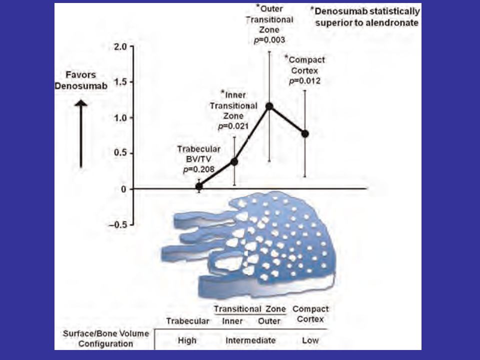
, Fellowship Diabetes/Endocrinology (AKUH), PG Dip Diab (UK) Consultant Endocrinologist.>")

>")
>")


>")


 on Aromatase Inhibitor-Associated Bone Loss in Postmenopausal Women with Early Breast Cancer Receiving Adjuvant Letrozole:>")