Download presentation
Presentation is loading. Please wait.

2
Update of the 2007 AASM Manual for the Scoring of Sleep
By Ahmad Younis Professor of Thoracic Medicine Mansoura Faculty of Medicine

3
Technical Considerations for Adult
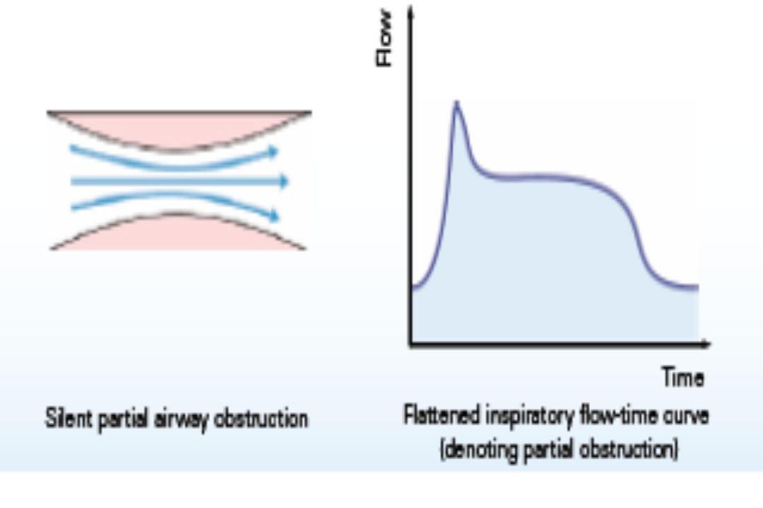
The 2007 scoring manual did not specify the sensor for detection of apnea and hypopnea during positive airway pressure (PAP) titration. PAP devices used for titration during PSG have the ability to output an analog or digital signal from the internal flow sensor . Use of this signal to detect apnea and hypopnea during PAP titration is recommended in both CPAP and NPPV titration clinical guidelines. Flattening of the inspiratory portion of the flow waveform provides evidence of airflow limitation and increased upper airway resistance.
 titration. PAP devices used for titration during PSG have the ability to output an analog or digital signal from the internal flow sensor . Use of this signal to detect apnea and hypopnea during PAP titration is recommended in both CPAP and NPPV titration clinical guidelines. Flattening of the inspiratory portion of the flow waveform provides evidence of airflow limitation and increased upper airway resistance.")
4
As the upper airway begins to collapse, the shape of the inspiratory flow-time curve changes and the central section flattens.

9
Based on consensus and clinical evidence, the task force recommends that the PAP device flow signal should be used to score apneas or hypopneas during PAP titration. Note, the magnitude of oral airflow, if present, during a PAP titration with a nasal mask is not estimated by the PAP flow signal.

10
Respiratory monitoring
The gold standard for airflow detection is a pneumotachograph, usually placed in the outlet of a mask over the nose and mouth, which measures the pressure drop across a linear resistance. This technology is not practical for clinical studies.

11
Pneumotachographs When air is blown down a partially obstructed tube there is a drop in air pressure beyond the obstruction The extent of the pressure drop is dependent on the rate of airflow In a differential pressure pneumotachograph air pressure is measured instantaneously before and after the obstruction and airflow and volume calculated from the pressure drop The two most commonly used are the Lilly and the Fleisch

13
Respiratory monitoring
Thermal sensors detect a change in temperature between inhaled and exhaled gas. Here thermal airflow sensors include thermistors, thermocouples, or polyvinylidene fluoride (PVDF) sensors. The task force found no evidence to change this recommendation for diagnostic sleep studies, although it broadened the definition of thermal sensors to include PVDF sensors.
 sensors. The task force found no evidence to change this recommendation for diagnostic sleep studies, although it broadened the definition of thermal sensors to include PVDF sensors.")
14
Thermistors and Thermocouple airflow sensors

15
Nasal airflow measurement using polyvinylidene fluoride (PVDF) nasal sensor.
 nasal sensor.")
16
Polyvinylidene fluoride sensor
The word piezoelectricity means electricity resulting from electrical polarization by mechanical stress. Certain materials such as zinc oxide, lead zirconate titanate, aluminum nitride, etc., when subjected to mechanical stress, develop surface electric charges. Thus , when suitable electrodes are provided, these charges will appear on these electrodes and hence a measurable voltage will be developed across them.

17
Polyvinylidene fluoride sensor
Piezoelectric materials are also available as polymeric films such as polyvinylidene fluoride . It is superior compared with other piezo materials in many ways: it exhibits high fidelity across a broad frequency range (nearly direct current [DC] to 1 GHz); it is very thin, light weight, flexible, and durable. PVDF has been used already in many medical applications related to cardiac activity and respiration.
; it is very thin, light weight, flexible, and durable. PVDF has been used already in many medical applications related to cardiac activity and respiration.")
18
Recommended sensors for routine respiratory monitoring

19
Nasal and oronasal pressure cannulas

20
Cannula for Simultaneous C02 and Pressure Monitoring

21
Simultaneous flow signals during hypopnea from a pneumotachograph, nasal prongs (nasal pressure), and linearized nasal prongs (square root transformation of nasal pressure). The nasal prongs signal decreases more than that of the pneumotachograph during a reduction in airflow. The linearized nasal prongs signal is very similar to that of the pneumotachograph.

22
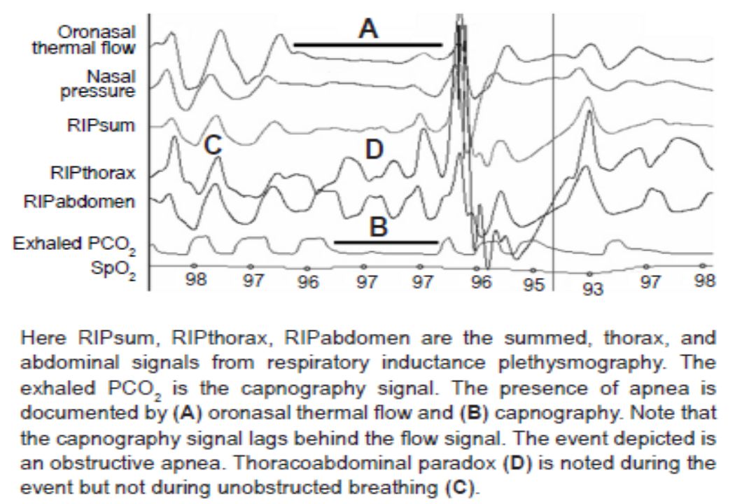
The nasal pressure signal shows an absence of airflow, whereas the nasal-oral thermal sensor shows continued airflow. This pattern of airflow is due to oral breathing. Nasal-oral thermal sensors are the recommended device for detecting apnea. SpO2 = pulse oximetry.

23
Obstructive apnea: Respiratory inductance plethysmography signals from the rib cage (RC) and abdominal bands (AB) are summed (RIPsum). The RIP sum is an estimate of tidal volume . Here, tidal volume is essentially zero (apnea) and RC and AB signals show paradox (moving in opposite directions).
 and RC and AB signals show paradox (moving in opposite directions)..")
24
Obstructive hypopnea: Respiratory inductance plethysmography signals from the rib cage (RC) and abdominal bands (AB) are summed (RIP sum). The RIP sum is an estimate of tidal volume. Here, tidal volume is reduced (hypopnea) and RC and AB signals show paradox (moving in opposite directions).
 and RC and AB signals show paradox (moving in opposite directions)..")
25
Respiratory monitoring
The 2007 scoring manual recommends somewhat different alternative sensors for apnea detection in adults (nasal pressure transducer or RIP) There is evidence that RIP flow or RIP sum or PVDF sum may be used as an alternative sensor for apnea detection with the understanding that most studies analyzed the combination of apneas and hypopneas. Calibration of RIP may improve the accuracy of RIP flow and RIP sum .
 There is evidence that RIP flow or RIP sum or PVDF sum may be used as an alternative sensor for apnea detection with the understanding that most studies analyzed the combination of apneas and hypopneas. Calibration of RIP may improve the accuracy of RIP flow and RIP sum .")
26
Respiratory monitoring
The available RIP signals include the dual RIP belt signals (thorax and abdomen),the RIPsum (sum of the thorax and abdomen belt signals) and the RIPflow (the time derivative of the RIPsum signal) . Deflections in the RIPsum signal provide an estimate of tidal volume when RIP is calibrated. In uncalibrated RIP , deflections in the RIP sum signal allow detection of a relative change in tidal volume compared to baseline breathing. If the RIP sum signal is not available, a reduction in tidal volume can be inferred if there is a reduction in the excursions of the thoracic and abdominal belts.
,the RIPsum (sum of the thorax and abdomen belt signals) and the RIPflow (the time derivative of the RIPsum signal) . Deflections in the RIPsum signal provide an estimate of tidal volume when RIP is calibrated. In uncalibrated RIP , deflections in the RIP sum signal allow detection of a relative change in tidal volume compared to baseline breathing. If the RIP sum signal is not available, a reduction in tidal volume can be inferred if there is a reduction in the excursions of the thoracic and abdominal belts.")
27
Respiratory monitoring
Note, the pattern of undiminished excursions in the signals from the thoracoabdominal belts that are out of phase during an event is also consistent with a reduction in tidal volume (RIP sum). The RIP flow signal is a semi-quantitative estimate of airflow in calibrated RIP, and relative airflow in uncalibrated RIP. Calibration of the RIP signal is usually not performed in routine clinical PSG unless the technology for calibration during natural breathing is available.
. The RIP flow signal is a semi-quantitative estimate of airflow in calibrated RIP, and relative airflow in uncalibrated RIP. Calibration of the RIP signal is usually not performed in routine clinical PSG unless the technology for calibration during natural breathing is available.")
28
Respiratory monitoring
During apnea, the RIPsum and RIPflow signals show absent or minimal excursions, and during hypopnea, the excursions are diminished compared to baseline breathing. Note, airflow limitation can be inferred from subtle qualitative changes in the inspiratory portion of the thorax RIP, abdominal RIP, and RIP sum signals, or from flattening of the inspiratory portion of the RIP flow waveform.

29
Alternative sensors for scoring respiratory events during diagnostic study
•

30
Respiratory monitoring
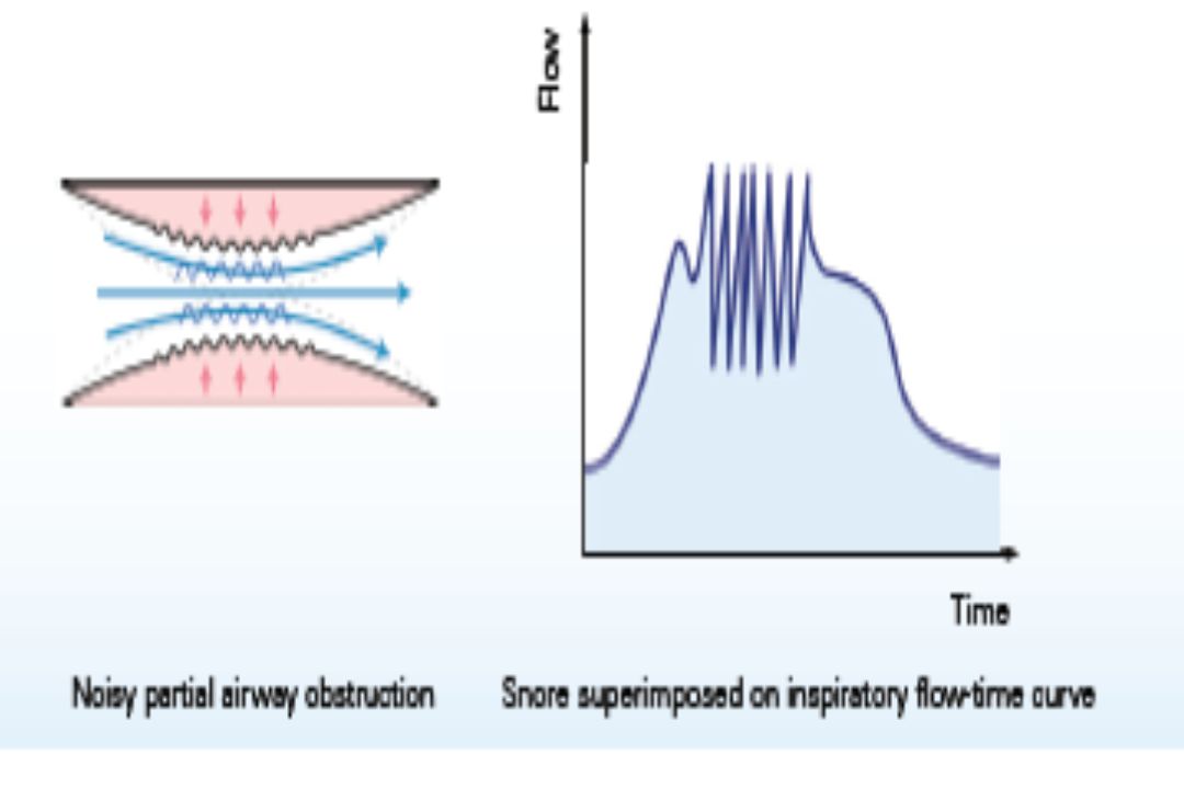
The 2007 scoring manual recommends use of the nasal pressure signal for scoring hypopnea in both adults and children. Flattening of the shape of the inspiratory nasal pressure waveform is a surrogate for airflow limitation and is included in the respiratory effort related arousal (RERA) rules in the 2007 scoring manual. Snoring can also be detected as oscillations superimposed on the unfiltered nasal pressure signal if an appropriate high-frequency filter setting is used (100Hz).
 rules in the 2007 scoring manual. Snoring can also be detected as oscillations superimposed on the unfiltered nasal pressure signal if an appropriate high-frequency filter setting is used (100Hz).")
31
Nasal pressure signal displayed as a DC signal and as an AC signal with various low frequency filter settings (Hz)
")
32
Other sensors for respiratory monitoring

33
Piezo snore sensor

34
Main-stream capnographs

35
Side-stream capnographs

36
Respiratory monitoring
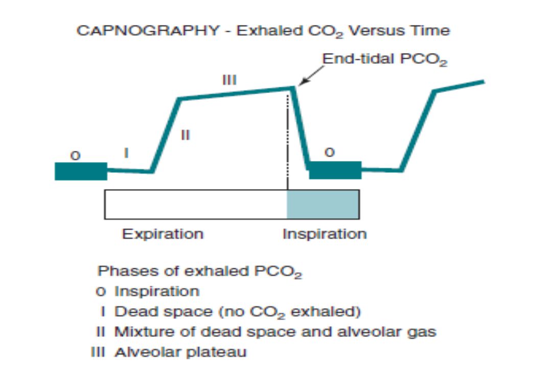
The end-tidal PCO2 , a more accurate description of the signal is exhaled PCO2, but the phrase end-tidal PCO2 monitoring is widely used. Monitoring of exhaled PCO2 is routinely performed during pediatric PSG, and the absence of signal deflections (no CO2 exhaled) has been used to score apneas. The side stream method is most commonly used and consists of gas suctioned via a nasal cannula to an external sensor at bedside. Mouth breathing and occlusion of the nasal cannula impair the ability of end-tidal PCO2 monitoring to detect apnea. The magnitude of signal excursion depends entirely on the highest value of PCO2 in the exhaled breath rather than the magnitude of tidal volume or flow.
 has been used to score apneas. The side stream method is most commonly used and consists of gas suctioned via a nasal cannula to an external sensor at bedside. Mouth breathing and occlusion of the nasal cannula impair the ability of end-tidal PCO2 monitoring to detect apnea. The magnitude of signal excursion depends entirely on the highest value of PCO2 in the exhaled breath rather than the magnitude of tidal volume or flow.")
38
Sensors for Detection of Respiratory Effort
The 2007 scoring manual recommends esophageal manometry or calibrated or uncalibrated respiratory inductance plethysmography for detection of respiratory effort in adults and children. Esophageal manometry is the gold standard for detection of respiratory effort, and the signal excursions provide an estimate of the magnitude of effort. Esophageal manometry is rarely used in clinical practice due to its invasiveness and patient discomfort. The detection of respiratory effort to differentiate central and obstructive apnea is the major concern. Failure to detect respiratory effort when present may result in the incorrect classification of an obstructive apnea as central.

39
Sensors for Detection of Respiratory Effort
Some patients have larger excursions in either the thorax or abdominal belts during the night, and this can vary with body position. If one effort belt fails, the other still provides information about respiratory effort. During biocalibration the polarity of belt signals is adjusted so that belt distension results in signal excursion in the same direction for both belts. Use of dual belts has the additional advantage of the ability to demonstrate paradoxical motion of the thorax and abdomen and adds the ability to identify events as obstructive .

40
Sensors for Detection of Respiratory Effort
The technology available for respiratory effort belts includes strain gauges, impedance plethysmography, inductance plethysmography (RIP), and belts with piezoelectric or PVDF sensors . An advantage of the RIP technology is that inductance of the band and ultimately the signal output depends on the entire surface area enclosed by the band. Effort belts with piezoelectric or PVDF sensors typically utilize a single sensor between belt material surrounding the thorax or abdomen .The signal depends on variations in the tension on the sensor which may or may not reflect the magnitude of thoraco-abdominal excursions.
, and belts with piezoelectric or PVDF sensors . An advantage of the RIP technology is that inductance of the band and ultimately the signal output depends on the entire surface area enclosed by the band. Effort belts with piezoelectric or PVDF sensors typically utilize a single sensor between belt material surrounding the thorax or abdomen .The signal depends on variations in the tension on the sensor which may or may not reflect the magnitude of thoraco-abdominal excursions.")
41
RIP mate Belt RIP Adult Kit

42
Respiratory inductance plethysmography

43
Piezo respiratory effort sensors

44
PVDF Effort Belts, Adult

45
Sensors for Detection of Respiratory Effort
Studies have shown that RIP belts are able to detect subtle changes in respiratory effort and out of phase (paradoxical) motion of the thorax and abdomen excursions is often noted during obstructive apnea or hypopnea. Calibration of RIP signals should improve the accuracy of this sensor technology for detection of respiratory effort. However , even calibrated RIP may not detect feeble respiratory effort in some patients with misclassification of obstructive apneas as central.
 motion of the thorax and abdomen excursions is often noted during obstructive apnea or hypopnea. Calibration of RIP signals should improve the accuracy of this sensor technology for detection of respiratory effort. However , even calibrated RIP may not detect feeble respiratory effort in some patients with misclassification of obstructive apneas as central.")
46
Esophageal pressure deflections increase during an obstructive apnea that might at first glance appear to be central apnea (absent inspiratory effort). In this patient, the chest and abdomen effort belt signals showed minimal deflections during obstructive apnea. The situation was corrected by increasing the gain of the signals while scoring respiratory events.

47
Sensors for Detection of Respiratory Effort
The average number of central apneas, obstructive apneas, hypopneas, and the overall AHI as determined using RIP versus PVDF belts for respiratory effort detection were almost identical and showed a high level of agreement as assessed by the κ statistic. Thus, PVDF sensor effort belts appear to adequately detect respiratory effort in adults .

48
Sensors for Detection of Respiratory Effort
The 2007 scoring manual includes surface diaphragmatic /intercostal electromyography (EMG) as an alternative sensor for detection of respiratory effort. Bursts of the diaphragmatic/ intercostal EMG signal are noted with each inspiration. Similar monitoring methods as those used for recording of anterior tibial muscle EMG can be used. Unlike leg EMG, the diaphragmatic/intercostal EMG signal is often contaminated with prominent electrocardiographic activity.
 as an alternative sensor for detection of respiratory effort. Bursts of the diaphragmatic/ intercostal EMG signal are noted with each inspiration. Similar monitoring methods as those used for recording of anterior tibial muscle EMG can be used. Unlike leg EMG, the diaphragmatic/intercostal EMG signal is often contaminated with prominent electrocardiographic activity.")
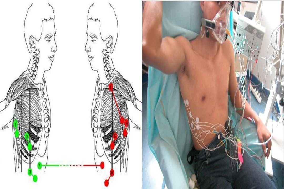
50
An obstructive apnea with respiratory effort monitored by both chest and abdominal respiratory inductance plethysmography (RIP) bands and right intercostal electromyogram (EMG). The right intercostal EMG signal shows bursts coincident with inspiratory effort (and movement of chest and abdomen).A blow up of one EMG burst is shown at the bottom of the figure in a raw form and with the electrocardiogram (ECG) artifact minimized. SpO2 = pulse oximetry.
.A blow up of one EMG burst is shown at the bottom of the figure in a raw form and with the electrocardiogram (ECG) artifact minimized. SpO2 = pulse oximetry. .")
51
Sensors for Detection of Respiratory Effort
Given the above considerations, the task force decided to uphold the 2007 manual recommendation that esophageal manometry or calibrated or uncalibrated dual thoraco-abdominal RIP belts be used for detection of respiratory effort in adults and children [Recommended] (Consensus). It was concluded that dual thoraco-abdominal PVDF belts may be used to detect respiratory effort in adult patients but with a lower level of recommendation due to limited published evidence [Acceptable] .
. It was concluded that dual thoraco-abdominal PVDF belts may be used to detect respiratory effort in adult patients but with a lower level of recommendation due to limited published evidence [Acceptable] .")
53
Detection of Blood Oxygen
The task force did not find evidence to change the 2007 recommended sensor for estimation of arterial oxygen saturation which is pulse oximetry (SpO2) with an appropriate averaging time [Recommended] It was noted that the presence of carboxyhemoglobin (e.g., in heavy smokers)may result in the SpO2 being higher than the true fraction of total hemoglobin bound to oxygen. Accurate measurement of the amount of carboxyhemoglobin requires use of a co-oximeter that uses the absorption of four or more wavelengths of light (compared to two wavelengths in routine oximetry).
 with an appropriate averaging time [Recommended] It was noted that the presence of carboxyhemoglobin (e.g., in heavy smokers)may result in the SpO2 being higher than the true fraction of total hemoglobin bound to oxygen. Accurate measurement of the amount of carboxyhemoglobin requires use of a co-oximeter that uses the absorption of four or more wavelengths of light (compared to two wavelengths in routine oximetry).")
54
A long averaging time impairs the ability of oximetry to detect arterial oxygen desaturation. SpO2 = pulse oximetry.

55
Apneas are followed by arterial oxygen desaturations
Apneas are followed by arterial oxygen desaturations. Longer apneas are associated with more severe desaturation. SpO2 = pulse oximetry.

56
Detection of Blood Oxygen
The presence of carboxyhemoglobin also shifts the oxygen hemoglobin saturation curve to the left causing a given SpO2 to be associated with a lower than expected partial pressure of oxygen.

57
Left, The shift in the oxygen hemoglobin dissociation curve left or right depends on the illustrated factors . Right, A shift to the right increases the arterial partial pressure of oxygen (PaO2) for a given arterial oxygen saturation (SaO2). 2,3-DPG = 2,3-diphosphoglycerate .
 for a given arterial oxygen saturation (SaO2). 2,3-DPG = 2,3-diphosphoglycerate . .")
58
Detection of Blood Oxygen
Because of the sigmoid shape of the oxyhemoglobin dissociation curve, a much greater drop in arterial partial pressure of oxygen (PaO2) occurs in the setting of a drop of 4% from a baseline saturation of 96% to 92% (PaO2 change ~18 mm Hg) compared to the same drop of 4% from a baseline saturation of 92% to 88% (PaO2 change ~ 9 mm Hg). Linking respiratory event definitions to a specific change in saturation for event detection requires a greater fall in the PaO2 for patients with a high baseline saturation (e.g., 98%) than those with lower baseline saturations.
 occurs in the setting of a drop of 4% from a baseline saturation of 96% to 92% (PaO2 change ~18 mm Hg) compared to the same drop of 4% from a baseline saturation of 92% to 88% (PaO2 change ~ 9 mm Hg). Linking respiratory event definitions to a specific change in saturation for event detection requires a greater fall in the PaO2 for patients with a high baseline saturation (e.g., 98%) than those with lower baseline saturations.")
59
Detection of Blood Oxygen
The desaturation associated with a respiratory event is defined as a drop from a baseline SpO2 preceding the event to the nadir in the SpO2 following the event. While identification of the nadir in the SpO2 following a respiratory events is usually straightforward, selecting a “baseline” SpO2 in a patient with back-to-back respiratory events is more difficult. The highest SpO2 following a respiratory event can exceed values present during stable breathing.Defining a “baseline SpO2” during sleep may be difficult in such patients.

60
Detection of Snoring The 2007 AASM scoring manual did not recommend a sensor for snoring. Optimal visualization of snoring requires a high frequency filter setting that permits recording/display of rapid oscillations (100 Hz in the 2007 scoring manual). Snoring may be visualized in the nasal pressure signal as high-frequency oscillations superimposed on the slower varying flow signal but is not seen in the PAP device flow signal which is either filtered or too under-sampled to show the high frequency vibrations. Snore sensors are typically piezoelectric sensors that detect vibration of the neck or microphones that record the sound of snoring.
. Snoring may be visualized in the nasal pressure signal as high-frequency oscillations superimposed on the slower varying flow signal but is not seen in the PAP device flow signal which is either filtered or too under-sampled to show the high frequency vibrations. Snore sensors are typically piezoelectric sensors that detect vibration of the neck or microphones that record the sound of snoring.")
61
Snoring noted both in the snore sensor (applied to the neck near the trachea) and as a vibration (oscillation) in the nasal pressure signal. Note that the nasal pressure signal also has a flattened shape. SpO2 = pulse oximetry

62
Detection of Snoring The AASM guidelines for CPAP and for NPPV titration both listed a snore signal as an option for recording. The task force recommends several sensors as options for snore detection: the unfiltered nasal pressure signal, piezoelectric sensors to detect vibration, or acoustic sensors (e.g., microphone) to record sound [Recommended](Consensus). The ability of the sensor to detect simulated snoring should be demonstrated before sleep recording. The task force concurs with the 2007 manual that whether or not to monitor snoring is at the discretion of the clinician or investigator [Optional] (Consensus).
 to record sound [Recommended](Consensus). The ability of the sensor to detect simulated snoring should be demonstrated before sleep recording. The task force concurs with the 2007 manual that whether or not to monitor snoring is at the discretion of the clinician or investigator [Optional] (Consensus).")
63
Detection of Hypoventilation
The gold standard method for documenting hypoventilation is the processing of an arterial sample for determination of PaCO2. Given the difficulty of drawing an arterial sample during sleep, the 2007 scoring manual states that finding an elevated PaCO2 obtained immediately after waking would provide evidence of hypoventilation during sleep. Regardless of whether this value underestimates the sleeping PaCO2, the ability to draw or process an arterial blood gas sample is rarely available in sleep centers.

64
Detection of Hypoventilation
Surrogate measures such as end-tidal PCO2 (PETCO2) and transcutaneous PCO2 (PTCCO2) are commonly used during PSG. A 2007 scoring manual note for the hypoventilation rule in adults states that there is insufficient evidence to allow specification of sensors for direct or surrogate measures of PaCO2. “both end-tidal CO2 and transcutaneous CO2 may be used as surrogate measures of PaCO2 if there is demonstration of reliability and validity within laboratory practices.”
 and transcutaneous PCO2 (PTCCO2) are commonly used during PSG. A 2007 scoring manual note for the hypoventilation rule in adults states that there is insufficient evidence to allow specification of sensors for direct or surrogate measures of PaCO2. both end-tidal CO2 and transcutaneous CO2 may be used as surrogate measures of PaCO2 if there is demonstration of reliability and validity within laboratory practices.")
65
Capnocheck® Sleep Capnograph / Oximeter

67
The CO2 tracing is delayed relative to exhaled airflow in the side stream method

68
The exhaled CO2 tracing shows continued deflections during “inspiratory apnea” due to small exhaled puffs of air rich in CO2

69
Documentation of an increased partial pressure of carbon dioxide (PCO2) during sleep. Note the alveolar plateaus (black circles). PCO2 = exhaled PCO2 waveform tracing; PETCO2 = most recent end-tidal reading; SpO2 = pulse oximetry.
. PCO2 = exhaled PCO2 waveform tracing; PETCO2 = most recent end-tidal reading; SpO2 = pulse oximetry..")
70
Detection of Hypoventilation
In most sleep centers, a bedside device containing the CO2 measuring sensor continuously suctions gas through a nasal cannula worn by the patient (side stream method). During inhalation, room air is suctioned (PCO2 = 0), and during exhalation , the PCO2 in the exhaled gas is measured. The end tidal PCO2 provides an estimate of the arterial value (usually PaCO2 > PETCO2). The PaCO2 – PETCO2 difference is usually 2 to 7 mm Hg, and the difference is higher in patients with lung disease. The PETCO2 is not an accurate estimate of the PaCO2 during mouth breathing or with low tidal volume and fast respiratory rates.
. During inhalation, room air is suctioned (PCO2 = 0), and during exhalation , the PCO2 in the exhaled gas is measured. The end tidal PCO2 provides an estimate of the arterial value (usually PaCO2 > PETCO2). The PaCO2 – PETCO2 difference is usually 2 to 7 mm Hg, and the difference is higher in patients with lung disease. The PETCO2 is not an accurate estimate of the PaCO2 during mouth breathing or with low tidal volume and fast respiratory rates.")
71
Detection of Hypoventilation
Some manufacturers make a sampling nasal cannula with a “mouth guide” to allow sampling of gas exhaled through the mouth. To be considered accurate, a definite plateau in the exhaled PETCO2 versus time waveform should be observed. End-tidal PCO2 measurements are often inaccurate during application of supplemental oxygen or during mask ventilation. The exhaled gas sample is diluted by supplemental oxygen flow or PAP device flow. Some clinicians use a small nasal cannula under the mask to sample exhaled gas at the nares in order to minimize dilution during PAP titration. However, the accuracy of measurements using this approach has not been documented.

72
Detection of Hypoventilation
Transcutaneous CO2 monitoring (PTCCO2) is also used during PSG to estimate the PaCO2, but the signal has a longer response time than the PETCO2 to acute changes in ventilation . In adults, one study using an earlobe sensor found that the PTCCO2 lagged behind PaCO2 by about 2 minutes. Newer transcutaneous device technology has allowed more rapid response time and enabled the devices to work at a lower skin temperature. The advantage of PTCCO2 monitoring compared to PETCO2 is that the accuracy of transcutaneous measurements is not degraded by mouth breathing, supplemental oxygen, or mask ventilation. PTCCO2 will not provide data for breath-by-breath changes, e.g., changes in the first few breaths after an apnea.
 is also used during PSG to estimate the PaCO2, but the signal has a longer response time than the PETCO2 to acute changes in ventilation . In adults, one study using an earlobe sensor found that the PTCCO2 lagged behind PaCO2 by about 2 minutes. Newer transcutaneous device technology has allowed more rapid response time and enabled the devices to work at a lower skin temperature. The advantage of PTCCO2 monitoring compared to PETCO2 is that the accuracy of transcutaneous measurements is not degraded by mouth breathing, supplemental oxygen, or mask ventilation. PTCCO2 will not provide data for breath-by-breath changes, e.g., changes in the first few breaths after an apnea.")
73
Carbon dioxide transcutaneous pressure monitor (tcPCO2, with SpO2)
")
74
Trends in the arterial oxygen saturation (SpO2) and transcutaneous partial pressure of carbon dioxide (TcPCO2) during the night. Note the simultaneous increase in transcutaneous PCO2 and the decrease in SpO2 during episodes of rapid eye movement (REM) sleep.
 sleep..")
75
Detection of Hypoventilation
Both PTCCO2 and PETCO2 have clinical utility as surrogates of PaCO2 during diagnostic studies . Use of both methodologies requires careful review of tracings to determine if artifacts are present. PTCCO2 is the preferred technology in patients with lung disease, significant mouth breathing , or those who are using supplemental oxygen or mask ventilation . PTCCO2 values can occasionally be quite spurious and clinical judgment is needed. When readings do not match the clinical setting, a change in sensor site or recalibration may be needed. PETCO2 is preferred where breath-to-breath changes in PaCO2 need to be detected. In this setting, the ability to detect an increase in PCO2 associated with a respiratory event is clinically useful.

76
Detection of Hypoventilation
The task force concurred with the 2007 manual that whether or not to monitor hypoventilation in adults during diagnostic study or PAP titration is at the discretion of the clinician or investigator [Optional] (Consensus). The task force recommends that arterial PCO2, transcutaneous PCO2, or end-tidal PCO2 be used for detecting hypoventilation during diagnostic study in both adults and children [Recommended] (Consensus). During PAP titration in both adults and children, either arterial PCO2 or transcutaneous PCO2 is the recommended method to detect hypoventilation [Recommended] (Consensus).
. The task force recommends that arterial PCO2, transcutaneous PCO2, or end-tidal PCO2 be used for detecting hypoventilation during diagnostic study in both adults and children [Recommended] (Consensus). During PAP titration in both adults and children, either arterial PCO2 or transcutaneous PCO2 is the recommended method to detect hypoventilation [Recommended] (Consensus).")
77
Caveats that accompany these recommendations:
Sensors should be properly calibrated according to manufacturer specifications. Clinical judgment is essential when assessing the accuracy of end-tidal PCO2 and transcutaneous PCO2 readings. The values should NOT be assumed to be accurate surrogates of the arterial PCO2 when the values do not fit the clinical picture. Transcutaneous PCO2 should be calibrated with a reference gas according to the manufacturer’s recommendations and when the accuracy of the reading is doubtful. End-tidal PCO2, to be accurate, a plateau in the PCO2 versus time wave form should be present. Validation of the surrogate PCO2 with a simultaneous PaCO2 or capillary gas is ideal but not required.

78
Event Duration Rules for Adult Patients
The 2007 scoring manual states that the event duration for scoring either apnea or hypopnea is measured from the nadir preceding the first breath that is clearly reduced to the beginning of the first breath that approximates baseline breathing amplitude. For apnea duration, the oronasal thermal sensor signal (diagnostic study) or PAP device flow signal (PAP titration study) should be used to determine the event duration [Recommended] (Consensus). For hypopnea event duration, the nasal pressure signal (diagnostic study) or PAP device flow signal (PAP titration study) should be utilized [Recommended](Consensus). If the recommended sensor fails or the signal is inaccurate an alternative sensor signal can be used.
 or PAP device flow signal (PAP titration study) should be used to determine the event duration [Recommended] (Consensus). For hypopnea event duration, the nasal pressure signal (diagnostic study) or PAP device flow signal (PAP titration study) should be utilized [Recommended](Consensus). If the recommended sensor fails or the signal is inaccurate an alternative sensor signal can be used.")
79
Event Duration Rules for Adult Patients
The ability to determine baseline breathing is a problem in patients that have nearly continuous events. The AASM “Chicago consensus states, “Baseline is defined as the mean amplitude of stable breathing and oxygenation in the 2 minutes preceding onset of the event (in individuals who have a stable breathing pattern during sleep) or the mean amplitude of the 3 largest breaths in the 2 minutes preceding onset of the event (in individuals without a stable breathing pattern).
 or the mean amplitude of the 3 largest breaths in the 2 minutes preceding onset of the event (in individuals without a stable breathing pattern).")
80
Event Duration Rules for Adult Patients
” The 2007 scoring manual states, “When baseline breathing amplitude cannot be easily determined (and when underlying breathing variability is large), events can be terminated when either there is a clear and sustained increased in breathing amplitude, or in the case where an oxygen desaturation has occurred, there is event-associated oxygen re-saturation of at least 2%. The task force recommends that the 2007 manual guideline for determining baseline breathing be upheld [Recommended] (Consensus).
, events can be terminated when either there is a clear and sustained increased in breathing amplitude, or in the case where an oxygen desaturation has occurred, there is event-associated oxygen re-saturation of at least 2%. The task force recommends that the 2007 manual guideline for determining baseline breathing be upheld [Recommended] (Consensus).")
81
Definition of RDI and ODI for Adult Patients
Both the 1999 Chicago consensus and the ICSD-2 recommended that the sum of apnea, hypopneas, and RERAs per hour of sleep be used to diagnose OSA (along with symptoms). The Centers for Medicare and Medicaid defines the term RDI as the number of apneas and hypopneas per hour of monitoring. The task force reached consensus on the definition of RDI as the sum of the AHI and RERA index. However, reporting of the RDI metric should be considered [Optional] (Consensus) as scoring RERAs is also considered [Optional].
. The Centers for Medicare and Medicaid defines the term RDI as the number of apneas and hypopneas per hour of monitoring. The task force reached consensus on the definition of RDI as the sum of the AHI and RERA index. However, reporting of the RDI metric should be considered [Optional] (Consensus) as scoring RERAs is also considered [Optional].")
82
Definitions of RDI and ODI [Recommended] (Consensus)
RDI = AHI + RERA index ODI = ≥ 3% arterial oxygen desaturations /hour
![Definitions of RDI and ODI [Recommended] (Consensus)](http://slideplayer.com/slide/6169512/18/images/82/Definitions+of+RDI+and+ODI+%5BRecommended%5D+%28Consensus%29.jpg "RDI = AHI + RERA index. ODI = ≥ 3% arterial oxygen desaturations /hour.")
83
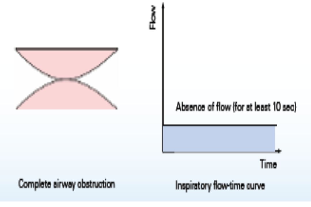
Apnea Rule for Adults The 2007 scoring manual rule for apnea does NOT require an associated arterial oxygen desaturation. The task force found no evidence to support a change in the 2007 scoring manual recommendation for a ≥ 90% drop in oronasal thermal flow lasting at least 10 seconds (adults). The basis for a 90% drop in the thermal sensor signal is entirely arbitrary, but is an attempt to operationalize the requirement of “absent or nearly absent airflow” that often appears in the literature for the definition of an apnea. The task force did clarify that the PAP device flow sensor be used for scoring apnea during PAP titration and name alternative apnea sensors for diagnostic study .
. The basis for a 90% drop in the thermal sensor signal is entirely arbitrary, but is an attempt to operationalize the requirement of absent or nearly absent airflow that often appears in the literature for the definition of an apnea. The task force did clarify that the PAP device flow sensor be used for scoring apnea during PAP titration and name alternative apnea sensors for diagnostic study .")
84
Apnea Rule for Adults The requirement that 90% of event duration must meet amplitude criteria was not a part of the Chicago consensus paper respiratory event definitions and does not appear in apnea definitions elsewhere in the literature prior to 2007. There are cases where there is a drop ≥ 90% of baseline in airflow that lasts for longer than 10 seconds but much less than 90% of the event duration . Using the 2007 apnea rule, one cannot score such an event as an apnea. This event may not meet hypopnea criteria and therefore cannot be scored as either an apnea or hypopnea.

85
Apnea Rule for Adults The length of a respiratory event as defined by the event duration rule may be more difficult to define than the duration of the drop in airflow meeting amplitude criteria. For these reasons, the task force eliminated requirement 3( at least 90% of events duration must meet amplitude reduction criteria) and added a note addressing the situation in which a shorter portion of a longer hypopnea event qualifies as an apnea.
 and added a note addressing the situation in which a shorter portion of a longer hypopnea event qualifies as an apnea.")
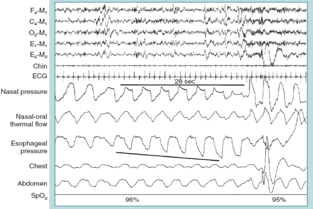
87
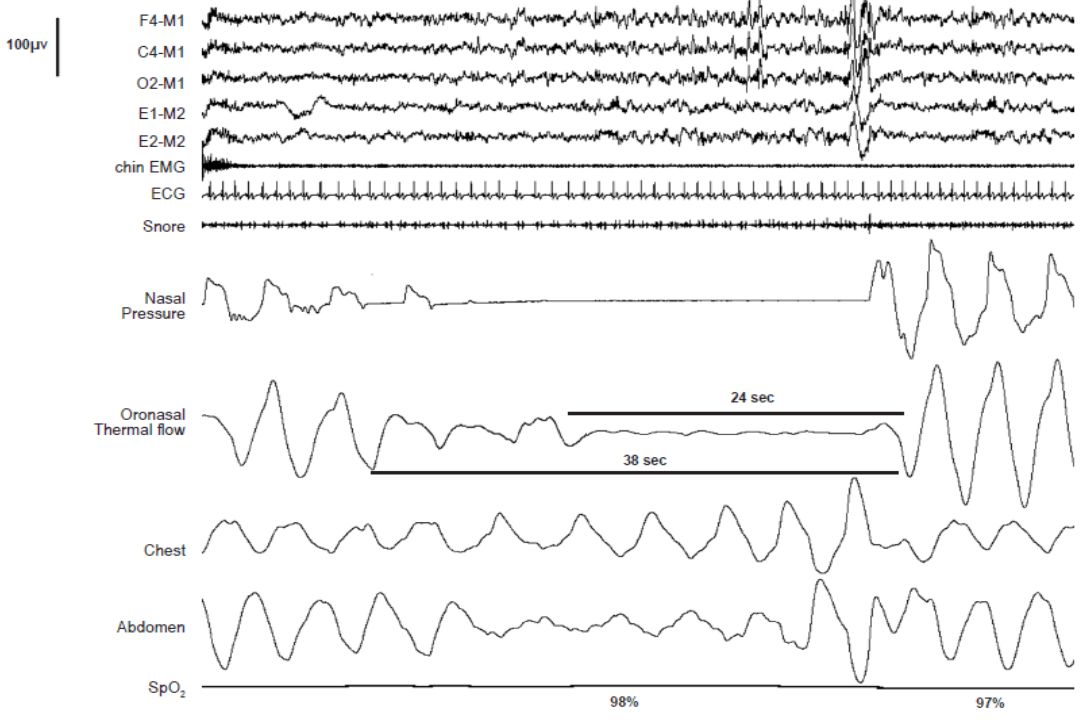
An apnea cannot be scored using the apnea rule in the 2007 scoring manual as 24 seconds (the duration of the ≥ 90% drop in oronasal flow) is not 90% ofthe event duration. A hypopnea cannot be scored based on the drop in nasal pressure, as there is no associated desaturation or arousal. Using the proposed revised apnea definition this event would be scored as an apnea as there is a ≥ 90% reduction in the peak excursions of the oronasal thermal signal compared to baseline that lasts ≥ 10 seconds. The respiratory effort (thoracoabdominal excursions) during the entire apnea indicates that this is an obstructive apnea.
 during the entire apnea indicates that this is an obstructive apnea.")
88
Score a respiratory event in adults as an apnea if both of the following are met:
1. There is a drop in the peak signal excursion by ≥ 90% of pre-event baseline using an oronasal thermal sensor (diagnostic study), positive airway pressure device flow (titration study), or an alternative apnea sensor. 2. The duration of the ≥ 90% drop in sensor signal is ≥ 10 seconds. Note: If a portion of a respiratory event that would otherwise meet criteria for a hypopnea meets criteria for apnea, the entire event should be scored as an apnea. The duration of the event is from the nadir in flow preceding the first breath that is clearly reduced to the start of the first breath that approximates baseline breathing.
, positive airway pressure device flow (titration study), or an alternative apnea sensor. 2. The duration of the ≥ 90% drop in sensor signal is ≥ 10 seconds. Note: If a portion of a respiratory event that would otherwise meet criteria for a hypopnea meets criteria for apnea, the entire event should be scored as an apnea. The duration of the event is from the nadir in flow preceding the first breath that is clearly reduced to the start of the first breath that approximates baseline breathing.")
89
Apnea Rule for Adults The task force also considered the possibility of combining both apneas and hypopneas into a single respiratory event. The numbers of both event types are ultimately combined to compute an apnea-hypopnea index. The classification of apnea as obstructive, mixed, or central in the 2007 scoring manual is based on respiratory effort. The task force considered combining obstructive and mixed events as well as a revision of the definition of central and mixed apneas to address events where there are only 1 or 2 obstructive breaths (see Figure 4).
.")
90
Apnea Rule for Adults If one changes the central apnea definitions to allow 1 or 2 obstructed breaths, then a change in the mixed apnea definition must follow. The choice of how many obstructive breaths to allow and still consider an event to be central seems entirely arbitrary. The task force recommended that the definitions of obstructive,central, and mixed apnea in the 2007 scoring manual not be revised (Consensus).
.")
91
A mixed apnea event is illustrated that contains a single obstructed breath at the end of what would otherwise be scored as a central apnea. The current scoring manual does not mandate that the obstructive portion have more than one obstructive breath.

92
Hypopnea Rules for Adults
The technology used included airflow detection by thermistors attached to mouth and lip and chest movement detection by impedance plethysmography. The 1999 consensus conference defined an apnea-hypopnea as a 10-second or longer event characterized by either a clear decrease (> 50%) of a valid measure of breathing or a clear amplitude reduction (but < 50% decrease) of a validated measure of breathing associated with either an arousal or ≥ 3% oxygen desaturation occurring near the termination of the putative event.
 of a valid measure of breathing or a clear amplitude reduction (but < 50% decrease) of a validated measure of breathing associated with either an arousal or ≥ 3% oxygen desaturation occurring near the termination of the putative event.")
93
The 2007 scoring manual provides two hypopnea definitions (recommended and alternative, also known as “4A” and “4B”). Centers of Medicare and Medicaid Services (CMS) currently accepts only the recommended definition. The recommended hypopnea definition requires a 30% or greater drop in flow for 10 seconds or longer associated with ≥ 4% oxygen desaturation . The alternative hypopnea definition requires a ≥ 50% in flow for 10 seconds or longer associated with a ≥ 3% oxygen desaturation OR an arousal.
 currently accepts only the recommended definition. The recommended hypopnea definition requires a 30% or greater drop in flow for 10 seconds or longer associated with ≥ 4% oxygen desaturation . The alternative hypopnea definition requires a ≥ 50% in flow for 10 seconds or longer associated with a ≥ 3% oxygen desaturation OR an arousal.")
94
The task force readdressed the hypopnea definition issue with respect to:
the required arterial oxygen desaturation, the inclusion of arousal criteria , and the qualifying drop in flow (30% versus 50%).
.")
95
3% Versus 4% Oxygen Desaturation
The use of ≥ 3% desaturation criterion yields an AHI that is as predictive of adverse outcomes as an AHI based on ≥ 4% oxygen desaturation criterion. Recognizing the apparent equivalence of hypopnea definitions requiring ≥ 3% or ≥ 4% desaturation, the task force has recommended adoption of the 3% criterion. It should be noted that using ≥ 3% instead of ≥ 4% desaturation requirement for defining hypopnea does increase the AHI substantially, with median AHI in a general community sample being almost twice as great using a 3% as a 4% criterion .

96
Inclusion of Arousal in Hypopnea Definition
Whether or not to include arousal as part of the hypopnea definition remains controversial . Opponents of inclusion of arousal in the hypopnea definition cite the fact that the majority of studies have not found an association between arousal frequency and adverse cardiovascular outcomes (independent of arterial oxygen desaturation) Another argument against inclusion of arousal in the hypopnea definition is that the scoring of arousals is said to be less reliable and would consequently reduce the reliability of scoring respiratory events. The current Medicare/Medicaid hypopnea definition does not consider arousals and that a hypopnea definition based only on flow and oxygen saturation would be applicable to limited channel sleep testing (i.e., out-of-center sleep testing).
 Another argument against inclusion of arousal in the hypopnea definition is that the scoring of arousals is said to be less reliable and would consequently reduce the reliability of scoring respiratory events. The current Medicare/Medicaid hypopnea definition does not consider arousals and that a hypopnea definition based only on flow and oxygen saturation would be applicable to limited channel sleep testing (i.e., out-of-center sleep testing).")
97
Inclusion of Arousal in Hypopnea Definition
Proponents of inclusion of arousal in the hypopnea definition cite evidence that sleep fragmentation without arterial oxygen desaturation can be associated with symptoms (e.g., daytime sleepiness) and that treatment with CPAP can improve symptoms and objective sleepiness. For symptomatic patients with milder OSA and a significant proportion of events associated with arousal but not ≥ 3% desaturation, the AHI may be ≥ 5/hour using the alternative hypopnea definition but not the recommended definition. Of interest, if one uses a hypopnea definition based on desaturation or arousal there are relatively few RERA events.
 and that treatment with CPAP can improve symptoms and objective sleepiness. For symptomatic patients with milder OSA and a significant proportion of events associated with arousal but not ≥ 3% desaturation, the AHI may be ≥ 5/hour using the alternative hypopnea definition but not the recommended definition. Of interest, if one uses a hypopnea definition based on desaturation or arousal there are relatively few RERA events.")
98
Inclusion of Arousal in Hypopnea Definition
PSG would permit scoring of hypopneas based on arousal as well as arterial oxygen saturation. Respiratory events that cause arousal but are associated with relatively minor drops in the arterial oxygen saturation could be identified as hypopneas. This would allow identification and treatment of a wider spectrum of symptomatic patients. The interscorer reliability (intraclass correlations coefficients [ICC]) for the arousal index based on central derivations to be 0.80. In current practice ,arousals are scored based on frontal, central, and occipital derivations with montages typically showing respiratory channels. A hypopnea definition based only on desaturation would result in misdiagnosis of some patients in whom respiratory events fragment sleep but result in minor drops in the SpO2.
 for the arousal index based on central derivations to be In current practice ,arousals are scored based on frontal, central, and occipital derivations with montages typically showing respiratory channels. A hypopnea definition based only on desaturation would result in misdiagnosis of some patients in whom respiratory events fragment sleep but result in minor drops in the SpO2.")
99
30% Versus 50% Drop in Flow The 2007 scoring manual definitions of hypopnea for adults use either a 30% drop in flow recommended definition) or 50% drop (alternative definition). Given the difficulty of accurately measuring flow or tidal volume in clinical settings, linking a change in flow or tidal volume to a physiological consequence would help identify an event as physiologically relevant. The degree of oxygen desaturation for a given reduction in airflow varies widely between individuals and depends on baseline arterial oxygen desaturation, oxygen stores (lung volumes), obesity, and the presence or absence of lung disease . Less than 50% drop in flow could result in significant oxygen desaturations in some individuals , while in others a desaturation may not occur.
 or 50% drop (alternative definition). Given the difficulty of accurately measuring flow or tidal volume in clinical settings, linking a change in flow or tidal volume to a physiological consequence would help identify an event as physiologically relevant. The degree of oxygen desaturation for a given reduction in airflow varies widely between individuals and depends on baseline arterial oxygen desaturation, oxygen stores (lung volumes), obesity, and the presence or absence of lung disease . Less than 50% drop in flow could result in significant oxygen desaturations in some individuals , while in others a desaturation may not occur.")
100
30% Versus 50% Drop in Flow The task force also recognizes that the proposed definition of hypopnea is not currently accepted by the Centers for Medicare and Medicaid Services (CMS) reimbursement. Following the logic of the proposed revised apnea definition, the requirement that the qualifying drop in flow must occupy > 90% of the event duration was removed from the hypopnea definition.
 reimbursement. Following the logic of the proposed revised apnea definition, the requirement that the qualifying drop in flow must occupy > 90% of the event duration was removed from the hypopnea definition.")
101
Hypopnea Rule for Adults [Recommended] (Consensus) Score a respiratory event as a hypopnea if all of the following are met: 1. The peak signal excursions drop by ≥ 30% of pre-event baseline using nasal pressure (diagnostic study), PAP device flow (titration study), or an alternative hypopnea sensor. 2. The duration of the ≥ 30% drop in signal excursions is ≥ 10 seconds. 3. There is ≥ 3% oxygen desaturation from pre-event baseline or the event is associated with an arousal.
![Hypopnea Rule for Adults [Recommended] (Consensus) Score a respiratory event as a hypopnea if all of the following are met:](http://slideplayer.com/slide/6169512/18/images/101/Hypopnea+Rule+for+Adults+%5BRecommended%5D+%28Consensus%29+Score+a+respiratory+event+as+a+hypopnea+if+all+of+the+following+are+met%3A.jpg "1. The peak signal excursions drop by ≥ 30% of pre-event baseline using nasal pressure (diagnostic study), PAP device flow (titration study), or an alternative hypopnea sensor. 2. The duration of the ≥ 30% drop in signal excursions is ≥ 10 seconds. 3. There is ≥ 3% oxygen desaturation from pre-event baseline or the event is associated with an arousal.")
102
Classification of Hypopnea
In the 2007 scoring manual, the definition of Cheyne-Stokes breathing (CSB) does mention central hypopnea. The CMS criteria for reimbursement of a PAP device with a backup rate requires that 50% of events be central in nature. Some patients with CSB or complex sleep apnea have a large proportion of central hypopneas . Scoring central hypopneas would allow them to qualify for a device with a backup rate. Many of the publications on CSB in heart failure include central hypopneas in computing an AHI.
 does mention central hypopnea. The CMS criteria for reimbursement of a PAP device with a backup rate requires that 50% of events be central in nature. Some patients with CSB or complex sleep apnea have a large proportion of central hypopneas . Scoring central hypopneas would allow them to qualify for a device with a backup rate. Many of the publications on CSB in heart failure include central hypopneas in computing an AHI.")
103
Classification of Hypopnea
The Chicago consensus defined central apnea/hypopnea events as those events with a reduction in airflow and a clear reduction in esophageal pressure swings from baseline that parallels chronologically the reduction in airflow. The 2007 scoring manual only states that “classification of a hypopnea as obstructive, central, or mixed should not be performed without a quantitative assessment of ventilatory effort (esophageal manometry, calibrated RIP, or diaphragmatic /intercostal EMG). This presents a problem as calibrated RIP excursions do not always reflect the magnitude of respiratory effort (as measured by esophageal pressure excursions) and esophageal manometry is rarely used.
. This presents a problem as calibrated RIP excursions do not always reflect the magnitude of respiratory effort (as measured by esophageal pressure excursions) and esophageal manometry is rarely used.")
104
(A) central hypopnea is characterized by lack of flattening in the airflow (nasal pressure) and a reduction in respiratory effort (esophageal pressure excursions). The reduction in flow is chronologically parallel to the reduction in effort. (B) An obstructive hypopnea is characterized by airflow limitation (flattening of the nasal pressure waveform) and increasing respiratory effort without an increase in airflow (nasal pressure).
 An obstructive hypopnea is characterized by airflow limitation (flattening of the nasal pressure waveform) and increasing respiratory effort without an increase in airflow (nasal pressure)..")
105
Classification of Hypopnea
Obstructive hypopneas are usually associated with flattening of the inspiratory portion of the nasal pressure (or PAP device flow) waveform, often associated with snoring, and sometimes associated with thoracoabdominal paradox. Central hypopneas are typically characterized by absence of flattening of the inspiratory portion of the nasal pressure or PAP flow wave form (or flattening is present but unchanged from baseline breathing) and absence of thoracoabdominal paradox in the thoracic and abdominal RIP band excursions .
 waveform, often associated with snoring, and sometimes associated with thoracoabdominal paradox. Central hypopneas are typically characterized by absence of flattening of the inspiratory portion of the nasal pressure or PAP flow wave form (or flattening is present but unchanged from baseline breathing) and absence of thoracoabdominal paradox in the thoracic and abdominal RIP band excursions .")
106
obstructive hypopnea with snoring, flattening of the nasal pressure (NP) waveform , and paradoxical motion of the chest and abdominal (ABD)respiratory inductance plethysmography excursions. P denotes paradox during the hypopnea and no P the absence of paradox during unobstructed breathing.

107
A central hypopnea in a patient with Cheyne-Stokes breathing is illustrated. There is no evidence of snoring or thoracoabdominal paradox in the RIP bands (RIPthorax and RIPabdomen). There is no evidence of airflow limitation (flattening of the nasal pressure signal).
. There is no evidence of airflow limitation (flattening of the nasal pressure signal)..")
108
Classification of Hypopnea
Task force members pointed out that a decrease in RIP excursions cannot differentiate obstructive and central hypopneas because the excursions may decrease in both types of hypopneas . Although a disproportionate increase in effort when compared to flow can be indicative of obstruction, this is difficult to operationalize. Such a separation of hypopneas into central or obstructive is not clinically indicated in the majority of patients.

109
1. Snoring during the event
Classifying Hypopnea in Adults [Recommended] (Consensus) If electing to score obstructive hypopneas, score a hypopnea as obstructive if ANY of the following criteria are met: 1. Snoring during the event 2. Increased inspiratory flattening of the nasal pressure or PAP device flow signal compared to baseline breathing 3. Associated thoracoabdominal paradox occurs during the event but not during pre-event breathing.
 If electing to score obstructive hypopneas, score a hypopnea as obstructive if ANY of the following criteria are met: 1. Snoring during the event. 2. Increased inspiratory flattening of the nasal pressure or PAP device flow signal compared to baseline breathing. 3. Associated thoracoabdominal paradox occurs during the event but not during pre-event breathing.")
110
If electing to score central hypopneas, score a hypopnea as central if NONE of the following criteria are met: 1. Snoring during the event 2. Increased inspiratory flattening of the nasal pressure or PAP device flow signal compared to baseline breathing 3. Associated thoracoabdominal paradox occurs during the event but not during pre-event breathing.

111
Respiratory Effort-Related Arousal Rule for Adults
If a definition of hypopnea is used which requires an associated desaturation OR arousal , then there are relatively few events scored as RERAs. One change includes the use of PAP device flow flattening rather than nasal pressure flattening during PAP titration. “RERA”events are usually scored based on changes in nasal pressure (or PAP flow) rather than esophageal manometry. An increase in respiratory effort is inferred rather than being directly documented, leading some investigators to coin such an event as a “flow limitation arousal.” The term RERA is widely used, and the task force members did not feel a change in terminology was needed. Task force members recommend that the scoring of RERA events remains [Optional] (Consensus).
 rather than esophageal manometry. An increase in respiratory effort is inferred rather than being directly documented, leading some investigators to coin such an event as a flow limitation arousal. The term RERA is widely used, and the task force members did not feel a change in terminology was needed. Task force members recommend that the scoring of RERA events remains [Optional] (Consensus).")
112
Respiratory effort–related arousal (RERA)
Respiratory effort–related arousal (RERA). Flattening of nasal pressure for more than 10 seconds followed by an arousal. There is no desaturation so it would not qualify as a hypopnea (by the recommended definition). The drop in flow is not 30% of baseline so the event would not qualify as a hypopnea
. Flattening of nasal pressure for more than 10 seconds followed by an arousal. There is no desaturation so it would not qualify as a hypopnea (by the recommended definition). The drop in flow is not 30% of baseline so the event would not qualify as a hypopnea.")
114
RERA Rule for Adults [Recommended] (Consensus)
If electing to score respiratory effort-related arousals , score a respiratory event as a RERA if there is a sequence of breaths lasting at least 10 seconds characterized by increasing respiratory effort or by flattening of the inspiratory portion of the nasal pressure (diagnostic study) or PAP device flow (titration study) waveform leading to arousal from sleep when the sequence of breaths does not meet criteria for an apnea or hypopnea.
![RERA Rule for Adults [Recommended] (Consensus)](http://slideplayer.com/slide/6169512/18/images/114/RERA+Rule+for+Adults+%5BRecommended%5D+%28Consensus%29.jpg "If electing to score respiratory effort-related arousals , score a respiratory event as a RERA if there is a sequence of breaths lasting at least 10 seconds characterized by increasing respiratory effort or by flattening of the inspiratory portion of the nasal pressure (diagnostic study) or PAP device flow (titration study) waveform leading to arousal from sleep when the sequence of breaths does not meet criteria for an apnea or hypopnea.")
115
Hypoventilation Rule for Adults
A common definition of awake hypoventilation is PaCO2 > 45 mm Hg. In choosing a respiratory definition of hypoventilation for sleep studies there are two considerations. The first consideration is to identify a greater than normal increase in PaCO2 from wake to sleep, that is, hypoventilation that is “sleep-related.” The second is to identify an abnormal PaCO2 during sleep. In the 1999 Chicago consensus , it was stated that the normal increase in PaCO2 from wakefulness to sleep was from 2 to 7 mm Hg based on studies of arterial PaCO2 during sleep in normal subjects. “Sleep hypoventilation” was defined as a ≥ 10 mm Hg increase in PaCO2 from wake to sleep.

116
Hypoventilation Rule for Adults
The 2007 scoring manual also defined hypoventilation as ≥ 10 mm Hg increase in PaCO2 during sleep compared to an awake supine value. In neither definition was a minimum duration for the increased PaCO2 specified. Three patients with possible “sleep hypoventilation”

117
Hypoventilation Rule for Adults
Patient A has a 10 mm Hg increase in PaCO2 and would meet the criteria for hypoventilation according to the 2007 AASM scoring manual. Patient B also has a 10 mm Hg increase, but many would not consider a PaCO2 of 45 mm Hg during sleep to represent hypoventilation. Patient C presents with awake hypoventilation and only a small increase in PaCO2 with sleep onset; yet, most would consider this patient to have hypoventilation during sleep.

118
Hypoventilation Rule for Adults
Medicare and Medicaid services (CMS) have recently added a hypoventilation category for patient qualification for a respiratory assist device (e.g., bilevel PAP, bilevel PAP with a backup rate). The criteria include a daytime PaCO2 ≥ 45 mm Hg, and either a PaCO2 during sleep or immediately on awakening that is ≥ 7 mm Hg greater than the awake PaCO2 or a facility-based PSG demonstrates SpO2 ≤ 88% for at least 5 minutes of nocturnal recording time (minimum 2 hours of recording time) not caused by obstructive upper airway events .
 have recently added a hypoventilation category for patient qualification for a respiratory assist device (e.g., bilevel PAP, bilevel PAP with a backup rate). The criteria include a daytime PaCO2 ≥ 45 mm Hg, and either a PaCO2 during sleep or immediately on awakening that is ≥ 7 mm Hg greater than the awake PaCO2 or a facility-based PSG demonstrates SpO2 ≤ 88% for at least 5 minutes of nocturnal recording time (minimum 2 hours of recording time) not caused by obstructive upper airway events .")
119
Hypoventilation Rule for Adults
End-tidal PCO2 and transcutaneous PCO2 rather than PaCO2 are usually measured in the sleep center. There are normative data for PETCO2 in pediatric patients . However, there is a paucity of normative data for adult PETCO2 and for PTCCO2 in all age groups. Based on data that normal individuals rarely have a PaCO2 >55 mm Hg during sleep, the task force chose this threshold for sleep hypoventilation, with a minimum duration of 10 minutes, based on consensus.

120
Hypoventilation Rule for Adults
The task force considered the addition of a change in PaCO2 (or surrogate PCO2) from wakefulness to sleep with the provison that the absolute sleeping PaCO2 (or surrogate) value should reach a value that clearly represents hypoventilation. The duration of 10 minutes is admittedly arbitrary ; however, normative data for the amount of total sleep time at different PaCO2 values does not exist in sleeping adults . Scoring hypoventilation during sleep in adults is at the discretion of the clinician or investigator [Optional]. If reporting hypoventilation, the duration of hypoventilation as a percentage of total sleep time should be reported.
 from wakefulness to sleep with the provison that the absolute sleeping PaCO2 (or surrogate) value should reach a value that clearly represents hypoventilation. The duration of 10 minutes is admittedly arbitrary ; however, normative data for the amount of total sleep time at different PaCO2 values does not exist in sleeping adults . Scoring hypoventilation during sleep in adults is at the discretion of the clinician or investigator [Optional]. If reporting hypoventilation, the duration of hypoventilation as a percentage of total sleep time should be reported.")
121
Hypoventilation Rule for Adults [Recommended] (Consensus) If electing to score hypoventilation, score hypoventilation during sleep if either of the below occur: 1-There is an increase in the arterial PaCO2 (or surrogate) to a value > 55 mm Hg for ≥ 10 minutes. 2. There is ≥ 10 mm Hg increase in PaCO2 (or surrogate) during sleep (in comparison to an awake supine value) to a value exceeding 50 mm Hg for ≥ 10 minutes. Note: [Recommended] surrogates include end-tidal PCO2 or transcutaneous PCO2 for diagnostic study or transcutaneous PCO2 for PAP titration study.
![Hypoventilation Rule for Adults [Recommended] (Consensus) If electing to score hypoventilation, score hypoventilation during sleep if either of the below occur:](http://slideplayer.com/slide/6169512/18/images/121/Hypoventilation+Rule+for+Adults+%5BRecommended%5D+%28Consensus%29+If+electing+to+score+hypoventilation%2C+score+hypoventilation+during+sleep+if+either+of+the+below+occur%3A.jpg "1-There is an increase in the arterial PaCO2 (or surrogate) to a value > 55 mm Hg for ≥ 10 minutes. 2. There is ≥ 10 mm Hg increase in PaCO2 (or surrogate) during sleep (in comparison to an awake supine value) to a value exceeding 50 mm Hg for ≥ 10 minutes. Note: [Recommended] surrogates include end-tidal PCO2 or transcutaneous PCO2 for diagnostic study or transcutaneous PCO2 for PAP titration study.")
123
Cheyne-Stokes breathing
Cheyne-Stokes breathing (CSB) is a specific form of periodic breathing (waxing and waning amplitude of flow or tidal volume) characterized by a crescendo-decrescendo pattern of respiration between central apneas or central hypopneas. The pattern of CSB is important to note as it may reflect unrecognized congestive heart failure and is a risk factor for early mortality or the need for heart transplant in patients with known heart failure. The 2007 scoring manual definition of CSB requires a minimum of 3 consecutive cycles for a run of central apneas or hypopneas to be considered CSB .An AHI ≥ 5/hour (duration of monitoring not specified) due to CSB OR a minimum duration of 10 consecutive minutes of this pattern of breathing was also required.
 is a specific form of periodic breathing (waxing and waning amplitude of flow or tidal volume) characterized by a crescendo-decrescendo pattern of respiration between central apneas or central hypopneas. The pattern of CSB is important to note as it may reflect unrecognized congestive heart failure and is a risk factor for early mortality or the need for heart transplant in patients with known heart failure. The 2007 scoring manual definition of CSB requires a minimum of 3 consecutive cycles for a run of central apneas or hypopneas to be considered CSB .An AHI ≥ 5/hour (duration of monitoring not specified) due to CSB OR a minimum duration of 10 consecutive minutes of this pattern of breathing was also required.")
124
Differentiate CSB from other forms of cyclic central apnea.
A longer cycle length as well as the crescendo-decrescendo breathing pattern differentiate CSB from other forms of cyclic central apnea. Definition of cycle length is the time from beginning of a central apnea to the end of the next crescendo-decrescendo respiratory phase (start of the next apnea). Given the requirement of at least 3 consecutive central apneas, the task force adopted this definition of cycle length. If central hypopneas occur, the cycle length may be more ambiguous but can be defined as the time from the zenith in the respiratory phase preceding the central hypopnea to the zenith of the next respiratory phase.
. Given the requirement of at least 3 consecutive central apneas, the task force adopted this definition of cycle length. If central hypopneas occur, the cycle length may be more ambiguous but can be defined as the time from the zenith in the respiratory phase preceding the central hypopnea to the zenith of the next respiratory phase.")
125
(A) Schematic of Cheyne-Stokes breathing (airflow shown) with a minimum of 3 consecutive central apneas (effort not shown) separated by a crescendo-decrescendo pattern of breathing. (B) Cheyne-Stokes breathing with central apneas (only airflow shown) with a long cycle time of 80 seconds. (C) Cheyne-Stokes breathing with central hypopneas (airflow shown). Although respiratory effort is not shown, these are central hypopneas with no evidence of airflow limitation (no flattening). As it is difficult to identify a beginning or end of the hypopnea, cycle time is defined as the time from one zenith in airflow during the respiratory phase to the next zenith in airflow.
 Cheyne-Stokes breathing with central apneas (only airflow shown) with a long cycle time of 80 seconds. (C) Cheyne-Stokes breathing with central hypopneas (airflow shown). Although respiratory effort is not shown, these are central hypopneas with no evidence of airflow limitation (no flattening). As it is difficult to identify a beginning or end of the hypopnea, cycle time is defined as the time from one zenith in airflow during the respiratory phase to the next zenith in airflow.")
126
Primary central sleep apnea and narcotic induced central apnea
Patients with a number of disorders including primary central sleep apnea and narcotic induced central apnea can exhibit periodic breathing with a waxing and waning of respiration. A typical pattern is central apnea – respiratory phase (breathing) – central apnea. Unlike CSB, the respiratory phase (between central apneas) of patients with primary central apnea or narcotic induced central apnea does NOT usually have a crescendo-decrescendo pattern, and the duration of the respiratory phase is typically shorter than in CSB. A minority of these patients may exhibit a respiratory phase with a crescendo-decrescendo pattern .
 – central apnea. Unlike CSB, the respiratory phase (between central apneas) of patients with primary central apnea or narcotic induced central apnea does NOT usually have a crescendo-decrescendo pattern, and the duration of the respiratory phase is typically shorter than in CSB. A minority of these patients may exhibit a respiratory phase with a crescendo-decrescendo pattern .")
127
Central apneas are separated by respiration that sometimes shows a crescendo-decrescendo pattern. However, three consecutive ventilatory periods with a crescendo-decrescendo pattern are not present. In addition, the cycle length is only about 26 seconds.

128
What cycle length or length of breathing between consecutive apneas (respiratory phase) is required to score as CSB ? As few as three breaths could show a crescendo- decrescendo pattern. When CSB is associated with systolic heart failure the respiratory phase is long and the cycle length is approximately 60 seconds. Comparing the patterns of respiration in patients with idiopathic central sleep apnea (primary CSA) and CSB due to systolic heart failure. Patients with CSB had a longer cycle length due to a longer respiratory phase between central apneas . The duration of central apnea was similar in the two groups of patients. A longer cycle length (and respiratory phase) was associated with more impaired cardiac function.
 and CSB due to systolic heart failure. Patients with CSB had a longer cycle length due to a longer respiratory phase between central apneas . The duration of central apnea was similar in the two groups of patients. A longer cycle length (and respiratory phase) was associated with more impaired cardiac function.")
129
What cycle length or length of breathing between consecutive apneas (respiratory phase) is required to score as CSB ? CSB has been described in patients after cerebrovascular accidents and in patients with diastolic heart failure (normal ejection fraction). In general, one might expect the cycle lengths to be shorter in these patients. While some might disagree with classifying the pattern of breathing exhibited by these groups of patients as CSB, there are no guidelines available regarding the minimum cycle length or respiratory phase duration to score CSB.
. In general, one might expect the cycle lengths to be shorter in these patients. While some might disagree with classifying the pattern of breathing exhibited by these groups of patients as CSB, there are no guidelines available regarding the minimum cycle length or respiratory phase duration to score CSB.")
130
What cycle length or length of breathing between consecutive apneas (respiratory phase) is required to score as CSB ? A study of patients with CSB and various degrees of left ventricular dysfunction found considerable variation in the cycle length. Those individuals with a normal left ventricular ejection fraction exhibited a mean cycle length of 49.1 ± 17.4 seconds. Based on the above data, one might choose a minimum cycle length of 40 seconds to score CSB (or at least 5 to 6 breaths in the respiratory phase between apneas or hypopneas).
.")
131
The CSB scoring rule in the 2007 scoring manual requires
at least 3 consecutive central apneas and/or central hypopneas interspersed with a CSB pattern of breathing and either a central AHI of 5/hour or 10 consecutive minutes of CSB. The monitoring period for computation of the AHI was not specified. The presence or amount of CSB could have some prognostic significance in patients with heart failure. In patients with chronic heart failure documented as having CSB by PSG. A central AHI > 30/hour was a bad prognostic sign for survival. Non-survivors had a greater portion of the night in periodic breathing. In patients with advanced systolic heart failure ,a longer duration of CSB was associated with higher mortality and a higher NT-proBNP, a marker of poor cardiac function. In this study the mean duration of CSB time was about one hour.

132
Score a respiratory event as Cheyne-Stokes breathing if both of the following are met:
1. There are episodes of at least 3 consecutive central apneas and/or central hypopneas separated by a crescendo and decrescendo change in breathing amplitude with a cycle length of at least 40 seconds (typically 45 to 90 seconds). 2. There are 5 or more central apneas and/or central hypopneas per hour associated with the crescendo / decrescendo breathing pattern recorded over a minimum of 2 hours of monitoring. Note: The duration of CSB (absolute or as a percentage of total sleep time) or the number of CSB events should be presented in the study report.
. 2. There are 5 or more central apneas and/or central hypopneas per hour associated with the crescendo / decrescendo breathing pattern recorded over a minimum of 2 hours of monitoring. Note: The duration of CSB (absolute or as a percentage of total sleep time) or the number of CSB events should be presented in the study report.")
133
Posbiopsy procedure

Similar presentations


 Testing>")


