Download presentation
Presentation is loading. Please wait.

1
Foot Anatomy

2
Bone Anatomy Tarsal Bones Calcaneus Cuboid Navicular 3 Cuneiforms
5 metatarsals 14 phalanges (proximal, middle, distal) Exception
 Exception.")
3
Mnemonic for Learning Tarsal Bones:
Tiger Cubs Need M I L C Navicular A boat It sails on the Cs Talus Medial cuneiform (1) Intermediate cuneiform (2) Superior View of Foot Mnemomic is for tarsals only; does not include metatarsals or phalanges Push slide show button at bottom R Lateral cuneiform (3) Calcaneus Cuboid Click R Button for Slideshow
 Intermediate. cuneiform (2) Superior View of Foot. Mnemomic is for tarsals only; does not include metatarsals or phalanges. Push slide show button at bottom R. Lateral. cuneiform (3) Calcaneus. Cuboid. Click R Button for Slideshow.")
4
Division of the Foot Rearfoot Midfoot Forefoot

5
Hindfoot (Rearfoot) Subtalor Joint Talus and calcaneus articulation
Inferior Talus

6
Midfoot Composed of Navicular 3 cuneiforms cuboid

7
Forefoot 5 MT’s Phalanges Proximally 1-3 articulate with cuneiforms
Proximally 4-5 articulate with cuboid Bases articulate with: Phalanges

8
Articulations and Ligamentous Support
Subtalor Joint Three facets Motions of the Subtalor Joint Inversion Eversion

9
Hindfoot Articulations and Ligamentous Support
Subtalor Joint Ligamentous Support Medial Deltoid Ligament Lateral ATF CF PTF Intra-articular Ligaments Interosseous Talocalcaneal Medial Talocalcaneal Lateral Talocalcaneal

10
Midfoot Articulations and Ligamentous Support
Six Joints Talocalcaneonavicular Calcaneocuboid Cuboideonavicular Intercuneiform Cuneocuboid Cuneonavicular

11
Midfoot Articulations and Ligamentous Support
Talocalcaneonavicular Joint Plantar Calcaneonavicular (Spring Ligament) Talonavicular Bifurcate Calcaneonavicular Calcaneocuboid Calcaneocuboid Joint Bifurcate Ligament Calcaneocuboid portion Plantar Calcaneocuboid Long Plantar Ligament
 Talonavicular. Bifurcate. Calcaneonavicular. Calcaneocuboid. Calcaneocuboid Joint. Bifurcate Ligament. Calcaneocuboid portion. Plantar Calcaneocuboid. Long Plantar Ligament.")
12
Midfoot Articulations and Ligamentous Support
Talocalcaneonacicular Joint Plantar Calcaneonavicular (Spring Ligament) Talonavicular Bifurcate Calcaneonavicular Calcaneocuboid Calcaneocuboid Joint Bifurcate Ligament Calcaneocuboid portion Plantar Calcaneocuboid Long Plantar Ligament
 Talonavicular. Bifurcate. Calcaneonavicular. Calcaneocuboid. Calcaneocuboid Joint. Bifurcate Ligament. Calcaneocuboid portion. Plantar Calcaneocuboid. Long Plantar Ligament.")
13
Midfoot Articulations and Ligamentous Support
Intercuneiform Joints Dorsal and Plantar Intercuneifrom Ligaments Cuneocuboid Plantar and Dorsal Cuneocuboid Ligaments Cuneonavicular Joints Plantar and Dorsal Cuneonavicular Ligaments

14
Midfoot Articulations and Ligamentous Support
Intercuneiform Joints Dorsal and Plantar Intercuneifrom Ligaments Cuneocuboid Plantar and Dorsal Cuneocuboid Ligaments Cuneonavicular Joints Plantar and Dorsal Cuneonavicular Ligaments

15
Forefoot Articulations and Ligamentous Support
Tarsometatarsal Joint (Lisfranc’s Joint) Intermetatarsal Joint Metatarsalphalangeal Joint (MTP) Interphalangeal Joint PIP DIP
 Intermetatarsal Joint. Metatarsalphalangeal Joint (MTP) Interphalangeal Joint. PIP. DIP.")
16
Forefoot Articulations and Ligamentous Support
Intermetatarsal Joint Plantar Metatarsal Lig Dorsal Metatarsal Lig MTP Joints Plantar Fascia Plantar Ligament MCL and LCL Interphalangeal Joints Plantar and dorsal joint capsule

17
Arches Ligaments in foot & ankle maintain arches
Two longitudinal arches Medial longitudinal arch - extends from calcaneus bone to talus, navicular, 3 cuneiforms, and proximal ends of 3 medial metatarsals Lateral longitudinal arch - extends from calcaneus to cuboid and proximal ends of 4th & 5th metatarsals Transverse arch extends across foot from 1st metatarsal to the 5th metatarsal

18
Arches of the Foot Medial Longitudinal Arch Calcaneus Talus Navicular
1-3 cuneiforms 1-3 MT’s

19
Arches of the Foot Medial Longitudinal Arch continued Ligament Support
Plantar Calcaneonavicular Long Plantar Lig Deltoid Plantar fascia

20
Arches of the Foot Medial Longitudinal Arch continued Ligament Support
Plantar Calcaneonavicular Long Plantar Lig Deltoid Plantar fascia

21
Arches of the Foot Medial Longitudinal Arch continued Ligament Support
Plantar Calcaneonavicular Long Plantar Lig Deltoid Plantar fascia

22
Arches of the Foot Medial Longitudinal Arch continued Ligament Support
Plantar Calcaneonavicular Long Plantar Lig Deltoid Plantar fascia

23
Arches of the Foot Medial Longitudinal Arch continued Muscular Support
Intrinsic Abductor Hallucis Flexor Hallucis Brevis Extrinsic Tibialis Posterior Flexor Hallucis Longus Flexor Digitorum Longus

24
Arches of the Foot Medial Longitudinal Arch continued Muscular Support
Intrinsic Abductor Hallucis Flexor Hallucis Brevis Extrinsic Tibialis Posterior Flexor Hallucis Longus Flexor Digitorum Longus

25
Arches of the Foot Lateral Longitudinal Arch Composed of
Calcaneus Cuboid 4-5th MT’s Ligament Support Long & Short Plantar Plantar Fascia

26
Arches of the Foot Lateral Longitudinal Arch continued Muscle Support
Intrinsic Abductor Digiti Minimi Flexor Digitorum Brevis Extrinisic Peroneus Longus, Brevis & Tertius

27
Arches of the Foot Transverse Arch Formed By: Ligament Support
Intermetatarsal Ligaments Plantar Fascia Muscle Support All intrinsic muscles Extrinisic Tibialis Posterior Tibialis Anterior Peroneus Longus

28
Plantar Fascia Once the skin of the sole of the foot has been removed, there is a very dense organized layer of deep fascia that runs down the middle of the sole; this is the plantar aponeurosis. The plantar aponeurosis is thought to help maintain the medial longitudinal arch of the foot.

29
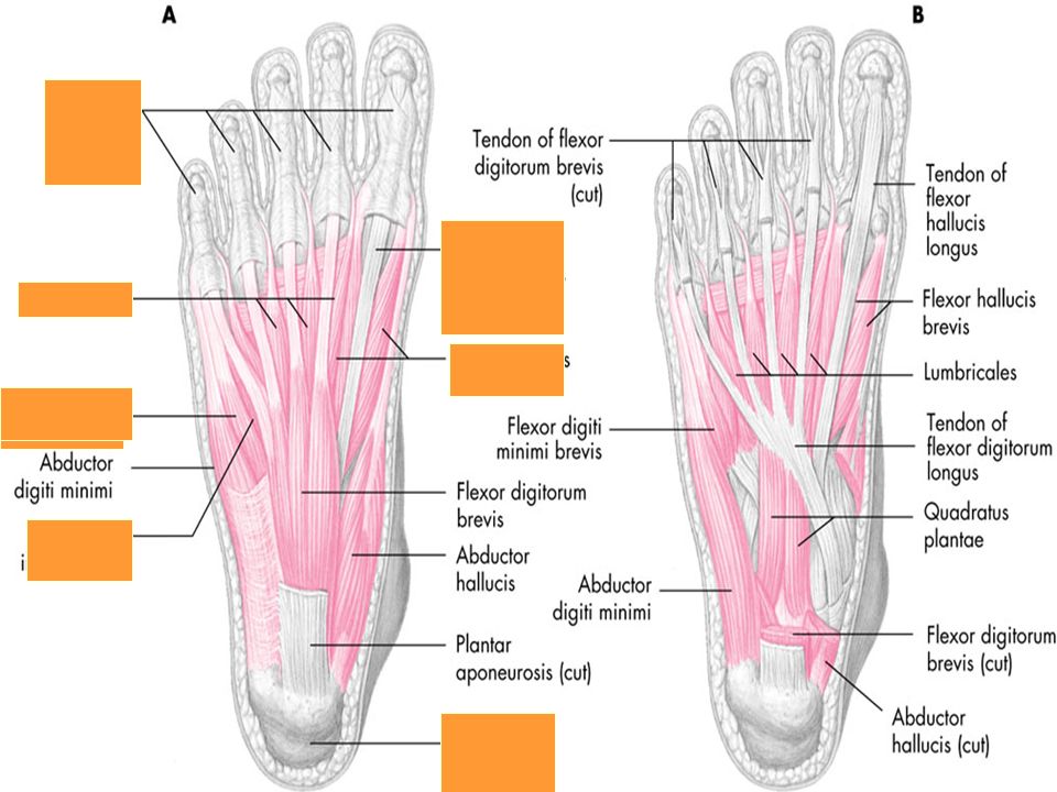
Foot Muscles – Plantar Surface
Superficial Layer Abductor Hallucis Abductor Digiti Minimi Flexor Digitorum Brevis

30
Foot Muscles – Plantar Surface
Middle Layer Quadratus Plantae Lumbricales

32
Foot Muscles – Plantar Surface
Deep Layer Flexor Hallucis Brevis Adductor Hallucis Transverse and Oblique Heads Flexor Digiti Minimi

33
Foot Muscles – Plantar Surface
Interosseus Layer Plantar Interossei Dorsal Interossei

35
Foot Muscles – Dorsal Surface
Extensor Digitorum Brevis Extensor Hallucis Brevis

Similar presentations