Download presentation
Presentation is loading. Please wait.

1
Prepared by : ANWAR ISSA RN-BSN-CCRT-ICU

3
P wave : is P wave normal ? PR interval : is PR interval normal ? QRS complex : is QRS normal ? P-QRS relation : what is the relation between P wave and QRS ? Rate : what is the rate ? How to analyze a rhythm strip ?

4
Are the P waves regular ? Is the P wave normal and upright in lead II ? Do all P waves look alike ? P WAVE

5
Are all the PRI ‘s constant? Is the PRI measurement within normal range? If the PRI varies, is there a pattern to the changing measurements? PR INTERVAL

6
Are all the QRS complexes of equal duration? What is the measurement of the QRS complex? Is the QRS measurement within normal limits? Do all the QRS complexes look alike? Are the unusual QRS complexes associated with ectopic beats? QRS COMPLEX

7
Is there a P for every QRS and vice versa? Are they related to each other? More P’s? More QRS’s ? P-QRS RELATION

8
What is the exact rate? Is the atrial rate the same as the ventricular rate? - If the rhythm is regular--- 300/# of big squares between RR. - - If the rhythm is irregular --- count the R waves on a 6 second strip and multiply by 10. HEART RATE

10
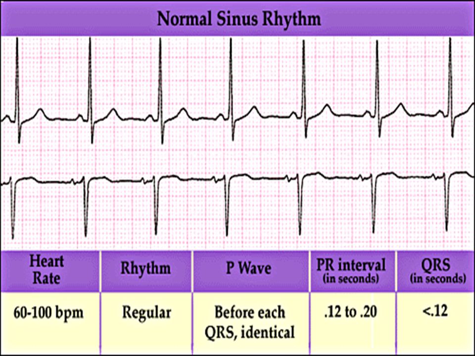
Normal sinus rhythm

13
* Condition where SA node stops firing, causing pause in electrical activity. * During the pause, atrial and ventricularcontraction do not occur.

16
MAT Multifocal Atrial Tachycardia Multifocal atrial tachycardia : narrow-complex tachycardia at 140 to 160 bpm with multiple P-wave morphologies (arrows)
")
19
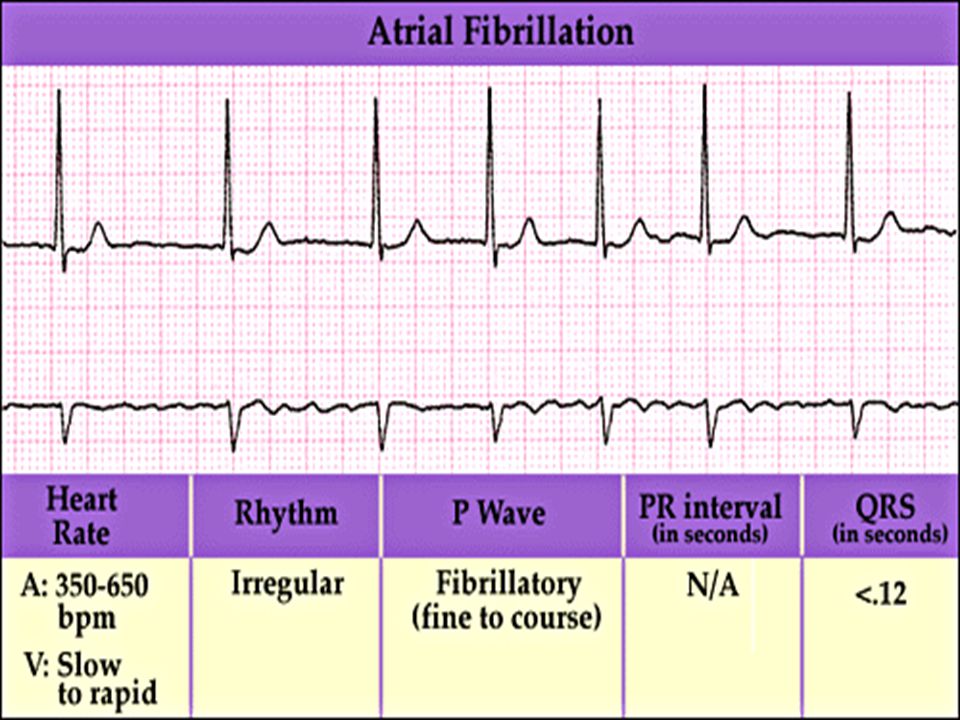
Atrial fibrillation / flutter Atrial fibrillation Atrial flutter

22
Junctional Tachycardia Rate > 100/min

23
SVT (SupraVentricular Tachycardia)
")
24
PSVT (Paroxysmal Supraventricular Tachycardia) Sinus rhythm with paroxysmal onset (arrow) of supraventricular tachycardia (PSVT)
 Sinus rhythm with paroxysmal onset (arrow) of supraventricular tachycardia (PSVT)")
26
Wolff-Parkinson-White Wolff-Parkinson-White syndrome: normal sinus rhythm with delta wave notching of positive upstroke of QRS complex

27
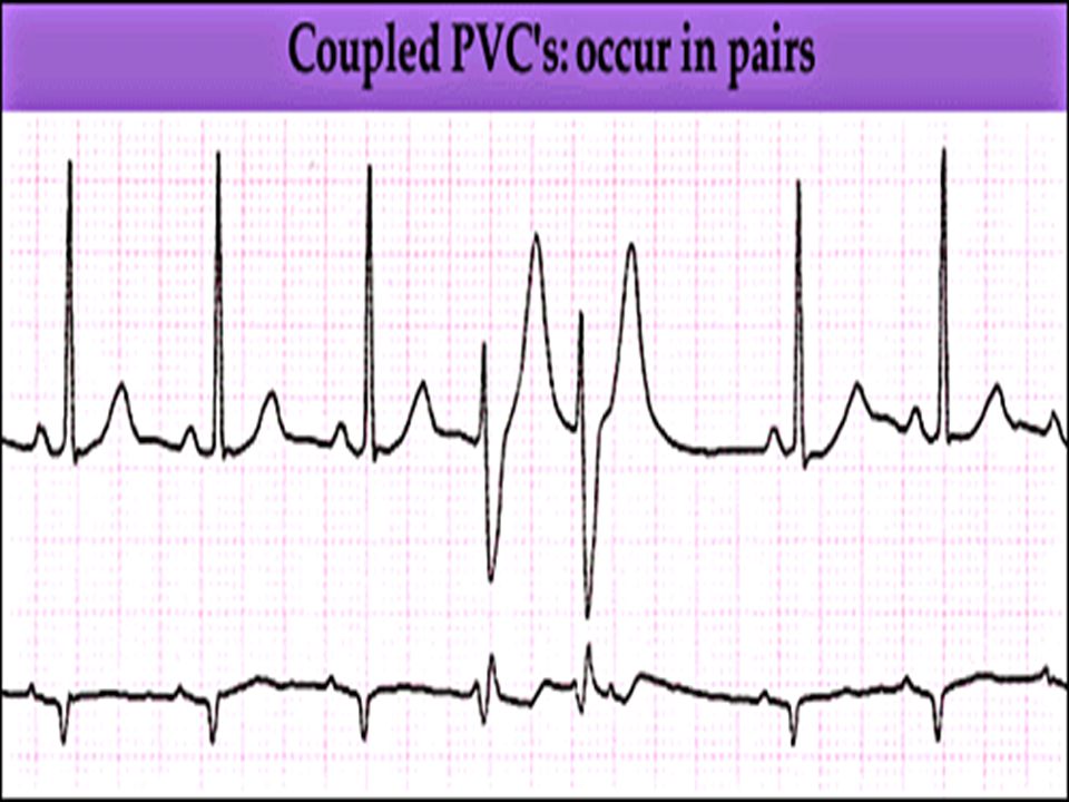
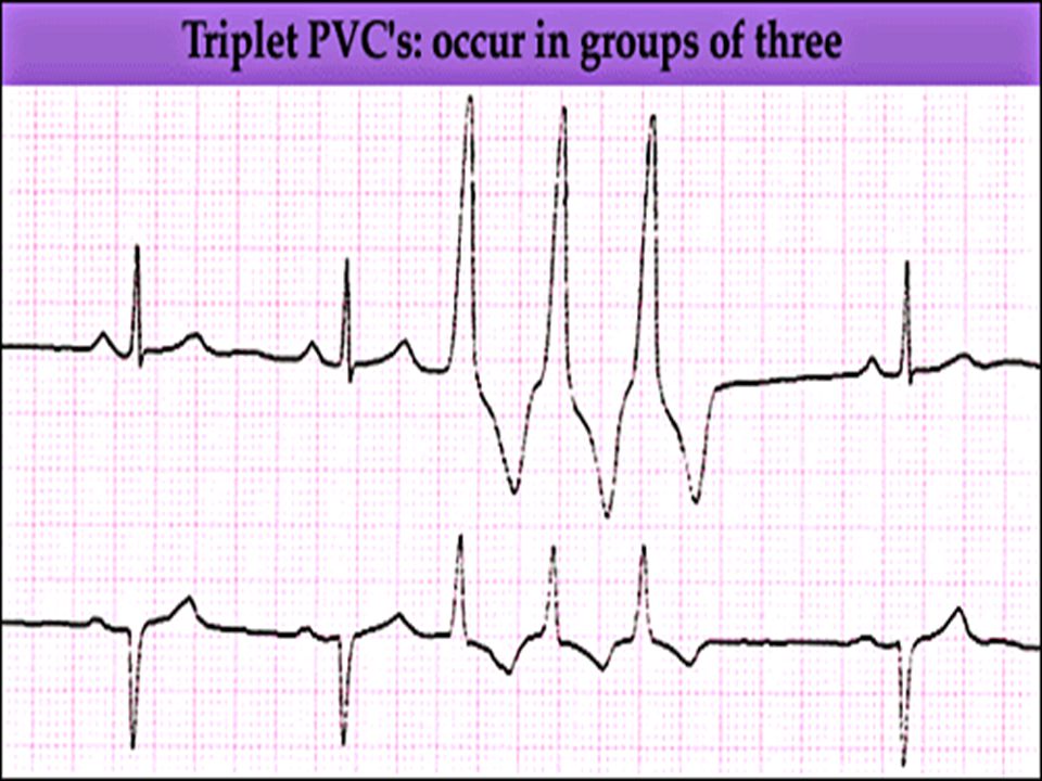
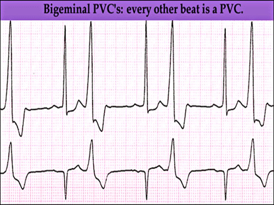
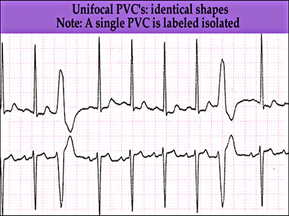
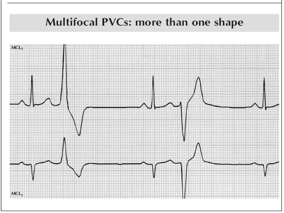
Premature Ventricular Contractions (PVC’s)
")
34
Ventricular Tachycardia Monomorphic ventricular tachycardia

35
Polymorphic Ventricular Tachycardia: Torsades de pointes Polymorphic ventricular tachycardia: QRS complexes display multiple morphologies (“polymorphic”)
")
37
Ventricular fibrillation Coarse VF Fine VF

42
Pacing above threshold (60 mA): with capture (QRS complex broad and ventricular; T wave opposite QRS) Pacing attempted: note pacing stimulus indicator (arrow) which is below threshold; no capture Pacing Rhythms
: with capture (QRS complex broad and ventricular; T wave opposite QRS) Pacing attempted: note pacing stimulus indicator (arrow) which is below threshold; no capture Pacing Rhythms")
44
2 1 Idio-Ventricular Rhythm

46
Asystole / Stand still

47
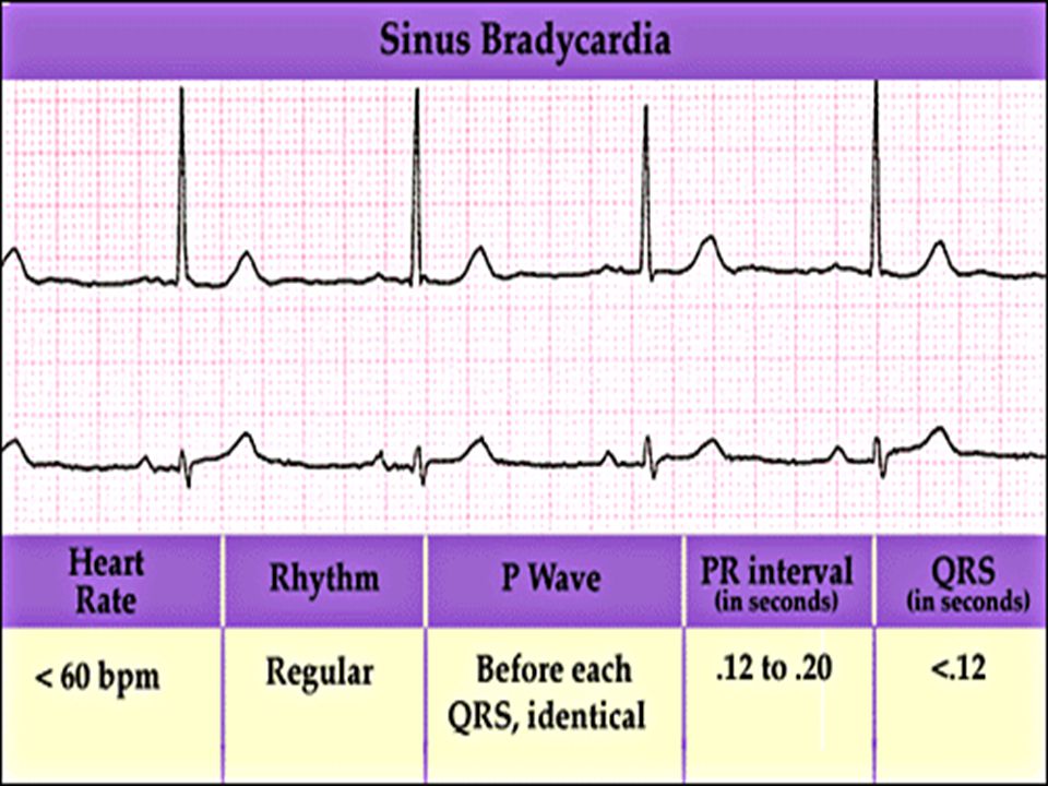
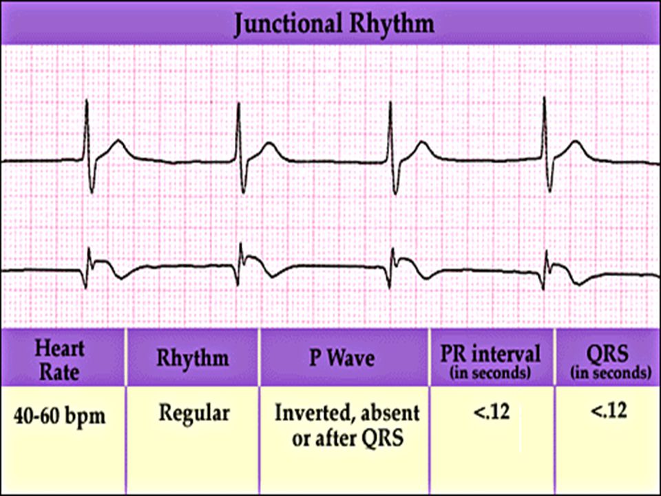
RATESINUS AV NODEVENTRICULAR > 100 Sinus tachycardia Junctional tachycardia Ventricular tachycardia 60-100 Normal Sinus Accelerated Junctional 40-60 Sinus Bradycardia Junctional rhythm Accelerated ventricular Rhythm 20-40 Idioventricular rhythm

Similar presentations