Download presentation
Presentation is loading. Please wait.

1
Clinical Assessment of Lower Urinary Tract Dysfunction Hann-Chorng Kuo Department of Urology Buddhist Tzu Chi General Hospital

2
Lower Urinary Tract Symptoms Storage symptoms Frequency, Urgency, Nocturia Incontinence Suprapubic fullness and pain Empty symptoms Hesitancy, Intermittency, Small caliber, Dysuria, Residual urine sensation

3
Urinary Incontinence Stress incontinence Urge incontinence Total incontinence Overflow incontinence Giggle incontinence Nocturnal enuresis

4
Voiding Diary

5
Physical Examination Abdominal physical examination Bladder, Operation scar Perineal examination Cystocele, Rectocele, Uterine prolapse Urine leakage on cough, fistula Vaginal mucosa, Vaginal tenderness Neurological examination B-C Reflex, PFM contractility, Anal tone

6
Sensory dermatomes of perineum & extremities

7
Clinical investigation of Lower urinary tract dysfunction Urethral sounding Prostatic fluid examination Ultrasound examination Pad weighing test Cystourethroscopy Potassium chloride test

8
Urethral Sounding

9
Prostatic Massage and Expressed prostatic secretion

10
Prostatitis Acute bacterial prostatitis Chronic bacterial prostatitis Abacterial prostatitis Prostatodynia (perineal pain syndrome) Using available symptom score or index to assess symptomatology
 Using available symptom score or index to assess symptomatology")
11
Symptomatology of Prostatitis Pelvic pain syndrome Disturbance in urination Disturbance in sexual function Depression Disturbance in intimate relationships

12
Diagnosis of Prostatitis Expressed prostatic secretions show numerous WBC and macrophage Abnormal EPS: WBC>10 or 15/HPF After massage U/A: WBC >10/HPF Calcification in prostatic ultrasound Elevated prostatic specific antigen Increased EPS PH (>7.8)
")
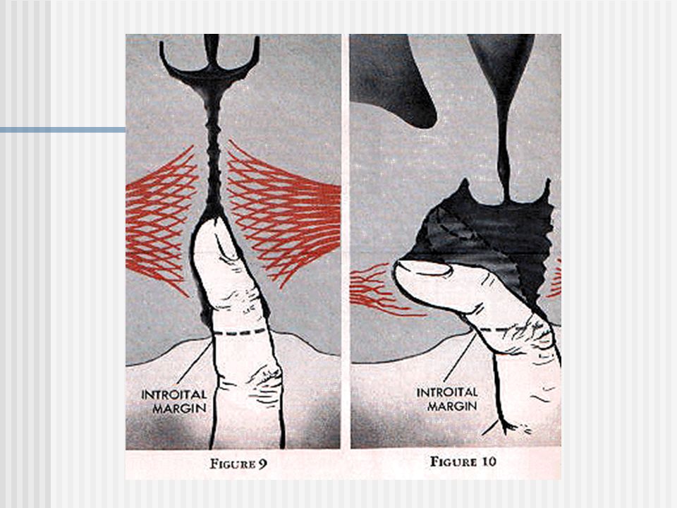
13
Ultrasound Examination in Male LUTS Prostate enlargement is not indicator of BOO in men with LUTS Transition zone index provides a better indicator for BOO Bladder neck dysfunction Trabeculated bladder Low residual urine

14
Prostatic Configuration in Transrectal ultrasound

15
Prostatic enlargement Benign prostatic enlargement Prostatic cancer

16
Correlation of TZI with Prostate volume & Qmax

17
Clinical Prostate Score Uroflowmetry (mL/s)Voided volume (mL) Qmax ≥ 15 ≥2500 10 < Qmax < 150 <2501 Qmax ≤ 101TPV (mL) Flow pattern ≤200 Normal >20 but <401 Compressive obstructive1 ≥402 Constrictive obstructive2TZI Intermittent2 ≤0.3 Residual urine (mL) >03 but 0.51 <1000 ≥0.52 ≥ 1002Median lobe enlargement Key:Abbreviation as in Tables I and Ⅲ Presence2 Absence0
Voided volume (mL) Qmax ≥ 15 ≥ < Qmax < 150 <2501 Qmax ≤ 101TPV (mL) Flow pattern ≤200 Normal >20 but <401 Compressive obstructive1 ≥402 Constrictive obstructive2TZI Intermittent2 ≤0.3 Residual urine (mL) >03 but 0.51 <1000 ≥0.52 ≥ 1002Median lobe enlargement Key:Abbreviation as in Tables I and Ⅲ Presence2 Absence0")
18
Urethral Ultrasound in SUI and Frequency Urgency Syndrome

19
Measurement of Bladder Neck Hypermobility in Frequency Urgency Syndrome in Women

20
Bladder Neck Descent in Women with LUTS NPVL(cm)PVA(degrees) RestingStrainingIncrementRestingStrainingIncrement* SUI 1912.05±0.692.20±0.480.15±0.5834.6±23.466.5±28.631.9±19.9 FUS 782.05±0.392.11±0.430.06±0.2018.4±19.237.4±29.119.0±17.6 ASYM 272.08±0.332.13±0.310.05±0.208.2±10.620.7±23.212.6±16.7 ANOVA NS P<0.05P0.05P<0.05
PVA(degrees) RestingStrainingIncrementRestingStrainingIncrement* SUI ± ± ± ± ± ±19.9 FUS ± ± ± ± ± ±17.6 ASYM ± ± ± ± ± ±16.7 ANOVA NS P<0.05P0.05P<0.05")
21
Bladder Neck Incompetence in Frequency Urgency Syndrome

22
Bladder Neck Incompetence and Hypermobility

23
Measurement of External Sphincter Volume in SUI

24
Different Urethral Structure

25
Urethral Ultrasound in ISD and Cystocele

26
Striated Urethral Sphincter in SUI and Cystocele PatientsN Cross-Sectional Area (mm 2 ) Smooth Muscle Component (mm 2 ) Striated Muscle Component (mm 2 ) A.Non-SUI 51104.4 ±35.646.1±22.558.3±27.3 B.SUI 6086.7 ±29.943.9±19.042.8±20.7 Cystocele* (9)75.7 ±23.137.9±12.237.8±22.8 Statistics A vs B: P =0.005NSA vs B: P =0.001
 Smooth Muscle Component (mm 2 ) Striated Muscle Component (mm 2 ) A.Non-SUI ± ± ±27.3 B.SUI ± ± ±20.7 Cystocele* (9)75.7 ± ± ±22.8 Statistics A vs B: P =0.005NSA vs B: P =0.001")
27
Female Urethral Incompetence Bladder neck incompetence Urethral incompetence

28
Assessing Pubococcygeus muscle function Inspection Perineum buldging downward Vaginal introitus opens Anus everted Performing straining or coughing Contraction of pubococcygeus m.

29
Cystocele and Prolapse

30
Assessing Pubococcygeus muscle function Palpation In normal vagina, resistance is met in all direction by finger palpation The atrophied pubococcygeus m. is not easily palpated with little resistance One third of women have a good voluntary contraction function

32
Voluntary Contraction of Pelvic Floor Muscles

33
Pad Weighing Test for Stress Urinary Incontinence Provide semi-objective measurement of urine loss 1 hr, 2 hr, 24 hr, 48 hr test Drink 500ml, walking & stair climbing 30 min, standing up 10x, coughing 10x, running 1 min, bending 5x, wash hands 1 min Pad weight gain by 1 gm

34
Laboratory examinations Urinalysis & urine culture- evidence of pus cells and bacteria in urine Blood chemistry, blood sugar- azotemia, diabetes may cause polyuria, detrusor underactivity KUB- a lower ureteral stone cause storage symptoms and empty symptoms

35
Office Urodynamic Study Uroflowmetry Postvoid residual urine (PVR) Cystometry with or without EMG Potassium chloride test
 Cystometry with or without EMG Potassium chloride test")
36
Uroflowmetry – Parameters

37
Uroflowmetry – Intermittent flow

38
Uroflowmetry – Straining flow

39
Uroflowmetry – Low contractility

40
Uroflowmetry – Obstructive flow

41
Voiding Cystometry (Pressure flow study) Filling cystometry cannot diagnose 24% of the patients with LUTS Patients with voiding symptoms should undergo pressure flow study Detrusor underactivity, bladder outlet obstruction, postvoid detrusor contraction, occult neuropathic detrusor overactivity
 Filling cystometry cannot diagnose 24% of the patients with LUTS Patients with voiding symptoms should undergo pressure flow study Detrusor underactivity, bladder outlet obstruction, postvoid detrusor contraction, occult neuropathic detrusor overactivity")
42
Multi-channel Pressure Flow Study

43
Relationship of Pressure & Flow

44
Cystometry – after contraction

45
Pressure flow study – DHIC

46
Pressure flow study – Cystocele and BOO in woman

47
Low contractility & low flow

48
SCI & NVD – Type 1 DESD

49
DI & voluntary PFM contraction

50
Idiopathic detrusor overactivity in Storage phase

51
Detrusor overactivity in contracted bladder

52
Neurogenic detrusor overactivity in CVA patient

53
Provoked Detrusor overactivity in storage phase

54
Potassium Test A test for urothelium leak syndrome 40mL of 0.4M KCL was infused into the bladder following normal saline Record the pain scale after KCl test: nil, burning, tingling, dull pain, sharp pain, urgency Acute and irradiation cystitis: 100% Interstitial cystitis: 80%

55
Increased Bladder sensation after KCl infusion

56
Potassium sensitivity test in women with frequency urgency and IC In 196 women with frequency urgency and/or pain, 138 had a positive KCl test (70.4%) 128 women with a positive KCl test, 44 (34.4%)proven IC and 84 non-IC A positive KCl test indicates urothelial leak but not characteristic IC, nor can bladder pain predict IC
 128 women with a positive KCl test, 44 (34.4%)proven IC and 84 non-IC A positive KCl test indicates urothelial leak but not characteristic IC, nor can bladder pain predict IC")
57
Postvoid Residual Volume Estimated immediately after voiding Transabdominal ultrasound provides accurate volume estimation Diuresis may falsely increase PVR Patient might not void completely due to embarrassment Do not forget PVR in clinical assessment of LUTS

Similar presentations