Download presentation
Presentation is loading. Please wait.

6
CORE CURRICULUM PCI Sandeep Gautam, M.D.

7
ACC/AHA/SCAI 2005 Guideline Update for Percutaneous Coronary Intervention
8
Contents 1. OUTCOMES – Definitions of PCI Success: Angiographic Success, Procedural Success, Clinical Success. Acute Outcome: Procedural Complications, Success Rates. Long-Term Outcome and Restenosis. Predictors of Success/Complications: Lesion Morphology and Classification, Clinical Factors (Left Main CAD, women, DM). Comparison With Bypass Surgery/ Medicine. 2. INSTITUTIONAL AND OPERATOR COMPETENCY 3. CLINICAL PRESENTATIONS – Asymptomatic Ischemia, CCS Class III Angina, UA/NSTEMI, STEMI, Prior CABG, Use of Adjunctive Technology. 4. MANAGEMENT OF PATIENTS UNDERGOING PCI – Evolution of Technologies: Acute and late term results. Antiplatelet and Antithrombotic Adjunctive Therapies. Post-PCI Management. 5. SPECIAL CONSIDERATIONS – Ad Hoc Angioplasty, Cardiac Transplant Patients, Clinical Restenosis, Cost-Effectiveness Analysis.
. Comparison With Bypass Surgery/ Medicine. 2. INSTITUTIONAL AND OPERATOR COMPETENCY. 3. CLINICAL PRESENTATIONS – Asymptomatic Ischemia, CCS Class III Angina, UA/NSTEMI, STEMI, Prior CABG, Use of Adjunctive Technology. 4. MANAGEMENT OF PATIENTS UNDERGOING PCI – Evolution of Technologies: Acute and late term results. Antiplatelet and Antithrombotic Adjunctive Therapies. Post-PCI Management. 5. SPECIAL CONSIDERATIONS – Ad Hoc Angioplasty, Cardiac Transplant Patients, Clinical Restenosis, Cost-Effectiveness Analysis.")
10
Background More than PCI procedures are performed yearly in the United States, and it has been estimated that nearly procedures are performed annually worldwide. Presently, PTCA alone is used in less than 30% cases, compared to PCI with stenting in greater than 70% cases. Atherectomy devices and stenting continue to be applied to a wider patient domain that includes multivessel disease and complex coronary anatomy. However, strong evidence (level A data from multiple randomized clinical trials) is primarily available for stenting over PTCA in selected patients undergoing single-vessel PCI.
 is primarily available for stenting over PTCA in selected patients undergoing single-vessel PCI.")
11
Approved Devices Balloon expandable stents, DES, extraction atherectomy, directional coronary atherectomy, rotational atherectomy, rheolytic thrombectomy catheter, proximal and distal embolic protection devices, excimer laser coronary atherectomy, and local radiation devices to reduce in-stent restenosis (ISR).
.")
12
Laskey WK, Kimmel S, Krone RJ
Laskey WK, Kimmel S, Krone RJ. Contemporary trends in coronary intervention: a report from the Registry of the Society for Cardiac Angiography and Interventions. Catheter Cardiovasc Interv 2000;49:19-22.

13
Outcomes Outcomes are measured in terms of success and complications - These are related to a) mechanisms of the employed devices, and b) the clinical and anatomic patient-related factors. Complications can be divided into 2 categories: (a) those common to all arterial catheterization procedures and (b) those related to the specific technology used for the coronary procedure.
 mechanisms of the employed devices, and b) the clinical and anatomic patient-related factors. Complications can be divided into 2 categories: (a) those common to all arterial catheterization procedures and (b) those related to the specific technology used for the coronary procedure.")
14
Definitions of PCI Success
Angiographic Success Procedural Success Clinical Success

15
Angiographic Success The consensus definition for PTCA was the achievement of a minimum stenosis diameter reduction to less than 50% in the presence of grade 3 Thrombolysis In Myocardial Infarction (TIMI) flow. However, with the advent of coronary stents, a minimum stenosis diameter reduction to less than 20% has been the clinical benchmark of an optimal angiographic result. There may be a disparity between the visual assessment and computer-aided quantitative stenosis measurement, and, thus, the determination of success may be problematic when success rates are self-reported.
 flow. However, with the advent of coronary stents, a minimum stenosis diameter reduction to less than 20% has been the clinical benchmark of an optimal angiographic result. There may be a disparity between the visual assessment and computer-aided quantitative stenosis measurement, and, thus, the determination of success may be problematic when success rates are self-reported.")
16
Procedural Success A successful PCI should achieve angiographic success without major clinical complications (e.g., death, MI, emergency coronary artery bypass surgery) during hospitalization. Criteria for procedure-related MI: 1) Development of Q waves 2) CK-MB elevations 3 to 5 times the upper limit of normal. 3) Greater than 5 times elevations in Troponin T or I. The timing of the peak elevation after PCI is unclear. The conventional definition of MI requires 2 of the following: a) prolonged chest discomfort or its equivalent; b) development of pathologic Q waves; and c) rise in serum cardiac biomarkers above a critical level.
 during hospitalization. Criteria for procedure-related MI: 1) Development of Q waves. 2) CK-MB elevations 3 to 5 times the upper limit of normal. 3) Greater than 5 times elevations in Troponin T or I. The timing of the peak elevation after PCI is unclear. The conventional definition of MI requires 2 of the following: a) prolonged chest discomfort or its equivalent; b) development of pathologic Q waves; and c) rise in serum cardiac biomarkers above a critical level.")
17
Clinical Success The patient should have persistent relief of signs and symptoms of myocardial ischemia for more than 6 months after the procedure. Restenosis is the principal cause of lack of long-term clinical success. This is not considered a complication but rather an associated response to vascular injury.

18
Procedural Complications - Death
Death as a result of PCI is directly related to the occurrence of coronary artery occlusion and is most frequently associated with pronounced LV failure. Reported rates for death after diagnostic cath range from 0.08% to 0.14%, whereas overall unadjusted in-hospital rates for PCI range from 0.4% to 1.9%. The highest mortality rate is seen in patients with STEMI and cardiogenic shock. The clinical and angiographic variables associated with increased mortality include advanced age, female gender, diabetes, prior MI, periprocedural stroke, multivessel disease, left main or equivalent coronary disease, a large area of myocardium at risk, pre-existing impairment of LV or renal function, post-PCI worsening of renal function, and collateral vessels supplying significant areas of myocardium that originate distal to the segment to be dilated.

19
Procedural Complications - MI
Rates of periprocedural MI have ranged from 0.4% to 4.9%. More than 70% of patients exhibit elevated troponin values after an otherwise successful intervention. One study has suggested a postprocedural increase in troponin T of 5 times normal is predictive for adverse events at 6 years. The long-term prognostic significance of smaller postprocedural troponin T elevations awaits further investigation.

22
Procedural Complications - CABG
Typically, CABG is performed as a rescue revascularization procedure to treat acute ischemia or infarction resulting from PCI-induced acute coronary occlusion. In the era of balloon angioplasty, the rate of emergency CABG was 3.7%. With the availability of stents, the reported rate was 0.4% among a similar cohort of patients.

23
Procedural Complications - Bleeding
A frequently used definition for bleeding developed by the TIMI group includes classification as major, moderate, or minor. Major bleeding is defined as intracranial, intraocular, or retroperitoneal hemorrhage or any hemorrhage requiring a transfusion or surgical intervention or that results in a hematocrit decrease of greater than 15% or hemoglobin decrease of greater than 5 g per dL. Episodes of hemorrhage of lesser magnitude would fall into the moderate/minor categories.

25
Acute Outcome: Success Rates
The chance of dilating a chronic total occlusion averages 65%. The success rates for total occlusions associated with STEMI are over 90%. Procedural success rates have risen from a range of 80% to 85% to a range of 90% to 95%.

26
Long-Term Outcome and Restenosis
Defined as greater than 50% diameter stenosis at follow-up angiography. Ten-year follow-up of the initial cohort of patients treated with PTCA revealed an 89.5% survival rate (95% in patients with single-vessel disease, 81% in patients with multivessel disease). DM - In randomized patients with treated diabetes undergoing PTCA in BARI, the 5-year survival was 65.5%, and the cardiac mortality rate was 20.6% compared with 5.8% in patients without treated diabetes. Women - In the NHLBI PTCA Registry, 4- year survival was significantly lower in women (89.2%) than in men (93.4%).
. DM - In randomized patients with treated diabetes undergoing PTCA in BARI, the 5-year survival was 65.5%, and the cardiac mortality rate was 20.6% compared with 5.8% in patients without treated diabetes. Women - In the NHLBI PTCA Registry, 4- year survival was significantly lower in women (89.2%) than in men (93.4%).")
27
Long-Term Outcome and Restenosis
Pathogenesis of restenosis - A combination of growth factor stimulation, smooth muscle cell migration and proliferation, organization of thrombus, platelet deposition, and elastic recoil. Clinical factors: Diabetes, unstable angina/NSTEMI, STEMI, and prior restenosis. Angiographic factors: Proximal left anterior descending artery, small vessel diameters, total occlusion, long lesion length, and saphenous vein grafts. Procedural factors: Higher postprocedure percent diameter stenosis, smaller minimal lumen diameter, and smaller acute gain. The most promising potential approaches to favorably impact the restenosis process are DES and catheter-based radiation.

29
Predictors of Success/Complications
Lesion Morphology and Classification: Descriptions of a High-Risk Lesion (Type C Lesion) Diffuse (length greater than 2 cm) Excessive tortuosity of proximal segment Extremely angulated segments, greater than 90° Total occlusions more than 3 months old and/or bridging collaterals* Inability to protect major side branches Degenerated vein grafts with friable lesions* *The high risk with these criteria is for technical failure and increased restenosis, not for acute complications.
 Diffuse (length greater than 2 cm) Excessive tortuosity of proximal segment. Extremely angulated segments, greater than 90° Total occlusions more than 3 months old and/or bridging collaterals* Inability to protect major side branches. Degenerated vein grafts with friable lesions* *The high risk with these criteria is for technical failure and increased restenosis, not for acute complications.")
30
SCAI Lesion Classification System: Characteristics of Class I-IV Lesions
Type I lesions (highest success expected, lowest risk) (1) Does not meet criteria for C lesion (2) Patent Type II lesions (1) Meets any of these criteria for ACC/AHA C lesion Diffuse (greater than 2 cm length) Excessive tortuosity of proximal segment Extremely angulated segments, greater than 90° Inability to protect major side branches Degenerated vein grafts with friable lesions Type III lesions (2) Occluded Type IV lesions (1) Meets any of the criteria for ACC/AHA C lesion Occluded for more than 3 months
 (1) Does not meet criteria for C lesion. (2) Patent. Type II lesions. (1) Meets any of these criteria for ACC/AHA C lesion. Diffuse (greater than 2 cm length) Excessive tortuosity of proximal segment. Extremely angulated segments, greater than 90° Inability to protect major side branches. Degenerated vein grafts with friable lesions. Type III lesions. (2) Occluded. Type IV lesions. (1) Meets any of the criteria for ACC/AHA C lesion. Occluded for more than 3 months.")
31
Left Main CAD CABG has long been considered the “gold standard” for revascularization of lesions in the unprotected left main (ULM) coronary artery. The experience with BMS for ULM PCI in the multicenter ULTIMA registry suggested a high early mortality (2% per month among hospital survivors over the first 6 months). Studies using DES have reported 6- month or 1-year mortality ranging from 0% to 14%. Some studies have reported performing routine angiography 4 to 8 months after PCI or earlier if clinically indicated. Guidelines continue to recommend PCI only in cases unsuitable for CABG.
. Studies using DES have reported 6- month or 1-year mortality ranging from 0% to 14%. Some studies have reported performing routine angiography 4 to 8 months after PCI or earlier if clinically indicated. Guidelines continue to recommend PCI only in cases unsuitable for CABG.")
32
Women An estimated 33% of the PCIs performed in the United States are in women. In several large-scale registries, in-hospital and long term mortality is significantly higher in women (Odds Ratio ). Compared with men, women undergoing PCI are older with a higher incidence of HTN, DM, hyperlipidemia, and comorbid disease, but have similar epicardial coronary disease. Gender differences in mortality have persisted for patients treated with stents both in the setting of acute and nonacute MI. Smaller vessel size, hypertensive heart disease, and diastolic dysfunction in women have been thought to play a role. A few studies have noted that gender is not an independent predictor of mortality after adjusting for body surface area. IVUS studies have not detected any gender-specific differences in plaque morphology or luminal dimensions after adjustment for BSA. Women tend to have increased bleeding and vascular complications. These have decreased with the use of smaller sheath sizes and early sheath removal, weight-adjusted heparin dosing, and less aggressive anticoagulation regimens. An increased rate of minor bleeding has been reported in women treated with abciximab.
. Compared with men, women undergoing PCI are older with a higher incidence of HTN, DM, hyperlipidemia, and comorbid disease, but have similar epicardial coronary disease. Gender differences in mortality have persisted for patients treated with stents both in the setting of acute and nonacute MI. Smaller vessel size, hypertensive heart disease, and diastolic dysfunction in women have been thought to play a role. A few studies have noted that gender is not an independent predictor of mortality after adjusting for body surface area. IVUS studies have not detected any gender-specific differences in plaque morphology or luminal dimensions after adjustment for BSA. Women tend to have increased bleeding and vascular complications. These have decreased with the use of smaller sheath sizes and early sheath removal, weight-adjusted heparin dosing, and less aggressive anticoagulation regimens. An increased rate of minor bleeding has been reported in women treated with abciximab.")
33
The Elderly Patient Age greater than 75 years is one of the major risk factor for complications. Octogenarians undergoing PCI have a higher incidence of prior MI, lower LV ejection fraction, and more frequent CHF. A separate category has not been created in these guidelines for the elderly, except for primary PCI for cardiogenic shock in patients greater than 75 years of age. However, their higher incidence of comorbidities and risk for bleeding complications should be taken into account when considering the need for PCI.

34
Diabetes Mellitus The efficacy of stenting with GP IIb/IIIa inhibitors was assessed in the diabetic population compared with those without diabetes in a substudy of the EPISTENT (Evaluation of IIb/IIIa Platelet Inhibitor for Stenting) trial. The combination of stenting and abciximab among diabetics resulted in a significant reduction in 6-month rates of death and target-vessel revascularization compared with stent/placebo or PTCA/abciximab therapy. In the BARI trial, the benefit of bypass surgery in diabetic patients was greater in those patients with more extensive disease (e.g., more than 4 lesions). This advantage was largely due to a lower mortality for subsequent MI. At 3 years of follow-up, the survival rates of the diabetic subsets treated with CABG and PCI were not significantly different in either ARTS (Arterial Revascularization Therapies Study) or AWESOME (Angina With Extremely Serious Operative Mortality Evaluation). The sum effect of DES and GP IIb/IIIa inhibitors will be assessed against contemporary CABG in multivessel-disease patients with diabetes in the upcoming NIH–sponsored FREEDOM trial.
 trial. The combination of stenting and abciximab among diabetics resulted in a significant reduction in 6-month rates of death and target-vessel revascularization compared with stent/placebo or PTCA/abciximab therapy. In the BARI trial, the benefit of bypass surgery in diabetic patients was greater in those patients with more extensive disease (e.g., more than 4 lesions). This advantage was largely due to a lower mortality for subsequent MI. At 3 years of follow-up, the survival rates of the diabetic subsets treated with CABG and PCI were not significantly different in either ARTS (Arterial Revascularization Therapies Study) or AWESOME (Angina With Extremely Serious Operative Mortality Evaluation). The sum effect of DES and GP IIb/IIIa inhibitors will be assessed against contemporary CABG in multivessel-disease patients with diabetes in the upcoming NIH–sponsored FREEDOM trial.")
35
PCI After Coronary Artery Bypass Surgery
Patients having PCI of native vessels after prior CABG have nearly equivalent outcomes and complication rates compared with patients having similar interventions without prior surgery. For PCI of SVG, the rate of successful angioplasty exceeds 90%, the death rate is <1.2%, and the rate of Q-wave MI is <2.5%. The age of the SVG and duration and severity of myocardial ischemia should be considered. GP IIb/ IIIa blockers have not been shown to improve results of PCI in vein grafts. Preliminary studies of 2 different distal embolic protection devices (Percusurge and GuideWire) are associated with promising results. PCI of a protected left main stenosis with a patent and functional LAD or left circumflex coronary conduit can be considered as a palliative procedure with the potential to delay the ultimate application of repeat CABG surgery.
 are associated with promising results. PCI of a protected left main stenosis with a patent and functional LAD or left circumflex coronary conduit can be considered as a palliative procedure with the potential to delay the ultimate application of repeat CABG surgery.")
36
Coronary Perforation The incidence of coronary perforation has been reported at % with PTCA, % with directional coronary atherectomy, % with rotational atherectomy, % with extraction atherectomy, and 1.9-2% after excimer laser coronary angioplasty. Although 20% of perforations may be secondary to the coronary guidewire, most are related to the specific technology used. Perforation is usually (80% to 90%) evident at the time of the interventional procedure and is the primary differential diagnosis for cardiac tamponade manifest within 24 h of the procedure. Classification: Type I (extraluminal crater without extravasation), Type II (pericardial and myocardial blush without contrast jet extravasation) Type III (extravasation through a frank [1 mm] perforation)
 evident at the time of the interventional procedure and is the primary differential diagnosis for cardiac tamponade manifest within 24 h of the procedure. Classification: Type I (extraluminal crater without extravasation), Type II (pericardial and myocardial blush without contrast jet extravasation) Type III (extravasation through a frank [1 mm] perforation)")
37
Issues of Hemodynamic Support in High-Risk PCI
Hemodynamic compromise, defined as a decrease in SBP <90 mm Hg during balloon inflation, was associated with LVEF <35%, >50% of myocardium at risk, and PTCA performed on the last remaining vessel. IABP for high-risk PCI should be reserved only for patients patients with extremely depressed LV function and patients in cardiogenic shock. However, in patients with borderline hemodynamics, ongoing ischemia, or cardiogenic shock, insertion of an IABP just before coronary instrumentation has been associated with improved outcomes. It is also reasonable to obtain contralateral vascular access before the procedure in patients with a high risk of hemodynamic compromise. The decision to proceed with IABP before PCI remains a clinical judgment made by the physician based on the high-risk characteristics of coronary anatomy and overall status of the patient.

38
Comparison With Bypass Surgery
Generally speaking, the greater the extent of coronary atherosclerosis and its diffuseness, the more compelling the choice of coronary artery bypass surgery, particularly if LV function is depressed. In aggregate, trials comparing CABG and PCI have not shown a difference in terms of mortality or procedural MI among the populations studied, which have mostly included low-risk patients. Stents appear to have narrowed the late repeat revascularization difference that favored CABG in the balloon era. At this writing, no published studies are available comparing PCI with DES to CABG. Recent changes in patient management may influence CABG vs PCI decisions - Use of GP IIb/IIIa inhibitors, use of direct thrombin inhibitors during PCI, the more frequent use of IMA grafts, and the emergence of less invasive surgical approaches.

40
Comparison With Medicine
ACME (Angioplasty Compared to Medicine) pts with single- vessel disease, stable angina, and positive ETT to PTCA or medical therapy. PTCA provided better symptom control and exercise capacity. Death and MI were infrequent and similar. RITA-2 (Randomized Intervention Treatment of Angina) – crossover trial of 1018 pts with stable angina to PTCA or medical therapy, followed up for a mean of 7 years. PTCA resulted in better symptomatic improvement but was associated with a higher combined end point of death and periprocedural MI. 62% pts had multivessel CAD, and 34% had significant disease in the proximal segment of the LAD. AVERT (Atorvastatin Versus Revascularization Treatment) pts with stable CAD, nl LVEF, and class I or II angina to PTCA or atorvastatin 80 mg/d (mean LDL 77 mg/dl), followed for 18 months. 13% of the medical group had ischemic events compared with 21% of the PTCA group. Angina relief was greater in those treated with PTCA. MASS-II pts with stable angina, multivessel disease, and nl LVEF were randomized to 3 treatment groups: medical therapy, CABG, or PCI. Medical therapy had a low incidence of early events but was inferior to PCI and CABG for the control of angina. COURAGE (Clinical Outcomes Utilization Revascularization and Aggressive Drug Evaluation) - PCI plus intensive medical therapy VERSUS intensive medical therapy alone in pts with documented myocardial ischemia who meet an AHA task force Class I indication for PCI. BARI 2d - To compare revascularization in addition to aggressive medical therapy in patients with diabetes compared with aggressive medical therapy alone
 pts with single- vessel disease, stable angina, and positive ETT to PTCA or medical therapy. PTCA provided better symptom control and exercise capacity. Death and MI were infrequent and similar. RITA-2 (Randomized Intervention Treatment of Angina) – crossover trial of 1018 pts with stable angina to PTCA or medical therapy, followed up for a mean of 7 years. PTCA resulted in better symptomatic improvement but was associated with a higher combined end point of death and periprocedural MI. 62% pts had multivessel CAD, and 34% had significant disease in the proximal segment of the LAD. AVERT (Atorvastatin Versus Revascularization Treatment) pts with stable CAD, nl LVEF, and class I or II angina to PTCA or atorvastatin 80 mg/d (mean LDL 77 mg/dl), followed for 18 months. 13% of the medical group had ischemic events compared with 21% of the PTCA group. Angina relief was greater in those treated with PTCA. MASS-II pts with stable angina, multivessel disease, and nl LVEF were randomized to 3 treatment groups: medical therapy, CABG, or PCI. Medical therapy had a low incidence of early events but was inferior to PCI and CABG for the control of angina. COURAGE (Clinical Outcomes Utilization Revascularization and Aggressive Drug Evaluation) - PCI plus intensive medical therapy VERSUS intensive medical therapy alone in pts with documented myocardial ischemia who meet an AHA task force Class I indication for PCI. BARI 2d - To compare revascularization in addition to aggressive medical therapy in patients with diabetes compared with aggressive medical therapy alone.")
41
Patients With Asymptomatic Ischemia or CCS Class I or II Angina
Class IIa 1. PCI is reasonable in patients with asymptomatic ischemia or CCS class I or II angina and with 1 or more significant lesions in 1 or 2 coronary arteries suitable for PCI with a high likelihood of success and a low risk of morbidity and mortality. The vessels to be dilated must subtend a moderate to large area of viable myocardium or be associated with a moderate to severe degree of ischemia on noninvasive testing. (Level of Evidence: B). 2. PCI is reasonable for patients with asymptomatic ischemia or CCS class I or II angina, and recurrent stenosis after PCI with a large area of viable myocardium or high-risk criteria on noninvasive testing. (Level of Evidence: C) 3. Use of PCI is reasonable in patients with asymptomatic ischemia or CCS class I or II angina with significant left main CAD (greater than 50% diameter stenosis) who are candidates for revascularization but are not eligible for CABG. (Level of Evidence: B). Class IIb 1. The effectiveness of PCI for patients with asymptomatic ischemia or CCS class I or II angina who have 2- or 3-vessel disease with significant proximal LAD CAD who are otherwise eligible for CABG with 1 arterial conduit and who have treated diabetes or abnormal LV function is not well established. (Level of Evidence: B) 2. PCI might be considered for patients with asymptomatic ischemia or CCS class I or II angina with nonproximal LAD CAD that subtends a moderate area of viable myocardium and demonstrates ischemia on noninvasive testing. (Level of Evidence: C) Class III PCI is not recommended in patients with asymptomatic ischemia or CCS class I or II angina who do not meet the criteria as listed under the class II recommendations or who have 1 or more of the following: a. Only a small area of viable myocardium at risk (Level of Evidence: C) b. No objective evidence of ischemia. (Level of Evidence: C) c. Lesions that have a low likelihood of successful dilatation. (Level of Evidence: C) d. Mild symptoms that are unlikely to be due to myocardial ischemia. (Level of Evidence: C) e. Factors associated with increased risk of morbidity or mortality. (Level of Evidence: C) f. Left main disease and eligibility for CABG. (Level of Evidence: C) g. Insignificant disease (less than 50% coronary stenosis). (Level of Evidence: C)
. 2. PCI is reasonable for patients with asymptomatic ischemia or CCS class I or II angina, and recurrent stenosis after PCI with a large area of viable myocardium or high-risk criteria on noninvasive testing. (Level of Evidence: C) 3. Use of PCI is reasonable in patients with asymptomatic ischemia or CCS class I or II angina with significant left main CAD (greater than 50% diameter stenosis) who are candidates for revascularization but are not eligible for CABG. (Level of Evidence: B). Class IIb. 1. The effectiveness of PCI for patients with asymptomatic ischemia or CCS class I or II angina who have 2- or 3-vessel disease with significant proximal LAD CAD who are otherwise eligible for CABG with 1 arterial conduit and who have treated diabetes or abnormal LV function is not well established. (Level of Evidence: B) 2. PCI might be considered for patients with asymptomatic ischemia or CCS class I or II angina with nonproximal LAD CAD that subtends a moderate area of viable myocardium and demonstrates ischemia on noninvasive testing. (Level of Evidence: C) Class III. PCI is not recommended in patients with asymptomatic ischemia or CCS class I or II angina who do not meet the criteria as listed under the class II recommendations or who have 1 or more of the following: a. Only a small area of viable myocardium at risk (Level of Evidence: C) b. No objective evidence of ischemia. (Level of Evidence: C) c. Lesions that have a low likelihood of successful dilatation. (Level of Evidence: C) d. Mild symptoms that are unlikely to be due to myocardial ischemia. (Level of Evidence: C) e. Factors associated with increased risk of morbidity or mortality. (Level of Evidence: C) f. Left main disease and eligibility for CABG. (Level of Evidence: C) g. Insignificant disease (less than 50% coronary stenosis). (Level of Evidence: C)")
42
Patients With CCS Class III Angina
Class IIa 1. It is reasonable that PCI be performed in patients with CCS class III angina and single-vessel or multivessel CAD who are undergoing medical therapy and who have 1 or more significant lesions in 1 or more coronary arteries suitable for PCI with a high likelihood of success and low risk of morbidity or mortality. (Level of Evidence: B) 2. It is reasonable that PCI be performed in patients with CCS class III angina with single-vessel or multivessel CAD who are undergoing medical therapy with focal saphenous vein graft lesions or multiple stenoses who are poor candidates for reoperative surgery. (Level of Evidence: C) 3. Use of PCI is reasonable in patients with CCS class III angina with significant left main CAD (greater than 50% diameter stenosis) who are candidates for revascularization but are not eligible for CABG. (Level of Evidence: B) Class IIb 1. PCI may be considered in patients with CCS class III angina with single-vessel or multivessel CAD who are undergoing medical therapy and who have 1 or more lesions to be dilated with a reduced likelihood of success. (Level of Evidence: B) 2. PCI may be considered in patients with CCS class III angina and no evidence of ischemia on noninvasive testing or who are undergoing medical therapy and have 2- or 3-vessel CAD with significant proximal LAD CAD and treated diabetes or abnormal LV function. (Level of Evidence: B) Class III PCI is not recommended for patients with CCS class III angina with single-vessel or multivessel CAD, no evidence of myocardial injury or ischemia on objective testing, and no trial of medical therapy, or who have 1 of the following: a. Only a small area of myocardium at risk. (Level of Evidence: C) b. All lesions or the culprit lesion to be dilated with morphology that conveys a low likelihood of success. (Level of Evidence: C) c. Ahigh risk of procedure-related morbidity or mortality. (Level of Evidence: C) d. Insignificant disease (less than 50% coronary stenosis). (Level of Evidence: C) e. Significant left main CAD and candidacy for CABG. (Level of Evidence: C)
 2. It is reasonable that PCI be performed in patients with CCS class III angina with single-vessel or multivessel CAD who are undergoing medical therapy with focal saphenous vein graft lesions or multiple stenoses who are poor candidates for reoperative surgery. (Level of Evidence: C) 3. Use of PCI is reasonable in patients with CCS class III angina with significant left main CAD (greater than 50% diameter stenosis) who are candidates for revascularization but are not eligible for CABG. (Level of Evidence: B) Class IIb. 1. PCI may be considered in patients with CCS class III angina with single-vessel or multivessel CAD who are undergoing medical therapy and who have 1 or more lesions to be dilated with a reduced likelihood of success. (Level of Evidence: B) 2. PCI may be considered in patients with CCS class III angina and no evidence of ischemia on noninvasive testing or who are undergoing medical therapy and have 2- or 3-vessel CAD with significant proximal LAD CAD and treated diabetes or abnormal LV function. (Level of Evidence: B) Class III. PCI is not recommended for patients with CCS class III angina with single-vessel or multivessel CAD, no evidence of myocardial injury or ischemia on objective testing, and no trial of medical therapy, or who have 1 of the following: a. Only a small area of myocardium at risk. (Level of Evidence: C) b. All lesions or the culprit lesion to be dilated with morphology that conveys a low likelihood of success. (Level of Evidence: C) c. Ahigh risk of procedure-related morbidity or mortality. (Level of Evidence: C) d. Insignificant disease (less than 50% coronary stenosis). (Level of Evidence: C) e. Significant left main CAD and candidacy for CABG. (Level of Evidence: C)")
43
Operator and Institutional Volume
Class I 1. Elective PCI should be performed by operators with acceptable annual volume (at least 75 procedures) at high- olume centers (more than 400 procedures) with onsite cardiac surgery. (Level of Evidence: B) 2. Elective PCI should be performed by operators and institutions whose historical and current risk-adjusted outcomes statistics are comparable to those reported in contemporary national data registries. (Level of Evidence: C) 3. Primary PCI for STEMI should be performed by experienced operators who perform more than 75 elective PCI procedures per year and, ideally, at least 11 PCI procedures for STEMI per year. Ideally, these procedures should be performed in institutions that perform more than 400 elective PCIs per year and more than 36 primary PCI procedures for STEMI per year. (Level of Evidence B) Class IIa 1. It is reasonable that operators with acceptable volume (at least 75 PCI procedures per year) perform PCI at low-volume centers (200 to 400 PCI procedures per year) with onsite cardiac surgery. (Level of Evidence: BC) 2. It is reasonable that low-volume operators (fewer than 75 PCI procedures per year) perform PCI at high-volume centers (more than 400 PCI procedures per year) with onsite cardiac surgery. Ideally, operators with an annual procedure volume less than 75 should only work at institutions with an activity level of more than 600 procedures per year. Operators who perform fewer than 75 procedures per year should develop a defined mentoring relationship with a highly experienced operator who has an annual procedural volume of at least 150 procedures per year. (Level of Evidence: BC) Class IIb The benefit of primary PCI for STEMI patients eligible for fibrinolysis when performed by an operator who performs fewer than 75 procedures per year (or fewer than 11 PCIs for STEMI per year) is not well established. (Level of Evidence: C) Class III It is not recommended that elective PCI be performed by low- olume operators (fewer than 75 procedures per year) at low- olume centers (200 to 400) with or without onsite cardiac surgery. An institution with a volume of fewer than 200 procedures per year, unless in a region that is underserved because of geography, should carefully consider whether it should continue to offer this service. (Level of Evidence: BC)
 at high- olume centers (more than 400 procedures) with onsite cardiac surgery. (Level of Evidence: B) 2. Elective PCI should be performed by operators and institutions whose historical and current risk-adjusted outcomes statistics are comparable to those reported in contemporary national data registries. (Level of Evidence: C) 3. Primary PCI for STEMI should be performed by experienced operators who perform more than 75 elective PCI procedures per year and, ideally, at least 11 PCI procedures for STEMI per year. Ideally, these procedures should be performed in institutions that perform more than 400 elective PCIs per year and more than 36 primary PCI procedures for STEMI per year. (Level of Evidence B) Class IIa. 1. It is reasonable that operators with acceptable volume (at least 75 PCI procedures per year) perform PCI at low-volume centers (200 to 400 PCI procedures per year) with onsite cardiac surgery. (Level of Evidence: BC) 2. It is reasonable that low-volume operators (fewer than 75 PCI procedures per year) perform PCI at high-volume centers (more than 400 PCI procedures per year) with onsite cardiac surgery. Ideally, operators with an annual procedure volume less than 75 should only work at institutions with an activity level of more than 600 procedures per year. Operators who perform fewer than 75 procedures per year should develop a defined mentoring relationship with a highly experienced operator who has an annual procedural volume of at least 150 procedures per year. (Level of Evidence: BC) Class IIb. The benefit of primary PCI for STEMI patients eligible for fibrinolysis when performed by an operator who performs fewer than 75 procedures per year (or fewer than 11 PCIs for STEMI per year) is not well established. (Level of Evidence: C) Class III. It is not recommended that elective PCI be performed by low- olume operators (fewer than 75 procedures per year) at low- olume centers (200 to 400) with or without onsite cardiac surgery. An institution with a volume of fewer than 200 procedures per year, unless in a region that is underserved because of geography, should carefully consider whether it should continue to offer this service. (Level of Evidence: BC)")
44
Role of Onsite Cardiac Surgical Back-Up
Class I 1. Elective PCI should be performed by operators with acceptable annual volume (at least 75 procedures per year) at high-volume centers (more than 400 procedures annually) that provide immediately available onsite emergency cardiac surgical services. (Level of Evidence: B) 2. Primary PCI for patients with STEMI should be performed in facilities with onsite cardiac surgery. (Level of Evidence: B) Class III Elective PCI should not be performed at institutions that do not provide onsite cardiac surgery. (Level of Evidence: C)* *This recommendation may be subject to revision as clinical data and experience increase.
 at high-volume centers (more than 400 procedures annually) that provide immediately available onsite emergency cardiac surgical services. (Level of Evidence: B) 2. Primary PCI for patients with STEMI should be performed in facilities with onsite cardiac surgery. (Level of Evidence: B) Class III. Elective PCI should not be performed at institutions that do not provide onsite cardiac surgery. (Level of Evidence: C)* *This recommendation may be subject to revision as clinical data and experience increase.")
46
Patients With UA/NSTEMI
Class I An early invasive PCI strategy is indicated for pts with UA/NSTEMI who have no serious comorbidity and coronary lesions amenable to PCI. Pts must have any of the following high-risk features: a. Recurrent ischemia despite intensive anti-ischemic therapy. (Level of Evidence: A) b. Elevated troponin level. (Level of Evidence: A) c. New ST depression. (Level of Evidence: A) d. CHF symptoms or new or worsening MR. (Level of Evidence: A) e. Depressed LV systolic function. (Level of Evidence: A) f. Hemodynamic instability. (Level of Evidence: A) g. Sustained ventricular tachycardia. (Level of Evidence: A) h. PCI within 6 months. (Level of Evidence: A) i. Prior CABG. (Level of Evidence: A) Class IIa 1. It is reasonable that PCI be performed in patients with UA/NSTEMI and single-vessel or multivessel CAD who are undergoing medical therapy with focal saphenous vein graft lesions or multiple stenoses who are poor candidates for reoperative surgery. (Level of Evidence: C) 2. In the absence of high-risk features associated with UA/NSTEMI, it is reasonable to perform PCI in patients with amenable lesions and no contraindication for PCI with either an early invasive or early conservative strategy. (Level of Evidence: B) 3. Use of PCI is reasonable in patients with UA/NSTEMI with significant left main CAD (greater than 50% diameter stenosis) who are candidates for revascularization but are not eligible for CABG. (Level of Evidence: B) Class IIb 1. In the absence of high-risk features associated with UA/NSTEMI, PCI may be considered in patients with single-vessel or multivessel CAD who are undergoing medical therapy and who have 1 or more lesions to be dilated with reduced likelihood of success. (Level of Evidence: B) 2. PCI may be considered in patients with UA/NSTEMI who are undergoing medical therapy who have 2- or 3- essel disease, significant proximal LAD CAD, and treated diabetes or abnormal LV function. (Level of Evidence: B) Class III In the absence of high-risk features associated with UA/NSTEMI, PCI is not recommended for patients with UA/NSTEMI who have single-vessel or multivessel CAD and no trial of medical therapy, or who have 1 or more of the following: a. Only a small area of myocardium at risk. (Level of Evidence: C) b. All lesions or the culprit lesion to be dilated with morphology that conveys a low likelihood of success. (Level of Evidence: C) c. Ahigh risk of procedure-related morbidity or mortality. (Level of Evidence: C) d. Insignificant disease (less than 50% coronary stenosis). (Level of Evidence: C) e. Significant left main CAD and candidacy for CABG. (Level of Evidence: B)
 b. Elevated troponin level. (Level of Evidence: A) c. New ST depression. (Level of Evidence: A) d. CHF symptoms or new or worsening MR. (Level of Evidence: A) e. Depressed LV systolic function. (Level of Evidence: A) f. Hemodynamic instability. (Level of Evidence: A) g. Sustained ventricular tachycardia. (Level of Evidence: A) h. PCI within 6 months. (Level of Evidence: A) i. Prior CABG. (Level of Evidence: A) Class IIa. 1. It is reasonable that PCI be performed in patients with UA/NSTEMI and single-vessel or multivessel CAD who are undergoing medical therapy with focal saphenous vein graft lesions or multiple stenoses who are poor candidates for reoperative surgery. (Level of Evidence: C) 2. In the absence of high-risk features associated with UA/NSTEMI, it is reasonable to perform PCI in patients with amenable lesions and no contraindication for PCI with either an early invasive or early conservative strategy. (Level of Evidence: B) 3. Use of PCI is reasonable in patients with UA/NSTEMI with significant left main CAD (greater than 50% diameter stenosis) who are candidates for revascularization but are not eligible for CABG. (Level of Evidence: B) Class IIb. 1. In the absence of high-risk features associated with UA/NSTEMI, PCI may be considered in patients with single-vessel or multivessel CAD who are undergoing medical therapy and who have 1 or more lesions to be dilated with reduced likelihood of success. (Level of Evidence: B) 2. PCI may be considered in patients with UA/NSTEMI who are undergoing medical therapy who have 2- or 3- essel disease, significant proximal LAD CAD, and treated diabetes or abnormal LV function. (Level of Evidence: B) Class III. In the absence of high-risk features associated with UA/NSTEMI, PCI is not recommended for patients with UA/NSTEMI who have single-vessel or multivessel CAD and no trial of medical therapy, or who have 1 or more of the following: a. Only a small area of myocardium at risk. (Level of Evidence: C) b. All lesions or the culprit lesion to be dilated with morphology that conveys a low likelihood of success. (Level of Evidence: C) c. Ahigh risk of procedure-related morbidity or mortality. (Level of Evidence: C) d. Insignificant disease (less than 50% coronary stenosis). (Level of Evidence: C) e. Significant left main CAD and candidacy for CABG. (Level of Evidence: B)")
47
Patients With STEMI Class I General considerations:
1. If immediately available, primary PCI should be performed in patients with STEMI (including true posterior MI) or MI with new or presumably new left bundle- branch block who can undergo PCI of the infarct artery within 12 hours of symptom onset, if performed in a timely fashion (balloon inflation goal within 90 minutes of presentation) by persons skilled in the procedure (individuals who perform more than greater than or equal to 75 PCI procedures per year, ideally at least 11 PCI procedures per year for STEMI). The procedure should be supported by experienced personnel in an appropriate laboratory environment (one that performs more than 200 PCI procedures per year, of which at least 36 are primary PCI for STEMI, and that has cardiac surgery capability). (Level of Evidence: A) Primary PCI should be performed as quickly as possible, with a goal of a medical contact-to-balloon or door-to- alloon time within 90 minutes. (Level of Evidence: B) Specific Considerations: 2. Primary PCI should be performed for patients less than 75 years old with ST elevation or presumably new left bundle- ranch block who develop shock within 36 hours of MI and are suitable for revascularization that can be performed within 18 hours of shock, unless further support is futile because of the patient’s wishes or contraindications/unsuitability for further invasive care. (Level of Evidence: A) 3. Primary PCI should be performed in patients with severe congestive heart failure and/or pulmonary edema (Killip class 3) and onset of symptoms within 12 hours. The medical contact-to-balloon or door-toballoon time should be as short as possible (i.e., goal within 90 minutes). (Level of Evidence: B). Class IIa 1. Primary PCI is reasonable for selected patients 75 years or older with ST elevation or left bundle-branch block or who develop shock within 36 hours of MI and are suitable for revascularization that can be performed within 18 hours of shock. Patients with good prior functional status who are suitable for revascularization and agree to invasive care may be selected for such an invasive strategy. (Level of Evidence: B) 2. It is reasonable to perform primary PCI for patients with onset of symptoms within the prior 12 to 24 hours and 1 or more of the following: a. Severe congestive heart failure (Level of Evidence: C) b. Hemodynamic or electrical instability (Level of Evidence: C) c. Evidence of persistent ischemia (Level of Evidence: C) Class IIb The benefit of primary PCI for STEMI patients eligible for fibrinolysis when performed by an operator who performs fewer than 75 PCI procedures per year (or fewer than 11 PCIs for STEMI per year) is not well established. (Level of Evidence: C) Class III 1. Elective PCI should not be performed in a noninfarct- related artery at the time of primary PCI of the infarct related artery in patients without hemodynamic compromise. (Level of Evidence: C) 2. Primary PCI should not be performed in asymptomatic patients more than 12 hours after onset of STEMI who are hemodynamically and electrically stable. (Level of Evidence: C)
 or MI with new or presumably new left bundle- branch block who can undergo PCI of the infarct artery within 12 hours of symptom onset, if performed in a timely fashion (balloon inflation goal within 90 minutes of presentation) by persons skilled in the procedure (individuals who perform more than greater than or equal to 75 PCI procedures per year, ideally at least 11 PCI procedures per year for STEMI). The procedure should be supported by experienced personnel in an appropriate laboratory environment (one that performs more than 200 PCI procedures per year, of which at least 36 are primary PCI for STEMI, and that has cardiac surgery capability). (Level of Evidence: A) Primary PCI should be performed as quickly as possible, with a goal of a medical contact-to-balloon or door-to- alloon time within 90 minutes. (Level of Evidence: B) Specific Considerations: 2. Primary PCI should be performed for patients less than 75 years old with ST elevation or presumably new left bundle- ranch block who develop shock within 36 hours of MI and are suitable for revascularization that can be performed within 18 hours of shock, unless further support is futile because of the patient’s wishes or contraindications/unsuitability for further invasive care. (Level of Evidence: A) 3. Primary PCI should be performed in patients with severe congestive heart failure and/or pulmonary edema (Killip class 3) and onset of symptoms within 12 hours. The medical contact-to-balloon or door-toballoon time should be as short as possible (i.e., goal within 90 minutes). (Level of Evidence: B). Class IIa. 1. Primary PCI is reasonable for selected patients 75 years or older with ST elevation or left bundle-branch block or who develop shock within 36 hours of MI and are suitable for revascularization that can be performed within 18 hours of shock. Patients with good prior functional status who are suitable for revascularization and agree to invasive care may be selected for such an invasive strategy. (Level of Evidence: B) 2. It is reasonable to perform primary PCI for patients with onset of symptoms within the prior 12 to 24 hours and 1 or more of the following: a. Severe congestive heart failure (Level of Evidence: C) b. Hemodynamic or electrical instability (Level of Evidence: C) c. Evidence of persistent ischemia (Level of Evidence: C) Class IIb. The benefit of primary PCI for STEMI patients eligible for fibrinolysis when performed by an operator who performs fewer than 75 PCI procedures per year (or fewer than 11 PCIs for STEMI per year) is not well established. (Level of Evidence: C) Class III. 1. Elective PCI should not be performed in a noninfarct- related artery at the time of primary PCI of the infarct related artery in patients without hemodynamic compromise. (Level of Evidence: C) 2. Primary PCI should not be performed in asymptomatic patients more than 12 hours after onset of STEMI who are hemodynamically and electrically stable. (Level of Evidence: C)")
50
PCI in Fibrinolytic-Ineligible Patients
Class I Primary PCI should be performed in fibrinolytic-ineligible patients who present with STEMI within 12 hours of symptom onset. (Level of Evidence: C) Class IIa It is reasonable to perform primary PCI for fibrinolytic- ineligible patients with onset of symptoms within the prior 12 to 24 hours and 1 or more of the following: a. Severe congestive heart failure. (Level of Evidence: C) b. Hemodynamic or electrical instability. (Level of Evidence: C) c. Evidence of persistent ischemia. (Level of Evidence: C)
 Class IIa. It is reasonable to perform primary PCI for fibrinolytic- ineligible patients with onset of symptoms within the prior 12 to 24 hours and 1 or more of the following: a. Severe congestive heart failure. (Level of Evidence: C) b. Hemodynamic or electrical instability. (Level of Evidence: C) c. Evidence of persistent ischemia. (Level of Evidence: C)")
51
Facilitated PCI Facilitated PCI refers to a strategy of planned immediate PCI after an initial pharmacological regimen such as a fulldose fibrinolytic, a half-dose fibrinolytic, a GP IIb/IIIa inhibitor, or a combination of reduced-dose fibrinolytic therapy and a platelet GP IIb/IIIa inhibitor. Class IIb Facilitated PCI might be performed as a reperfusion strategy in higher-risk patients when PCI is not immediately available and bleeding risk is low. (Level of Evidence: B)
")
52
PCI After Failed Fibrinolysis (Rescue PCI)
Class I 1. Rescue PCI should be performed in patients less than 75 years old with ST elevation or left bundle-branch block who develop shock within 36 hours of MI and are suitable for revascularization that can be performed within 18 hours of shock, unless further support is futile because of the patient’s wishes or contraindications/ unsuitability for further invasive care. (Level of Evidence: B) 2. Rescue PCI should be performed in patients with severe congestive heart failure and/or pulmonary edema (Killip class 3) and onset of symptoms within 12 hours. (Level of Evidence: B) Class IIa 1. Rescue PCI is reasonable for selected patients 75 years or older with ST elevation or left bundle-branch block or who develop shock within 36 hours of MI and are suitable for revascularization that can be performed within 18 hours of shock. Patients with good prior functional status who are suitable for revascularization and agree to invasive care may be selected for such an invasive strategy. (Level of Evidence: B) 2. It is reasonable to perform rescue PCI for patients with 1 or more of the following: a. Hemodynamic or electrical instability. (Level of Evidence: C) b. Evidence of persistent ischemia. (Level of Evidence: C) Class III Rescue PCI in the absence of 1 or more of the above class I or IIa indications is not recommended. (Level of Evidence: CB)
 2. Rescue PCI should be performed in patients with severe congestive heart failure and/or pulmonary edema (Killip class 3) and onset of symptoms within 12 hours. (Level of Evidence: B) Class IIa. 1. Rescue PCI is reasonable for selected patients 75 years or older with ST elevation or left bundle-branch block or who develop shock within 36 hours of MI and are suitable for revascularization that can be performed within 18 hours of shock. Patients with good prior functional status who are suitable for revascularization and agree to invasive care may be selected for such an invasive strategy. (Level of Evidence: B) 2. It is reasonable to perform rescue PCI for patients with 1 or more of the following: a. Hemodynamic or electrical instability. (Level of Evidence: C) b. Evidence of persistent ischemia. (Level of Evidence: C) Class III. Rescue PCI in the absence of 1 or more of the above class I or IIa indications is not recommended. (Level of Evidence: CB)")
53
PCI After Successful Fibrinolysis or for Patients Not Undergoing Primary Reperfusion
Class I 1. In patients whose anatomy is suitable, PCI should be performed when there is objective evidence of recurrent MI. (Level of Evidence: C) 2. In patients whose anatomy is suitable, PCI should be performed for moderate or severe spontaneous or provocable myocardial ischemia during recovery from STEMI. (Level of Evidence: B) 3. In patients whose anatomy is suitable, PCI should be performed for cardiogenic shock or hemodynamic instability. (Level of Evidence: B) Class IIa 1. It is reasonable to perform routine PCI in patients with LV ejection fraction less than or equal to 0.40, CHF, or serious ventricular arrhythmias. (Level of Evidence: C) 1. It is reasonable to perform PCI when there is documented clinical heart failure during the acute episode, even though subsequent evaluation shows preserved LV function (LV ejection fraction greater than 0.40). (Level of Evidence: C) Class IIb PCI might be considered as part of an invasive strategy after fibrinolytic therapy. (Level of Evidence: C)
 2. In patients whose anatomy is suitable, PCI should be performed for moderate or severe spontaneous or provocable myocardial ischemia during recovery from STEMI. (Level of Evidence: B) 3. In patients whose anatomy is suitable, PCI should be performed for cardiogenic shock or hemodynamic instability. (Level of Evidence: B) Class IIa. 1. It is reasonable to perform routine PCI in patients with LV ejection fraction less than or equal to 0.40, CHF, or serious ventricular arrhythmias. (Level of Evidence: C) 1. It is reasonable to perform PCI when there is documented clinical heart failure during the acute episode, even though subsequent evaluation shows preserved LV function (LV ejection fraction greater than 0.40). (Level of Evidence: C) Class IIb. PCI might be considered as part of an invasive strategy after fibrinolytic therapy. (Level of Evidence: C)")
54
PCI for Cardiogenic Shock
Class I Primary PCI is recommended for patients less than 75 years old with ST elevation or left bundle-branch block who develop shock within 36 hours of MI and are suitable for revascularization that can be performed within 18 hours of shock, unless further support is futile because of the patient’s wishes or contraindications/ unsuitability for further invasive care. (Level of Evidence: A) Class IIa Primary PCI is reasonable for selected patients 75 years or older with ST elevation or left bundle-branch block who develop shock within 36 hours of MI and are suitable for revascularization that can be performed within 18 hours of shock. Patients with good prior functional status who are suitable for revascularization and agree to invasive care may be selected for such an invasive strategy. (Level of Evidence: B)
 Class IIa. Primary PCI is reasonable for selected patients 75 years or older with ST elevation or left bundle-branch block who develop shock within 36 hours of MI and are suitable for revascularization that can be performed within 18 hours of shock. Patients with good prior functional status who are suitable for revascularization and agree to invasive care may be selected for such an invasive strategy. (Level of Evidence: B)")
56
Young and Elderly Postinfarct Patients
Although not supported by randomized trials, routine cardiac catheterization after fibrinolytic therapy for STEMI has been a frequently performed strategy in all age groups. TIMI-IIB young (aged <50 years) and 859 older (aged years) pts randomly assigned to an invasive or conservative post- lytic management strategy. There was no difference in the 42-day rates of reinfarction or death among the older patient subgroup. Primary Angioplasty in Myocardial Infarction (PAMI) - reviewed 3362 patients with ST-elevation MI enrolled in the various PAMI trials. All underwent primary angioplasty. Hospital mortality was higher for older patients, but the improvement in survival was also significant. GUSTO-IIB - Irrespective of treatment, the risk of hospital mortality increased with age. For each 10-year increment in patient age, outcome was improved with angioplasty compared with fibrinolytic therapy. Given the current data, with the exception of patients presenting with cardiogenic shock, use of PCI should be determined by clinical need without special consideration of age.
 and 859 older (aged years) pts randomly assigned to an invasive or conservative post- lytic management strategy. There was no difference in the 42-day rates of reinfarction or death among the older patient subgroup. Primary Angioplasty in Myocardial Infarction (PAMI) - reviewed 3362 patients with ST-elevation MI enrolled in the various PAMI trials. All underwent primary angioplasty. Hospital mortality was higher for older patients, but the improvement in survival was also significant. GUSTO-IIB - Irrespective of treatment, the risk of hospital mortality increased with age. For each 10-year increment in patient age, outcome was improved with angioplasty compared with fibrinolytic therapy. Given the current data, with the exception of patients presenting with cardiogenic shock, use of PCI should be determined by clinical need without special consideration of age.")
57
Patients With Prior MI A prior MI is an independent predictor of death, reinfarction, and need for urgent coronary bypass surgery TIMI-II - Mortality tended to be lower among patients with a prior MI undergoing the invasive versus the conservative strategy, a benefit that persisted up to 1 year after study entry. In a registry involving patients with acute coronary syndromes, with and without ST-segment elevation, a history of prior MI caused no significant increase in relative risk for hospital mortality. The presence of prior MI places the patient in a higher-risk subset and should be considered in the PCI decision.

58
Percutaneous Intervention in Patients With Prior Coronary Bypass Surgery
Class I 1. When technically feasible, PCI should be performed in patients with early ischemia (usually within 30 days) after CABG. (Level of Evidence: B) 2. It is recommended that distal embolic protection devices be used when technically feasible in patients undergoing PCI to saphenous vein grafts. (Level of Evidence: B) Class IIa 1. PCI is reasonable in patients with ischemia that occurs 1 to 3 years after CABG and who have preserved LV function with discrete lesions in graft conduits. (Level of Evidence: B) 2. PCI is reasonable in patients with disabling angina secondary to new disease in a native coronary circulation after CABG. (If angina is not typical, objective evidence of ischemia should be obtained.) (Level of Evidence: B) 3. PCI is reasonable in patients with diseased vein grafts more than 3 years after CABG. (Level of Evidence: B) 4. PCI is reasonable when technically feasible in patients with a patent left internal mammary artery graft who have clinically significant obstructions in other vessels. (Level of Evidence: C). Class III 1. PCI is not recommended in patients with prior CABG for chronic total vein graft occlusions. (Level of Evidence: B) 2. PCI is not recommended in patients who have multiple target lesions with prior CABGand who have multivessel disease, failure of multiple SVGs, and impaired LV function unless repeat CABG poses excessive risk due to severe comorbid conditions. (Level of Evidence: B).
 after CABG. (Level of Evidence: B) 2. It is recommended that distal embolic protection devices be used when technically feasible in patients undergoing PCI to saphenous vein grafts. (Level of Evidence: B) Class IIa. 1. PCI is reasonable in patients with ischemia that occurs 1 to 3 years after CABG and who have preserved LV function with discrete lesions in graft conduits. (Level of Evidence: B) 2. PCI is reasonable in patients with disabling angina secondary to new disease in a native coronary circulation after CABG. (If angina is not typical, objective evidence of ischemia should be obtained.) (Level of Evidence: B) 3. PCI is reasonable in patients with diseased vein grafts more than 3 years after CABG. (Level of Evidence: B) 4. PCI is reasonable when technically feasible in patients with a patent left internal mammary artery graft who have clinically significant obstructions in other vessels. (Level of Evidence: C). Class III. 1. PCI is not recommended in patients with prior CABG for chronic total vein graft occlusions. (Level of Evidence: B) 2. PCI is not recommended in patients who have multiple target lesions with prior CABGand who have multivessel disease, failure of multiple SVGs, and impaired LV function unless repeat CABG poses excessive risk due to severe comorbid conditions. (Level of Evidence: B).")
59
Early Ischemia After CABG
Recurrent ischemia early (less than 30 days) postoperatively usually reflects graft failure, and may occur in both saphenous vein and arterial graft conduits. Etiology often includes thrombosis, incomplete revascularization and unbypassed native vessel stenoses or stenoses distal to a bypass graft anastomosis. Treatment options include emergency PCI, balloonn dilatation, intracoronary fibrinolysis, mechanical thrombectomy. If feasible, PCI of both bypass graft and native vessel offending stenoses should be attempted. IABP support and Adjunctive therapy with abciximab in the first week should be considered. When ischemia occurs 1 to 12 months after surgery, the cause is usually perianastomotic graft stenosis. Restenosis may be less frequent after angioplasty of SVGs dilated within 6 months of surgery compared with grafts of older age. Directional atherectomy or excimer laser coronary angioplasty may facilitate angioplasty and stent deployment in patients with aorto-ostial vein graft stenoses. Stenoses in the midportion or origin of the IMA graft are uncommon but respond to PCI. PCI has also been effective in relieving ischemia for patients with stenosis of the subclavian artery proximal to the origin of a patent left IMA bypass graft.
 postoperatively usually reflects graft failure, and may occur in both saphenous vein and arterial graft conduits. Etiology often includes thrombosis, incomplete revascularization and unbypassed native vessel stenoses or stenoses distal to a bypass graft anastomosis. Treatment options include emergency PCI, balloonn dilatation, intracoronary fibrinolysis, mechanical thrombectomy. If feasible, PCI of both bypass graft and native vessel offending stenoses should be attempted. IABP support and Adjunctive therapy with abciximab in the first week should be considered. When ischemia occurs 1 to 12 months after surgery, the cause is usually perianastomotic graft stenosis. Restenosis may be less frequent after angioplasty of SVGs dilated within 6 months of surgery compared with grafts of older age. Directional atherectomy or excimer laser coronary angioplasty may facilitate angioplasty and stent deployment in patients with aorto-ostial vein graft stenoses. Stenoses in the midportion or origin of the IMA graft are uncommon but respond to PCI. PCI has also been effective in relieving ischemia for patients with stenosis of the subclavian artery proximal to the origin of a patent left IMA bypass graft.")
60
Late Ischemia After CABG
Ischemia occurring more than 1 year postoperatively usually reflects the development of new stenoses in graft conduits and/or native vessels that may be amenable to PCI. At 3 years or more after SVG implantation, atherosclerotic plaque is frequently evident and is often progressive. Distal embolic protection devices have significantly reduced the occurrence of complications of embolization in SVGs and should be used when possible. ‘Slow flow’ may be ameliorated by intragraft administration of agents such as adenosine, diltiazem, nitroprusside, and verapamil. The adjunctive administration of abciximab during vein graft intervention was associated with a high incidence of death and nonfatal ischemic events. Final patency after PCI is greater for distal SVG lesions than for ostial or mid-SVG lesions, and stenosis location appears to be a better determinant of final patency than graft age or the type of interventional device used. Favorable results have been obtained with both local “targeted” and more prolonged infusion of fibrinolytic agents for nonocclusive intragraft thrombus

61
Early and Late Outcomes of PCI after CABG
The best long-term results are observed after PCI of distal SVG anastomotic stenoses within 1 year of operation, and in IMA distal anastomotic stenoses. Event-free survival is less favorable after PCI of totally occluded SVGs, ostial vein graft stenoses, or grafts with diffuse or multicentric disease. Coexistent multisystem disease may also influence long-term outcomes in this population. Another therapeutic option for patients with prior coronary bypass surgery grafting with the IMA through a “minimally invasive” surgical approach. This is particularly applicable to patients with chronic native-vessel LAD occlusion and friable atherosclerotic disease that involves a prior SVG to this vessel. In general, patients with multivessel disease, failure of multiple SVGs, and moderately impaired LV function derive the greatest benefit from re-CABG with arterial conduits.

62
Intravascular Ultrasound Imaging
Class IIa IVUS is reasonable for the following: a. Assessment of the adequacy of deployment of coronary stents, including the extent of stent apposition and determination of the minimum luminal diameter within the stent. (Level of Evidence: B) b. Determination of the mechanism of stent restenosis (inadequate expansion versus neointimal proliferation) and to enable selection of appropriate therapy (plaque ablationvascular brachytherapy versus repeat balloon expansion). (Level of Evidence: B) c. Evaluation of coronary obstruction at a location difficult to image by angiography in a patient with a suspected flow-limiting stenosis. (Level of Evidence: C) d. Assessment of a suboptimal angiographic result after PCI. (Level of Evidence: C) e. Establishment of the presence and distribution of coronary calcium in patients for whom adjunctive rotational atherectomy is contemplated. (Level of Evidence: C) f. Determination of plaque location and circumferential distribution for guidance of directional coronary atherectomy. (Level of Evidence: B). Class IIb IVUS may be considered for the following: a. Determination of the extent of atherosclerosis in patients with characteristic anginal symptoms and a positive functional study with no focal stenoses or mild CAD on angiography. (Level of Evidence: C) b. Preinterventional assessment of lesional characteristics and vessel dimensions as a means to select an optimal revascularization device. (Level of Evidence: C) c. Diagnosis of coronary disease after cardiac transplantation. (Level of Evidence: C) Class III IVUS is not recommended when the angiographic diagnosis is clear and no interventional treatment is planned. (Level of Evidence: C)
 b. Determination of the mechanism of stent restenosis (inadequate expansion versus neointimal proliferation) and to enable selection of appropriate therapy (plaque ablationvascular brachytherapy versus repeat balloon expansion). (Level of Evidence: B) c. Evaluation of coronary obstruction at a location difficult to image by angiography in a patient with a suspected flow-limiting stenosis. (Level of Evidence: C) d. Assessment of a suboptimal angiographic result after PCI. (Level of Evidence: C) e. Establishment of the presence and distribution of coronary calcium in patients for whom adjunctive rotational atherectomy is contemplated. (Level of Evidence: C) f. Determination of plaque location and circumferential distribution for guidance of directional coronary atherectomy. (Level of Evidence: B). Class IIb. IVUS may be considered for the following: a. Determination of the extent of atherosclerosis in patients with characteristic anginal symptoms and a positive functional study with no focal stenoses or mild CAD on angiography. (Level of Evidence: C) b. Preinterventional assessment of lesional characteristics and vessel dimensions as a means to select an optimal revascularization device. (Level of Evidence: C) c. Diagnosis of coronary disease after cardiac transplantation. (Level of Evidence: C) Class III. IVUS is not recommended when the angiographic diagnosis is clear and no interventional treatment is planned. (Level of Evidence: C)")
63
Coronary Artery Pressure and Flow: Use of Fractional Flow Reserve and Coronary Vasodilatory Reserve
Class IIa It is reasonable to use intracoronary physiologic measurements (Doppler ultrasound, fractional flow reserve) in the assessment of the effects of intermediate coronary stenoses (30% to 70% luminal narrowing) in patients with anginal symptoms. Coronary pressure or Doppler velocimetry may also be useful as an alternative to performing noninvasive functional testing (e.g., when the functional study is absent or ambiguous) to determine whether an intervention is warranted. (Level of Evidence: B) Class IIb 1. Intracoronary physiologic measurements may be considered for the evaluation of the success of PCI in restoring flow reserve and to predict the risk of restenosis. (Level of Evidence: C) 2. Intracoronary physiologic measurements may be considered for the evaluation of patients with anginal symptoms without an apparent angiographic culprit lesion. (Level of Evidence: C) Class III Routine assessment with intracoronary physiologic measurements such as Doppler ultrasound or fractional flow reserve to assess the severity of angiographic disease in patients with a positive, unequivocal noninvasive functional study is not recommended. (Level of Evidence: C)
 in the assessment of the effects of intermediate coronary stenoses (30% to 70% luminal narrowing) in patients with anginal symptoms. Coronary pressure or Doppler velocimetry may also be useful as an alternative to performing noninvasive functional testing (e.g., when the functional study is absent or ambiguous) to determine whether an intervention is warranted. (Level of Evidence: B) Class IIb. 1. Intracoronary physiologic measurements may be considered for the evaluation of the success of PCI in restoring flow reserve and to predict the risk of restenosis. (Level of Evidence: C) 2. Intracoronary physiologic measurements may be considered for the evaluation of patients with anginal symptoms without an apparent angiographic culprit lesion. (Level of Evidence: C) Class III. Routine assessment with intracoronary physiologic measurements such as Doppler ultrasound or fractional flow reserve to assess the severity of angiographic disease in patients with a positive, unequivocal noninvasive functional study is not recommended. (Level of Evidence: C)")
64
Antiplatelet and Antithrombotic Adjunctive Therapies for PCI
Class I 1. Patients already taking daily chronic aspirin therapy should take 75 to 325 mg of aspirin before the PCI procedure is performed. (Level of Evidence: A) 2. Patients not already taking daily chronic aspirin therapy should be given 300 to 325 mg of aspirin at least 2 hours and preferably 24 hours before the PCI procedure is performed. (Level of Evidence: C) 3. After the PCI procedure, in patients with neither aspirin resistance, allergy, nor increased risk of bleeding, aspirin 325 mg daily should be given for at least 1 month after bare-metal stent implantation, 3 months after sirolimus-eluting stent implantation, and 6 months after paclitaxel-eluting stent implantation, after which daily chronic aspirin use should be continued indefinitely at a dose of 75 to 162 mg. (Level of Evidence: B) 4. A loading dose of clopidogrel should be administered before PCI is performed. (Level of Evidence: A) An oral loading dose of 300 mg, administered at least 6 hours before the procedure, has the best established evidence of efficacy. (Level of Evidence: B) 5. In patients who have undergone PCI, clopidogrel 75 mg daily should be given for at least 1 month after bare- etal stent implantation (unless the patient is at increased risk of bleeding; then it should be given for a minimum of 2 weeks), 3 months after sirolimus stent implantation, and 6 months after paclitaxel stent implantation, and ideally up to 12 months in patients who are not at high risk of bleeding. (Level of Evidence: B) Class IIa 1. If clopidogrel is given at the time of procedure, supplementation with GP IIb/IIIa receptor antagonists can be beneficial to facilitate earlier platelet inhibition than with clopidogrel alone. (Level of Evidence: B) 2. For patients with an absolute contraindication to aspirin, it is reasonable to give a 300-mg loading dose of clopidogrel, administered at least 6 hours before PCI, and/or GP IIb/IIIa antagonists, administered at the time of PCI. (Level of Evidence: C) 3. When a loading dose of clopidogrel is administered, a regimen of greater than 300 mg is reasonable to achieve higher levels of antiplatelet activity more rapidly, but the efficacy and safety compared with a 300- mg loading dose are less established. (Level of Evidence: C) 4. It is reasonable that patients undergoing brachytherapy be given daily clopidogrel 75 mg indefinitely and daily aspirin 75 to 325 mg indefinitely unless there is significant risk for bleeding. (Level of Evidence: C) Class IIb In patients in whom subacute thrombosis may be catastrophic or lethal (unprotected left main, bifurcating left main, or last patent coronary vessel), platelet aggregation studies may be considered and the dose of clopidogrel increased to 150 mg per day if less than 50% inhibition of platelet aggregation is demonstrated. (Level of Evidence: C)
 2. Patients not already taking daily chronic aspirin therapy should be given 300 to 325 mg of aspirin at least 2 hours and preferably 24 hours before the PCI procedure is performed. (Level of Evidence: C) 3. After the PCI procedure, in patients with neither aspirin resistance, allergy, nor increased risk of bleeding, aspirin 325 mg daily should be given for at least 1 month after bare-metal stent implantation, 3 months after sirolimus-eluting stent implantation, and 6 months after paclitaxel-eluting stent implantation, after which daily chronic aspirin use should be continued indefinitely at a dose of 75 to 162 mg. (Level of Evidence: B) 4. A loading dose of clopidogrel should be administered before PCI is performed. (Level of Evidence: A) An oral loading dose of 300 mg, administered at least 6 hours before the procedure, has the best established evidence of efficacy. (Level of Evidence: B) 5. In patients who have undergone PCI, clopidogrel 75 mg daily should be given for at least 1 month after bare- etal stent implantation (unless the patient is at increased risk of bleeding; then it should be given for a minimum of 2 weeks), 3 months after sirolimus stent implantation, and 6 months after paclitaxel stent implantation, and ideally up to 12 months in patients who are not at high risk of bleeding. (Level of Evidence: B) Class IIa. 1. If clopidogrel is given at the time of procedure, supplementation with GP IIb/IIIa receptor antagonists can be beneficial to facilitate earlier platelet inhibition than with clopidogrel alone. (Level of Evidence: B) 2. For patients with an absolute contraindication to aspirin, it is reasonable to give a 300-mg loading dose of clopidogrel, administered at least 6 hours before PCI, and/or GP IIb/IIIa antagonists, administered at the time of PCI. (Level of Evidence: C) 3. When a loading dose of clopidogrel is administered, a regimen of greater than 300 mg is reasonable to achieve higher levels of antiplatelet activity more rapidly, but the efficacy and safety compared with a 300- mg loading dose are less established. (Level of Evidence: C) 4. It is reasonable that patients undergoing brachytherapy be given daily clopidogrel 75 mg indefinitely and daily aspirin 75 to 325 mg indefinitely unless there is significant risk for bleeding. (Level of Evidence: C) Class IIb. In patients in whom subacute thrombosis may be catastrophic or lethal (unprotected left main, bifurcating left main, or last patent coronary vessel), platelet aggregation studies may be considered and the dose of clopidogrel increased to 150 mg per day if less than 50% inhibition of platelet aggregation is demonstrated. (Level of Evidence: C)")
65
Glycoprotein IIb/IIIa Inhibitors
Class I In patients with UA/NSTEMI undergoing PCI without clopidogrel administration, a GP IIb/IIIa inhibitor (abciximab, eptifibatide, or tirofiban) should be administered. (Level of Evidence: A)* Class IIa 1. In patients with UA/NSTEMI undergoing PCI with clopidogrel administration, it is reasonable to administer a GP IIb/IIIa inhibitor (abciximab, eptifibatide, or tirofiban). (Level of Evidence: B)* 2. In patients with STEMI undergoing PCI, it is reasonable to administer abciximab as early as possible. (Level of Evidence: B) 3. In patients undergoing elective PCI with stent placement, it is reasonable to administer a GP IIb/IIIa inhibitor (abciximab, eptifibatide, or tirofiban). (Level of Evidence: B) Class IIb In patients with STEMI undergoing PCI, treatment with eptifibatide or tirofiban may be considered. (Level of Evidence: C)
 should be administered. (Level of Evidence: A)* Class IIa. 1. In patients with UA/NSTEMI undergoing PCI with clopidogrel administration, it is reasonable to administer a GP IIb/IIIa inhibitor (abciximab, eptifibatide, or tirofiban). (Level of Evidence: B)* 2. In patients with STEMI undergoing PCI, it is reasonable to administer abciximab as early as possible. (Level of Evidence: B) 3. In patients undergoing elective PCI with stent placement, it is reasonable to administer a GP IIb/IIIa inhibitor (abciximab, eptifibatide, or tirofiban). (Level. of Evidence: B) Class IIb. In patients with STEMI undergoing PCI, treatment with eptifibatide or tirofiban may be considered. (Level of Evidence: C)")
68
Unfractionated Heparin, Low-Molecular- Weight Heparin, and Bivalirudin
Class I 1. Unfractionated heparin should be administered to patients undergoing PCI. (Level of Evidence: C) 2. For patients with heparin-induced thrombocytopenia, it is recommended that bivalirudin or argatroban be used to replace heparin. (Level of Evidence: B) Class IIa 1. It is reasonable to use bivalirudin as an alternative to unfractionated heparin and glycoprotein IIb/IIIa antagonists in low-risk patients undergoing elective PCI. (Level of Evidence: B) 2. Low-molecular-weight heparin is a reasonable alternative to unfractionated heparin in patients with UA/NSTEMI undergoing PCI. (Level of Evidence: B) Class IIb Low-molecular-weight heparin may be considered as an alternative to unfractionated heparin in patients with STEMI undergoing PCI. (Level of Evidence: B)
 2. For patients with heparin-induced thrombocytopenia, it is recommended that bivalirudin or argatroban be used to replace heparin. (Level of Evidence: B) Class IIa. 1. It is reasonable to use bivalirudin as an alternative to unfractionated heparin and glycoprotein IIb/IIIa antagonists in low-risk patients undergoing elective PCI. (Level of Evidence: B) 2. Low-molecular-weight heparin is a reasonable alternative to unfractionated heparin in patients with UA/NSTEMI undergoing PCI. (Level of Evidence: B) Class IIb. Low-molecular-weight heparin may be considered as an alternative to unfractionated heparin in patients with STEMI undergoing PCI. (Level of Evidence: B)")
69
Heparin Dosing Guidelines
In pts who do not receive GP IIb/IIIa inhibitors, sufficient UFH should be given during PCI to achieve an ACT of 250 to 300 s with the HemoTec device and 300 to 350 s (200,201) with the Hemochron device. A weight-adjusted bolus heparin (70 to 100 IU per kg) can be used to avoid excess anticoagulation. Early sheath removal should be performed when the ACT falls to less than 150 to 180. The UFH bolus should be reduced to IU/kg when GP IIb/IIIa inhibitors are given in order to achieve a target ACT of 200 s. The currently recommended target ACT for eptifibatide and tirofiban is less than 300 s during PCI. Postprocedural heparin infusions are not recommended during GP IIb/IIIa therapy. In patients who received the last SQ administered dose of enoxaparin within 8 h, no additional anticoagulant therapy is needed before PCI is performed. In pts who received the last dose of enoxaparin between 8 and 12 h before PCI, an additional 0.3 mg/kg should be administered intravenously before PCI. Alternatively, supplemental anticoagulation with UFH can be used. UFH 50 U per kg (with a target ACT of 200 to 250 s) may be administered in those patients to be treated with a GP IIb/IIIa inhibitor; 60 U per kg (with a target ACT of 250 to 300 s) may be administered in those pts who are not treated with a GP IIb/IIIa inhibitor. Sheath removal when followed by manual groin compression may be performed 4 h after the last intravenous dose of enoxaparin or 6 to 8 h after the last subcutaneous dose of enoxaparin.
 with the Hemochron device. A weight-adjusted bolus heparin (70 to 100 IU per kg) can be used to avoid excess anticoagulation. Early sheath removal should be performed when the ACT falls to less than 150 to 180. The UFH bolus should be reduced to IU/kg when GP IIb/IIIa inhibitors are given in order to achieve a target ACT of 200 s. The currently recommended target ACT for eptifibatide and tirofiban is less than 300 s during PCI. Postprocedural heparin infusions are not recommended during GP IIb/IIIa therapy. In patients who received the last SQ administered dose of enoxaparin within 8 h, no additional anticoagulant therapy is needed before PCI is performed. In pts who received the last dose of enoxaparin between 8 and 12 h before PCI, an additional 0.3 mg/kg should be administered intravenously before PCI. Alternatively, supplemental anticoagulation with UFH can be used. UFH 50 U per kg (with a target ACT of 200 to 250 s) may be administered in those patients to be treated with a GP IIb/IIIa inhibitor; 60 U per kg (with a target ACT of 250 to 300 s) may be administered in those pts who are not treated with a GP IIb/IIIa inhibitor. Sheath removal when followed by manual groin compression may be performed 4 h after the last intravenous dose of enoxaparin or 6 to 8 h after the last subcutaneous dose of enoxaparin.")
Similar presentations


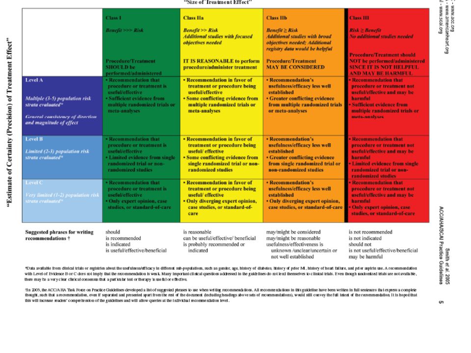

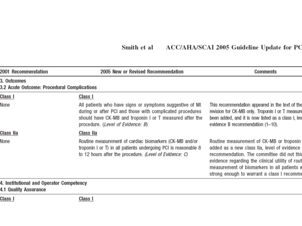
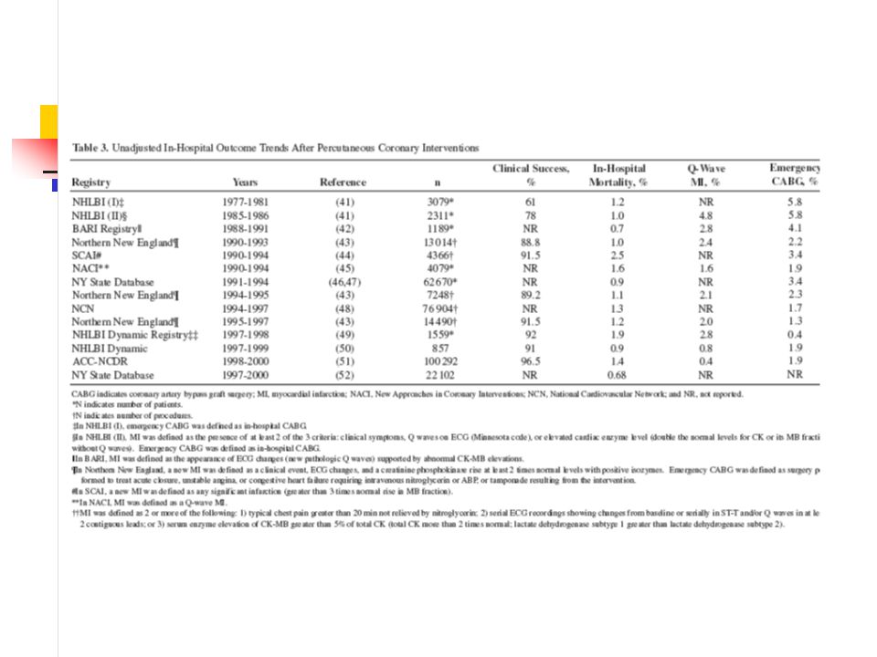
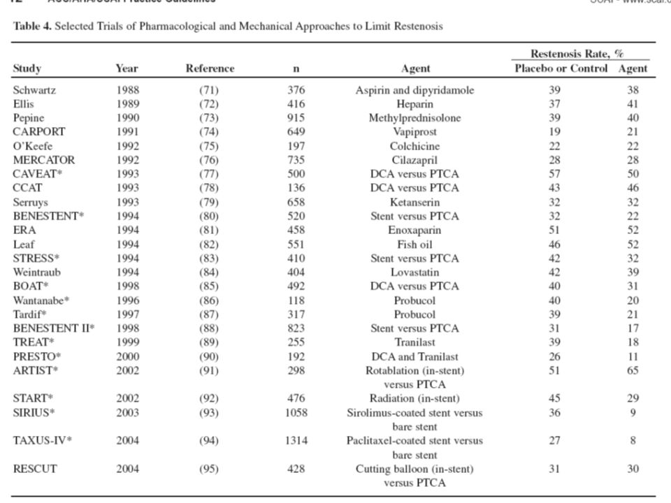


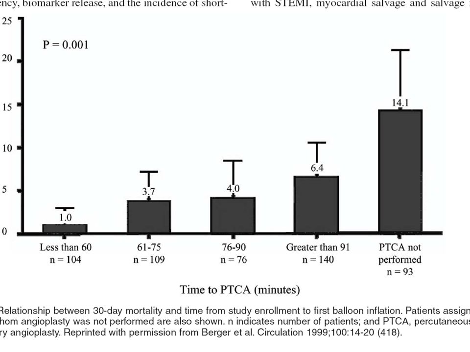
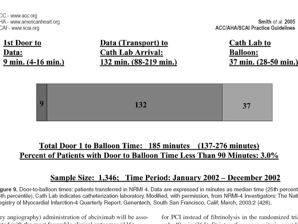
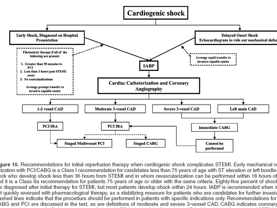
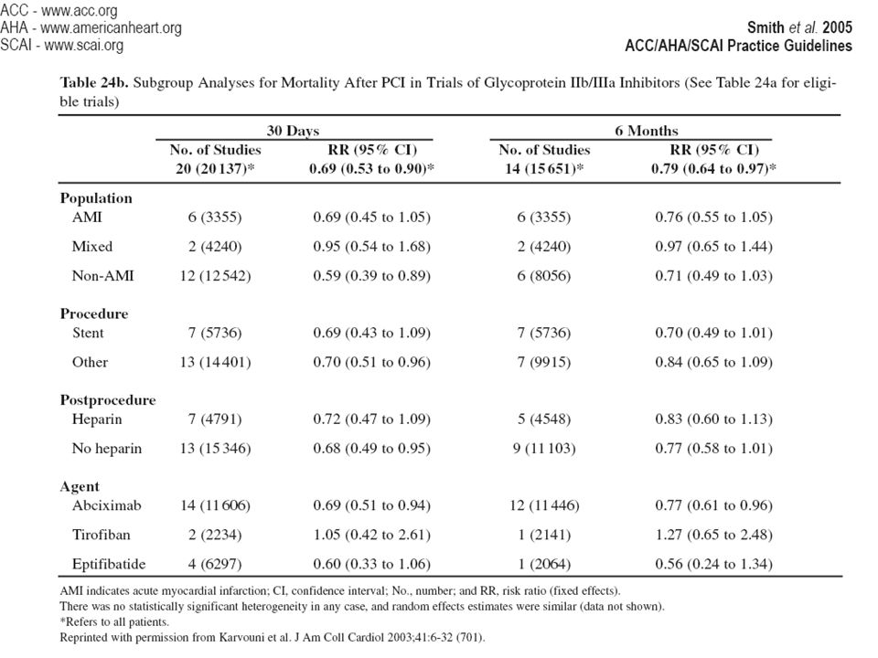
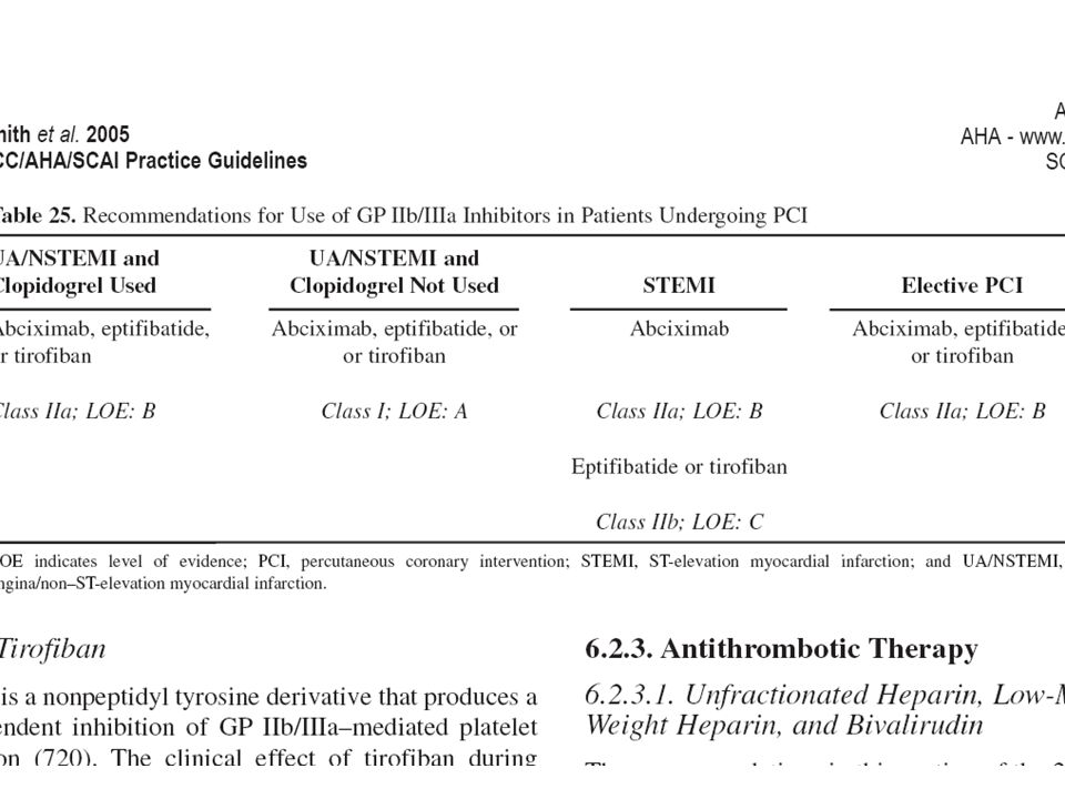

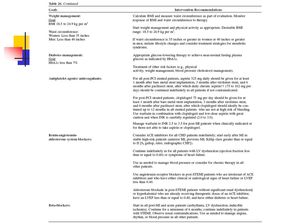


>")


