Download presentation
Presentation is loading. Please wait.

1
Co-registration and Spatial Normalisation
Martin Chadwick and Catherine Sebastian

2
(Co-registration and) Spatial
Overview Motion correction Smoothing kernel (Co-registration and) Spatial normalisation Standard template fMRI time-series Statistical Parametric Map General Linear Model Design matrix Parameter Estimates
 Spatial. normalisation. Standard. template. fMRI time-series. Statistical Parametric Map. General Linear Model. Design matrix. Parameter Estimates.")
3
Preprocessing Steps Realignment Coregistration Normalisation Smoothing
Motion correction: Adjust for movement between slices Coregistration Overlay structural and functional images: Link functional scans to anatomical scan Normalisation Warp images to fit to a standard template brain Smoothing To increase signal-to-noise ratio Extras (optional) Slice timing correction; unwarping
 Slice timing correction; unwarping.")
4
Co-registration Refers to any method for realigning images
Realignment for motion correction (last week) Aligning or overlaying images from different modalities T2* EPI image (low resolution) T1 structural MR image (high resolution)
 Aligning or overlaying images from different modalities. T2* EPI image (low resolution) T1 structural MR image (high resolution)")
5
Why Co-register structural and functional images?
Can overlay functional activations onto an individual’s own anatomy Can overlay group-level functional activations onto an average structural Gives you a better spatial image for later normalisation step, as warps derived from the higher resolution structural image can be applied to the functional image 1 and 2: Therefore gives you a more accurate picture of where activation is 3. This doesn’t always make much difference and means the pre-processing is slower, so you wouldn’t do this for every study

6
Recap: realignment parameters
Like motion correction (realignment), co-registration makes use of 6 parameters… Yaw Pitch Roll Z X Y Translation Rotation
, co-registration makes use of 6 parameters… Yaw. Pitch. Roll. Z. X. Y. Translation. Rotation.")
7
Differences between realignment and co-registration
Images may not be quite the same shape (distortion of EPI images, especially in phase encode direction) Structural and functional images do not have the same signal intensity in the same areas. Therefore there are additional steps No direct voxel to voxel match No direct voxel to voxel match
 Structural and functional images do not have the same signal intensity in the same areas. Therefore there are additional steps. No direct voxel to voxel match. No direct voxel to voxel match.")
8
The Normalised Mutual Information Approach
Different material will have different intensities within a scan modality (e.g. air will have a consistent brightness, and this will differ from other materials such as white matter). When looking between modalities, these consistencies can be used to compare images
. When looking between modalities, these consistencies can be used to compare images.")
9
An example not aligned aligned Joint histogram shows little noise
More noise: hard to define structures with certainty

10
Additional points In some studies structurals are not taken – it is possible to conduct fMRI analysis without co-registering to a structural Sometimes when you co-register, you have to reslice the data Reslicing is when you change the image dimensions (e.g. 3x3x3 to 2x2x2), which often involves interpolation as well. You can also change the apparent collection direction of the data (e.g. from axial to coronal). It's useful if you have two images that have very different voxel sizes (used to happen a lot with PET data). E.g. change image dimensions from 3x3x3 to 2x2x2, or change apparent direction of data collection from axial to coronal Useful if two images have very different voxel sizes Often involves interpolation Often used with PET data
, which often involves interpolation as well. You can also change the apparent collection direction of the data (e.g. from axial to coronal). It s useful if you have two images that have very different voxel sizes (used to happen a lot with PET data). E.g. change image dimensions from 3x3x3 to 2x2x2, or change apparent direction of data collection from axial to coronal. Useful if two images have very different voxel sizes. Often involves interpolation. Often used with PET data.")
11
Co-registration in SPM

12
Co-registration in SPM
Make selection NB Why would you reslice? What does this mean? Explains each option

13
Template: image that remains stationary
Image that is ‘jiggled about’ to match template Defaults used by SPM for estimating the match, including Normalised Mutual Information Run Reslice options: choose from the menu for each of the three options (usually just defaults)
")
14
Preprocessing Steps Realignment Coregistration Normalisation Smoothing
Motion correction: Adjust for movement between slices Coregistration Overlay structural and functional images: Link functional scans to anatomical scan Normalisation Warp images to fit to a standard template brain Smoothing To increase signal-to-noise ratio Extras (optional) Slice timing correction; unwarping
 Slice timing correction; unwarping.")
15
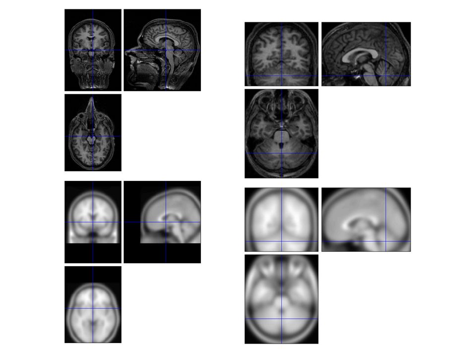
What is Normalisation? Warps images from different participants onto a template brain Matthew Brett

16
Why Normalise? We can average the signal across participants, allowing us to derive group statistics. This can allow us to: Improve the statistical power of the analysis Generalise findings to the population level Identify commonalities and differences between groups (e.g. patient vs. healthy) Report results in standard co-ordinate system (e.g. Talairach)
 Report results in standard co-ordinate system (e.g. Talairach)")
17
SPM: Spatial Normalisation
SPM uses a voxel-intensity-based approach to normalisation. adopts a two-stage procedure : Step 1: Linear transformation (12-parameter affine). This step accounts for the major differences in head shape and position, but there will be remaining smaller-scale differences. Step 2: Non-linear transformation (warping). The non-linear step is designed to take care of the smaller-scale differences in brain anatomy. Alternatives – anatomy based approaches e.g. FreeSurfer
. This step accounts for the major differences in head shape and position, but there will be remaining smaller-scale differences. Step 2: Non-linear transformation (warping). The non-linear step is designed to take care of the smaller-scale differences in brain anatomy. Alternatives – anatomy based approaches e.g. FreeSurfer.")
18
Step 1: Affine Transformation
Determines the optimum 12-parameter affine transformation to match the size and position of the images 12 parameters = 3 translations and 3 rotations (rigid-body) + 3 shears and 3 zooms Rotation Shear Translation Zoom
 + 3 shears and 3 zooms. Rotation. Shear. Translation. Zoom.")
19
Step 2: Non-linear Registration
The model for defining nonlinear warps uses deformations consisting of a linear combination of low-frequency periodic basis functions.

20
Over-fitting and Regularisation
Affine registration Template image Non-linear registration using regularisation. Non-linear registration without regularisation.

21
Caveats Impossible to make a meaningful perfect structural match between subjects, due to individual differences in anatomy Even if anatomy is well-matched, it does not guarantee that functionally homologous areas are spatially aligned – we don’t know the extent to which individuals may vary in their structure-function relationships. Pathology creates a particular problem here, as even relatively confined abnormalities or lesions can cause mis-registration in widespread areas of the brain, due to the global nature of the normalisation process. Need to bare this in mind in patient studies. Solution A partial solution for any remaining small-scale differences in anatomical or functional location is offered by the next stage of pre-processing, where the images are spatially smoothed.

22
Normalisation in SPM NB Why would you reslice? What does this mean?

23
Select Option Estimate will calculate what warps are needed to get from your selected image to the template, and will generate a file containing these images ending in “sn.mat”. Estimate and Write will do the same, and then apply these warps to your selected image, producing a new image file with the same filename, but with an additional “w” at the front (standing for “warped”). Note that you can select multiple images to apply the warps to – so that you can normalize a set of coregistered images in one step. Write can be used if you already have your specified sn.mat file from previously normalized data
. Note that you can select multiple images to apply the warps to – so that you can normalize a set of coregistered images in one step. Write can be used if you already have your specified sn.mat file from previously normalized data.")
24
Select image to be matched to template
Select image(s) to be warped using the sn.mat calculated from the Source Image Select SPM template: Structural – spm5\templates\T1.nii Functional - spm5\templates\EPI.nii Select voxel sizes for warped output images What is the best voxel size to choose here? Particularly is you are using structural images to warp your functionals.
 to be warped using the sn.mat calculated from the Source Image. Select SPM template: Structural – spm5\templates\T1.nii Functional - spm5\templates\EPI.nii. Select voxel sizes for warped output images. What is the best voxel size to choose here Particularly is you are using structural images to warp your functionals.")
26
Sources: http://www.fil.ion.ucl.ac.uk/spm/doc/intro/
Friston, K. J. Introduction: Experimental design and statistical parametric mapping Ashburner & Friston “Rigid Body Registration” Chapter 2, Human Brain Function, 2nd ed.; Ashburner & Friston “Spatial Normalization Using Basis Functions” Chapter 3, Human Brain Function, 2nd ed.; Rik Henson’s Preprocessing Slides: Previous MfD Slides

Similar presentations


>")


