Download presentation
Presentation is loading. Please wait.

1
Lupus Anticoagulants Dr.hani

2
An acquired autoimmune thrombophilia, characterized by: a) vascular thrombosis. b) recurrent pregnancy losses. c) thrombocytopenia. d) laboratory evidence for: -antibodies against phospholipids or phospholipid-binding protein cofactors.
 recurrent pregnancy losses. c) thrombocytopenia. d) laboratory evidence for: -antibodies against phospholipids or phospholipid-binding protein cofactors..")
3
Sapporo Criteria (Updated) International Consensus Statement on Classification Criteria for APS. –Clinical criteria. Vascular thrombosis. Pregnancy morbidity. –Laboratory criteria. Lupus anticoagulant. Anticardiolipin IgG or IgM antibody. Anti- 2 glycoprotein I IgG or IgM antibody.

4
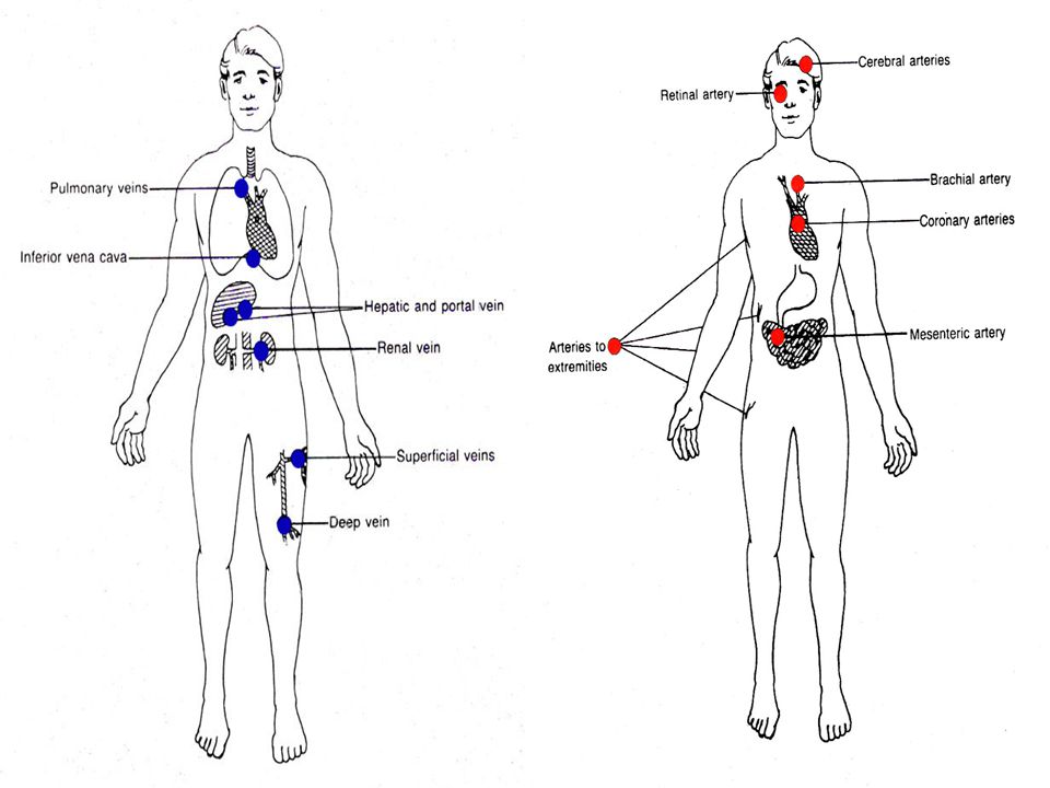
Clinical criteria for APS –Vascular thrombosis*. Venous thromboembolic disease (DVT, PE). Arterial thromboembolic disease. Small vessel thrombosis. * “Coexisting inherited or acquired thrombotic risk factors are not reasons for excluding patients from a diagnosis of APS trials.”

6
Clinical criteria for APS Pregnancy morbidity. –One or more unexplained deaths of a morphologically normal fetus at or beyond10 th week of gestation. –Three or more unexplained spontaneous abortions at or prior to 10 th week of gestation. –One or more premature births at or before the 34 th week of gestation due to eclampsia or placental insufficiency.

7
APS morbidity APS is the most common cause of acquired thrombophilia. Prevalence in general population: 2-4% 15-20% of all DVT with or without PE. 1/3 of new strokes in patients < 50 years age. 10-15% women with recurrent pregnancy losses. APS: significant proportion of thromboembolic disease and pregnancy loss in SLE. APL Abs present in 30-40% SLE. One third of those patients have clinical manifestations of APS. aCL positivity may precede a more severe form of SLE.

8
Pathogenic mechanisms of thrombosis The activation of cellular components (endothelial cells, platelets and monocytes) activation of the coagulation cascade inhibition of the fibrinolytic system inhibition of natural anticoagulant pathways activation of the complement system.
 activation of the coagulation cascade inhibition of the fibrinolytic system inhibition of natural anticoagulant pathways activation of the complement system.")
9
Multiple Strokes in a Young Woman (Brain MRI) Occlusion of Right Middle Cerebral Artery In a 3 Years Old Child with Severe Headache and Hemiparesis With aCL Antibodies +
 Occlusion of Right Middle Cerebral Artery In a 3 Years Old Child with Severe Headache and Hemiparesis With aCL Antibodies +")
10
“Non-criteria” APS findings Thrombocytopenia and/or hemolytic anemia. Transverse myelopathy or myelitis. Livido reticularis. Cardiac valve disease. Nephropathy. Non-thrombotic neurologic manifestations, including multiple sclerosis-like syndrome, chorea, or migraine headaches.

11
Laboratory criteria for APS Lupus anticoagulant: defined by a functional, –clot-based assay using the ISTH guidelines. Prolongation of a phospholipid-dependent screening assay; –Evidence of inhibitory activity; –Evidence that inhibitory activity is phospholipid-dependent; and, –Distinction from other ‘coagulopathies’… Anticardiolipin IgG or IgM antibody. Anti- 2 glycoprotein I IgG or IgM antibody. -- Measured on 2 or more occasions at least 12 weeks apart. -- The positive laboratory criteria and the clinical criteria should occur within 5 years of each other.

12
Lupus Anticoagulants Lupus anticoagulants are antibodies directed against several types of phospholipid-protein complexes. Lupus anticoagulants impair the in vitro phospholipid dependent activation of factor X, factor IX, and prothrombin. Consequently, the APTT is prolonged in the presence of a lupus anticoagulant. For this reason, most laboratories use the APTT as their primary lupus anticoagulant screen. Because they have a variety of targets.

13
Lupus Anticoagulants lupus anticoagulants are called nonspecific inhibitors. Chronic lupus anticoagulants confer a 30% risk of arterial or venous thrombosis. The presence of an LA is usually not associated with a bleeding problem unless accompanied by 1.thrombocytopenia, 2.factor II deficiency, 3.platelet dysfunction 4.drug administration (e.g., aspirin).
..")
14
criteria for the laboratory diagnosis of Lupus anticoagulants * IN order to make a diagnosis of LA; sample should show each of the following: 1.Prolongation of at least one phospholipid – dependent clotting assay. 2.Evidence of inhibitory activity shown by the effect of patient plasma on pooled normal plasma. 3.Evidence that the inhibitory activity is dependent on concentration of phospholipid. This may be achieved by addition or alteration of phospholipids/ hexagonal phase phospholipids/ platelet/ platelet vesicles in the test system. 4.LAs must be carefully distinguished from other coagulopathies that may give similar laboratory results or may occur concurrently with LAs. Specific factor assays and the clinical history may be helpful in differentiating LAs from these other possibilities.

15
Laboratory Diagnosis of Lupus Anticoagulants test: 3 group : Screening test Tests for Rule out other coagulopathies Confirmatory test

17
Laboratory Diagnosis of Lupus Anticoagulants The traditional screening methods used for the laboratory diagnosis of Lupus Anticoagulants can be broadly classified as follows: A.Immunological assays B.Clot based assays incorporating phospholipids in the reagent sysytem

18
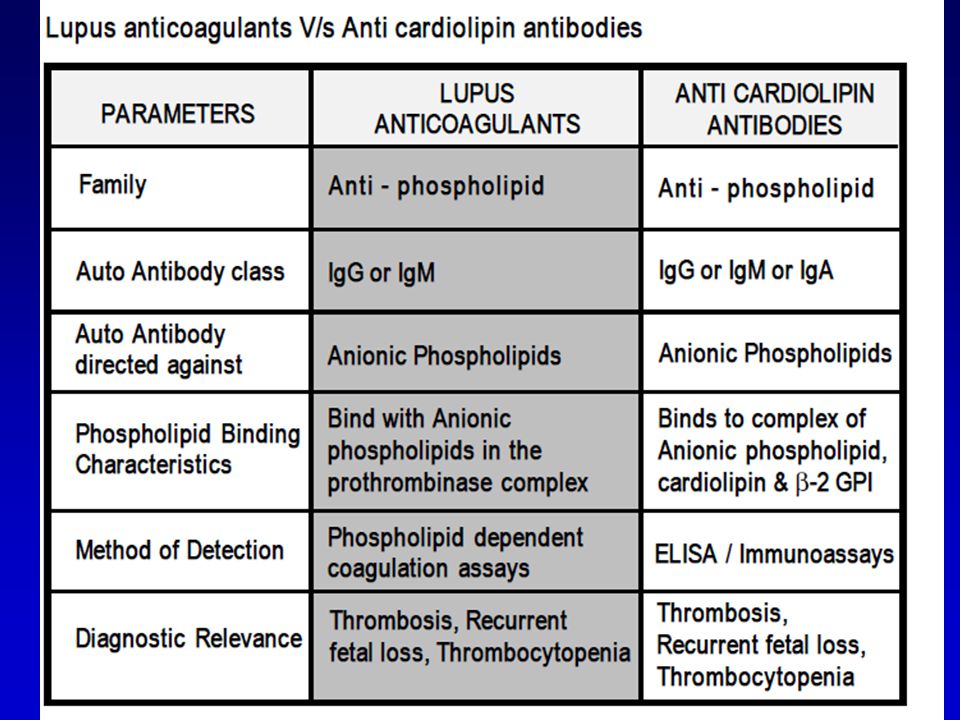
A. Immunological Assays Anti-Phospholipid Antibody AssaysPrinciple Antiphospholipid antibodies are a heterogeneous group of autoantibodies including: –ACA, LA, beta-2 glycoprotein-l (β 2 GPI), antiprothrombin, and antiphosphatidylserine (APTS). Anti-beta-2 glycoprotein I (anti-β 2 GPI) and Anticardiolipin antibodies (ACAs) are antiphospholipid immunoglobulins that are IgG, IgM, IgA, or a combination. Some patients with have an LA.Some patients with elevated anti- β 2 GPI and/or ACAs have been reported to also have an LA.
, antiprothrombin, and antiphosphatidylserine (APTS). Anti-beta-2 glycoprotein I (anti-β 2 GPI) and Anticardiolipin antibodies (ACAs) are antiphospholipid immunoglobulins that are IgG, IgM, IgA, or a combination. Some patients with have an LA.Some patients with elevated anti- β 2 GPI and/or ACAs have been reported to also have an LA..")
20
A. Immunological Assays Individuals with Lupus anticoagulants may also show the presence of other anti phospholipid antibodies(combination). The most frequent finding is the presence of Anti- cardiolipin antibodies. The commonly employed method is the ELISA technique where the solid phase is coated with cardiolipin and β-2 GPI (as a cofactor). The ELISA method detects IgM, IgG and IgA class of anti-cardiolipin antibodies. An important point to note is that Lupus anticoagulants and anti-cardiolipin antibodies differ in their phospholipid-binding characteristic hence detection of anti cardiolipin antibodies is not specific for the presence of Lupus anticoagulants though they may be present in the same patient.
. The most frequent finding is the presence of Anti- cardiolipin antibodies. The commonly employed method is the ELISA technique where the solid phase is coated with cardiolipin and β-2 GPI (as a cofactor). The ELISA method detects IgM, IgG and IgA class of anti-cardiolipin antibodies. An important point to note is that Lupus anticoagulants and anti-cardiolipin antibodies differ in their phospholipid-binding characteristic hence detection of anti cardiolipin antibodies is not specific for the presence of Lupus anticoagulants though they may be present in the same patient..")
21
B. Clot based assays 1.APTT ( Activated Partial Thromboplastin Time) 2.TTI (Tissue Thromboplastin Inhibition test) 3.KCT (Kaolin Clotting Time) 4.PNP (Platelet Neutralization procedure) 5.Hexagonal Phase Phospholipids (Staclot-LA) 6.dilute Russell's viper venom time (dRVVT)
 2.TTI (Tissue Thromboplastin Inhibition test) 3.KCT (Kaolin Clotting Time) 4.PNP (Platelet Neutralization procedure) 5.Hexagonal Phase Phospholipids (Staclot-LA) 6.dilute Russell s viper venom time (dRVVT).")
22
APTT ( Activated Partial Thromboplastin Time) substances are: kaolin (hydrated aluminum silicate) and cephalin. Kaolin serves to activate the contact-dependent Factor XII, and cephalin substitutes for platelet phospholipids.

23
APTT ( Activated Partial Thromboplastin Time) Since Lupus anticoagulants bind to the phospholipid complex they do prolong phospholipid based coagulation assays. Logically the activated partial thromboplastin time (APTT) is prolonged and this property has been used for the detection of LA using APTT reagents. In the context of LA detection, the APTT test has certain shortcomings: An important variable related to the suitability of APTT reagents in the detection of Lupus anticoagulants is the composition of phospholipids used in the reagent system. Different reagents have varying sensitivity for the presence of Lupus anticoagulants on the basis of their phospholipid composition.
 is prolonged and this property has been used for the detection of LA using APTT reagents. In the context of LA detection, the APTT test has certain shortcomings: An important variable related to the suitability of APTT reagents in the detection of Lupus anticoagulants is the composition of phospholipids used in the reagent system. Different reagents have varying sensitivity for the presence of Lupus anticoagulants on the basis of their phospholipid composition..")
24
APTT ( Activated Partial Thromboplastin Time) The APTT test is affected by the presence of inhibitors to Factor VIII, IX and XI. In order to rule out the presence of inhibitors usually mixing studies are performed. In mixing studies a 1:1 or 1:4 ( Normal plasma: Patient plasma) is used. Failure to correct the prolongation of clotting time using the mixing studies indicates the presence of Lupus anticoagulants. The APTT test is also the test of choice for monitoring heparin therapy. This reduces the specificity of the APTT to LA. The presence of heparin can be ruled out by using a thrombin time test. If thrombin time test shows normal values, then the sample does not contain heparin. But if the thrombin time test is abnormal, then heparin neutralization test is used which includes protamine sulphate. In this test, various concentrations of protamine sulphate are added to plasma before the addition of thrombin reagent. When protamine sulphate neutralizes all the heparin present, the clotting time reverts to normal value.
 is used. Failure to correct the prolongation of clotting time using the mixing studies indicates the presence of Lupus anticoagulants. The APTT test is also the test of choice for monitoring heparin therapy. This reduces the specificity of the APTT to LA. The presence of heparin can be ruled out by using a thrombin time test. If thrombin time test shows normal values, then the sample does not contain heparin. But if the thrombin time test is abnormal, then heparin neutralization test is used which includes protamine sulphate. In this test, various concentrations of protamine sulphate are added to plasma before the addition of thrombin reagent. When protamine sulphate neutralizes all the heparin present, the clotting time reverts to normal value..")
25
TTI (Tissue Thromboplastin Inhibition test) The Tissue Thromboplastin Inhibition test utilizes a diluted PT reagent. thromboplastin [Tissue Factor + Phospholipid] is diluted 1:50 and 1:500. The diluted thromboplastin is then incubated 1:1 with normal or test plasma at 37°C after which calcium chloride is added and the time to clot formation recorded. The ratio of the clotting time with the dilute thromboplastin [1:500] divided by the normal reagent[1:50] is compared to a normal control. A PT ratio ≥1.3 suggests the presence of a LA whilst a ratio ≤1.1 is considered normal

26
TTI (Tissue Thromboplastin Inhibition test) The TTI test is affected by numerous variables: 1.Species and tissue origin of thromboplastin can affect the test results as different sources of thromboplastin have varying sensitivity and responsiveness. 2.Choice of “Normal” reference plasma is the most critical variable, because depending on the laboratory the choice of reference plasma could be lyophilized plasma, a frozen plasma pool or fresh plasma. The ratio of patient to normal can therefore change according to the choice of “normal” plasma. 3.Some IgM Lupus anticoagulants do not prolong the TTI test

27
KCT (Kaolin Clotting Time) KCT is similar to APTT, the difference being that KCT reagent is devoid of phospholipids as contact activator. most sensitive test for the detection of circulating anticoagulants. The KCT detects all class of inhibitors The test is performed on a range of mixture of normal and patient’s plasma. Different patterns of response are obtained indicating the presence of Lupus anticoagulants or the deficiency of one or more coagulation factors.

28
KCT (Kaolin Clotting Time) The KCT test though sensitive is not specific for LA, additionally: It cannot be automized The test shows prolonged results with factor VIII, IX, XI & XII deficiency or corresponding inhibitors The test is also highly sensitive for the presence of heparin.
 The KCT test though sensitive is not specific for LA, additionally: It cannot be automized The test shows prolonged results with factor VIII, IX, XI & XII deficiency or corresponding inhibitors The test is also highly sensitive for the presence of heparin.")
29
KCT (Kaolin Clotting Time) Method Normal PlasmaPatient Plasma 100%0% 90%10% 80%20% 50% 20%80% 10%90% caolin is added and then calcium to initiate coagulation. The KCT is the time from adding calcium to clot formation. Kaolin suspension is turbid.

30
KCT (Kaolin Clotting Time) Method Normal PlasmaPatient Plasma 100%0% 90%10% 80%20% 50% 20%80% 10%90% initial steep slope of the KCT is very important and allows the test to be simplified and used as a screening test. A ratio of ≥1.2 is considered positive for a lupus anticoagulant

31
KCT (Kaolin Clotting Time) Method(mixing study) sensitivity of the test could be improved [whilst maintaining the specificity] by using a 4:1 mix of test plasma to control plasma [this avoids 'swamping' a weak LA with normal plasma] and appears to improve both specificity and sensitivity: The Chang index is converted into a percentage correction by multiplying the final result by 100. 1. In the original test using a 1:1 [50:50] mix of test plasma and control plasma, a positive result [i.e. lupus anticoagulant present] was a percentage correction >70% and a borderline result between as between 58-70%. 2. In the modified test using a 4:1 mix of test plasma and control plasma, a test was considered positive if the percentage correction was >50%.
![KCT (Kaolin Clotting Time) Method(mixing study) sensitivity of the test could be improved [whilst maintaining the specificity] by using a 4:1 mix of test plasma to control plasma [this avoids swamping a weak LA with normal plasma] and appears to improve both specificity and sensitivity: The Chang index is converted into a percentage correction by multiplying the final result by 100.](http://images.slideplayer.com/18/6082711/slides/slide_31.jpg "1. In the original test using a 1:1 [50:50] mix of test plasma and control plasma, a positive result [i.e. lupus anticoagulant present] was a percentage correction >70% and a borderline result between as between 58-70%. 2. In the modified test using a 4:1 mix of test plasma and control plasma, a test was considered positive if the percentage correction was >50%..")
32
Lupus anticoaagulant present i.e. a positive result. The KCT of normal plasma is the 0% dilution on the X axis i.e. no patient plasma is present. This should be in excess of 60s otherwise the normal plasma contains too high a concentration of PL and looses sensitivity. Remember in the KCT the only source of PL in the test

33
Lupus anticoagulant present + a clotting factor deficiency

34
Lupus anticoagulant present but deficient in β2- GPI i.e. an anti-β2-GPI antibody is present

35
No lupus anticoagulant present i.e. a negative result

36
4. PNP (Platelet Neutralization procedure) Principle The platelet neutralization procedure (PNP) is based on the ability of platelets to significantly correct in vitro coagulation abnormalities. The disrupted platelet membranes present in the freeze- thawed platelet suspension neutralize phospholipid antibodies present in the plasma of patients with LA. After the patient plasma is mixed with the freeze-thawed platelet suspension, the APTT will be shortened when compared with the original baseline APTT. But if an inhibitor is directed against specific coagulation factor, the clotting time is not shortened.
 Principle The platelet neutralization procedure (PNP) is based on the ability of platelets to significantly correct in vitro coagulation abnormalities. The disrupted platelet membranes present in the freeze- thawed platelet suspension neutralize phospholipid antibodies present in the plasma of patients with LA. After the patient plasma is mixed with the freeze-thawed platelet suspension, the APTT will be shortened when compared with the original baseline APTT. But if an inhibitor is directed against specific coagulation factor, the clotting time is not shortened..")
37
4. PNP (Platelet Neutralization procedure) Interpretation A correction of the baseline APTT of a defined amount of time (i.e., 3 to 5 seconds or more) by the platelet suspension as compared with the control is indicative of the presence of an LA.NOTE The PNP test though useful did not gain wide usage : Due to limited stability the platelet preparations loose their activities on storage hence do not show reproducible results. They cannot differentiate between Lupus anticoagulants and Factor VIII inhibitors.
 Interpretation A correction of the baseline APTT of a defined amount of time (i.e., 3 to 5 seconds or more) by the platelet suspension as compared with the control is indicative of the presence of an LA.NOTE The PNP test though useful did not gain wide usage : Due to limited stability the platelet preparations loose their activities on storage hence do not show reproducible results. They cannot differentiate between Lupus anticoagulants and Factor VIII inhibitors..")
38
Hexagonal Phospholipid Neutralization Assay Principle The hexagonal phospholipid neutralization assay uses the same principle as the PNP assay, normalization of the aPTT in the presence of added phospholipid, but this assay specifically uses a phospholipid in a hexagonal conformation, Neutralization by this hexagonal form in an assay with a very lupus-anticoagulant sensitive aPTT reagent, is a more sensitive confirmation test than the PNP.

39
Confirmatory Tests for Lupus Anticoagulants Confirmatory tests to identify an LA include 1.those that utilize a low concentration of phospholipid in the test system, thereby increasing the LA effect such as –the tissue thromboplastin inhibition test (TTIT), –dilute Russell's viper venom time (dRVVT), –and the kaolin clotting time (KCT) 2.those that increase the phospholipid, thereby neutralizing the LA effect, such as the platelet neutralization
, –dilute Russell s viper venom time (dRVVT), –and the kaolin clotting time (KCT) 2.those that increase the phospholipid, thereby neutralizing the LA effect, such as the platelet neutralization")
40
D R V V T D ilute R ussell’s V iper V enom T ime (DRVVT) dRVVT : The test of choice for screening and confirmation of LA –Indicating the phospholipid dependence of LA –Achieving maximum sensitivty for the precence of LA’s. In general dRVVT based tests comprise of:In general dRVVT based tests comprise of: –SCREENING REAGENT, containing limited amount of phospholipids with RVV (Russell’s Viper Venom) –CONFIRMATION REAGENT, containing additional phospholipids with same amount of Russell’s Viper Venom, to confirm the presence of phospholipid dependent Lupus anticoagulants.
 –CONFIRMATION REAGENT, containing additional phospholipids with same amount of Russell’s Viper Venom, to confirm the presence of phospholipid dependent Lupus anticoagulants..")
41
Principle of dRVVT for LA detection Russell’s Viper Venom directly activates Factor V and X in presence of phospholipid and calcium ions, bypassing Factor VII of the extrinsic pathway and the contact and antihaemophilic factors of the intrinsic pathway. In normal plasma, in the absence of lupus anticoagulants, Factor V and X is directly activated by Russell’s Viper Venom, which in presence of phospholipid and calcium ion leads to clot formation.

42
Principle of dRVVT for LA detection In patients with LA, autoantibodies bind the epitopes of reagent phospholipids thereby preventing the activation of prothrombinase complex. This results in a prolongation of clotting time with SCREENING reagent. The CONFIRMATION reagent incorporates additional phospholipids to neutralize the LA. Once LA are neutralized clot formation proceeds relatively uninterrupted achieving a lower clotting time, to prove the phospholipid dependence of the autoantibodies.

43
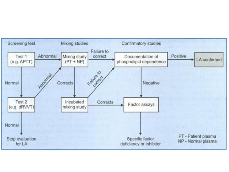
Interpretation of results with dRVVT test If SCREEN TIME is prolonged, to confirm the presence of lupus anticoagulants the plasma sample is tested with CONFIRMATION REAGENT. If CONFIRM TIME results shows a lower clotting time as compared to SCREEN TIME, it indicates the presence of phospholipid dependant Lupus Anticoagulants. Also the results can be expressed as ratio, The results expressed, as ratio is further useful in classifying the patient as normal, moderate, high and very high LA. If results of the ratio are borderline, mixing studies may be done further with the sample specimen. The mixing studies should be carried out with a 50:50 mixture of test plasma and normal plasma.

44
An algorithm for identification of APS

45
aia-360 immunoassay analyzer Eni kuestion ??

Similar presentations


 Had RE photophobia and pain month back Similar.>")


 is produced by a shift in the balance between.>")
 Myakis et al. J Thromb Haemost 2006;4:295-306 Vascular thrombosis one or more clinical.>")


