Download presentation
Presentation is loading. Please wait.

1
Course Introduction: The Brain, chemistry, neural signaling Jerome Feldman CS182/Ling109/CogSci110 Spring 2007 feldman@icsi.berkeley.edu

2
Learning I hear and I forget I see and I remember I do and I understand attributed to Confucius 551-479 B.C. There is no erasing in the brain

3
Instructor Access Instructor : Jerry Feldman Office Hours : Monday, Thursday 1 – 2 Soda 739 Email: jfeldman@cs.berkeley.edufeldman@cs.berkeley.edu TA: Leon Barrett Office Hours :TBA, Soda 739 Email: barrett@icsi.berkeley.edubarrett@icsi.berkeley.edu

4
The Neural Theory of Language and Thought This is a course on the current status of interdisciplinary studies that seek to answer the following questions: How is it possible for the human brain, which is a highly structured network of neurons, to think and to learn, use, and understand language? How are language and thought related to perception, motor control, and our other neural systems, including social cognition? How do the computational properties of neural systems and the specific neural structures of the human brain shape the nature of thought and language? What are the applications of neural computing?

5
Tinbergen’s Four Questions How does it work? How does it improve fitness? How does it develop and adapt? How did it evolve?

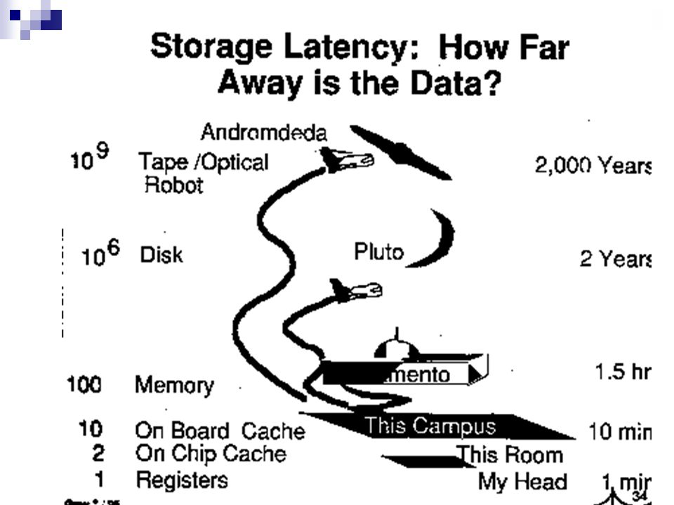
8
Brains ~ Computers 1000 operations/sec 100,000,000,000 units 10,000 connections/ graded, stochastic embodied fault tolerant evolves learns 1,000,000,000 ops/sec 1-100 processors ~ 4 connections binary, deterministic abstract crashes designed programmed

9
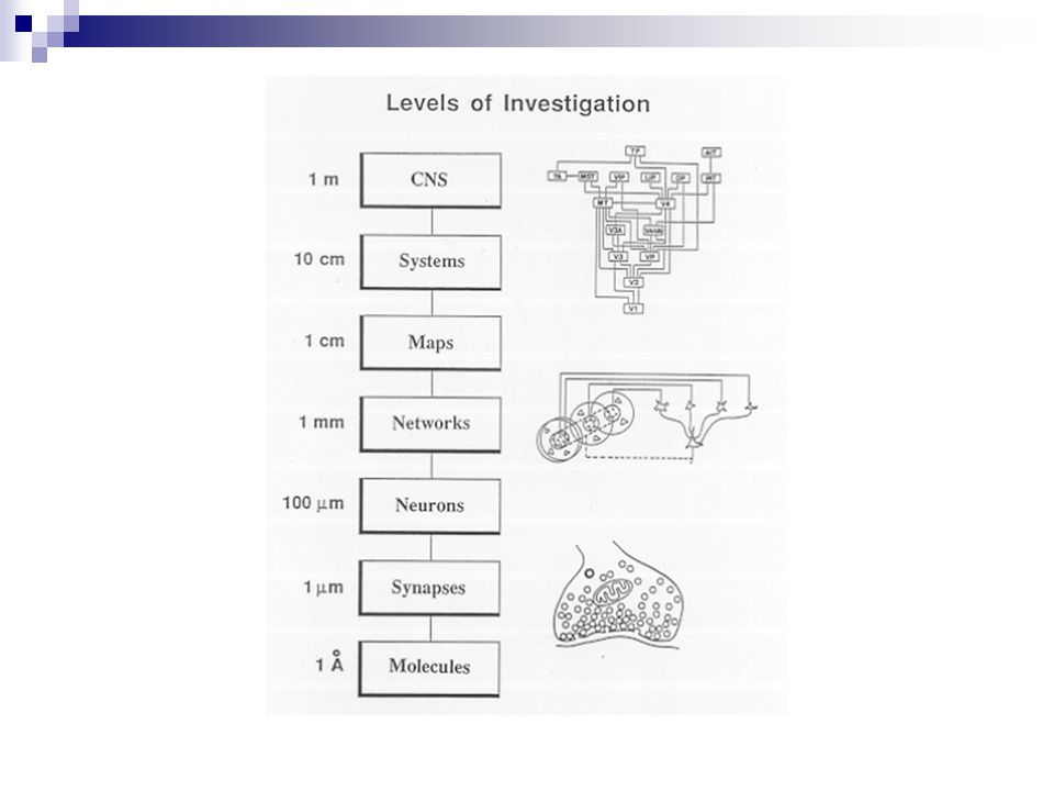
How does it all work?

12
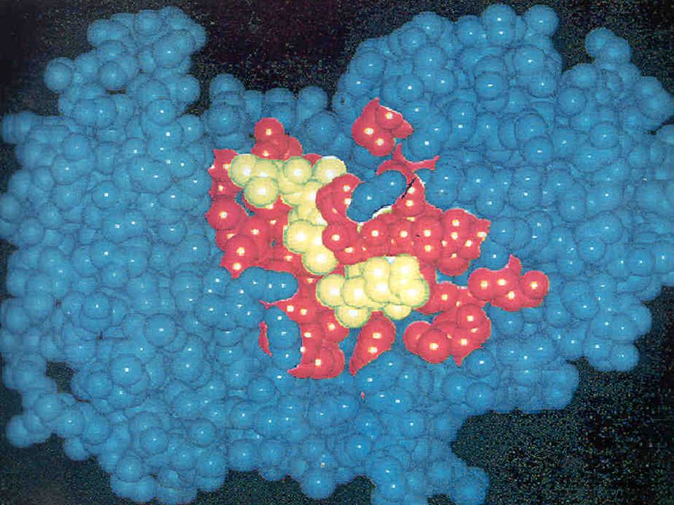
Artist’s rendition of the Actin molecule structure

13
Single Cell (Protozoan) Behaviors No Nervous System Foraging Behavior (swim toward food) Positive chemotaxis Defensive/Avodiance Behavior Negative chemotaxis Reproduction Asexual and Sexual reproduction using chemical messenger proteins (pheromones)
 Behaviors No Nervous System Foraging Behavior (swim toward food) Positive chemotaxis Defensive/Avodiance Behavior Negative chemotaxis Reproduction Asexual and Sexual reproduction using chemical messenger proteins (pheromones)")
15
Artist’s rendition of a typical cell membrane

16
Earliest Nervous Systems Hydra, jellyfish, corals, sea anemones Basic neural cell (Neuron) Early differentiation into 3 types of neurons STIMULUS Sensory Neuron Inter- Neuron Motor Neuron Effector
 Early differentiation into 3 types of neurons STIMULUS Sensory Neuron Inter- Neuron Motor Neuron Effector")
17
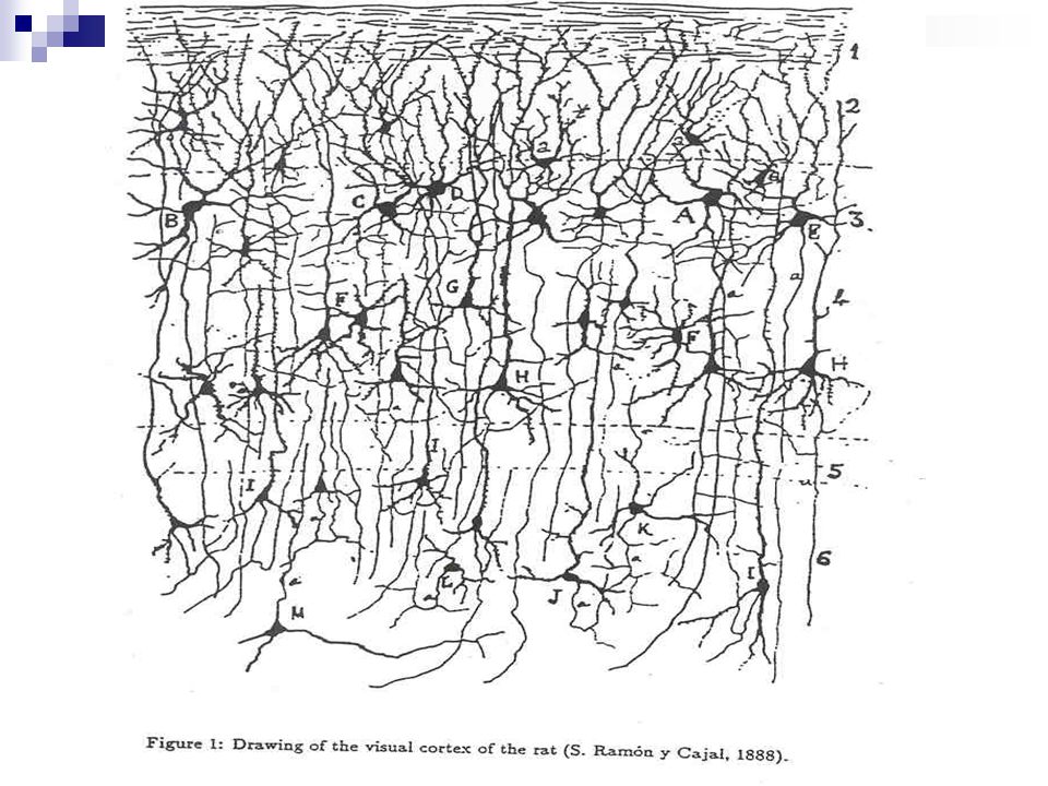
Neural Processing

18
Neurons cell body – metabolism, protein synthesis dendrites (input structure) receive inputs from other neurons perform spatio-temporal integration of inputs relay information to the cell body axon (output structure) a branching fiber that carries the message (spikes) from the cell to other neurons
 receive inputs from other neurons perform spatio-temporal integration of inputs relay information to the cell body axon (output structure) a branching fiber that carries the message (spikes) from the cell to other neurons")
19
postsynaptic neuron science-education.nih.gov

20
Synapse site of communication between two cells formed when an axon of a presynaptic cell “connects” with the dendrites of a postsynaptic cell

21
Synapse axon of presynaptic neuron dendrite of postsynaptic neuron bipolar.about.com/library

22
Synapse a synapse can be excitatory or inhibitory arrival of activity at an excitatory synapse depolarizes the local membrane potential of the postsynaptic cell and makes the cell more prone to firing – usually connects on dendrite arrival of activity at an inhibitory synapse hyperpolarizes the local membrane potential of the postsynaptic cell and makes it less prone to firing – usually connects on cell body the greater the synaptic strength, the greater the depolarization or hyperpolarization

24
UNIPOLARBIPOLAR MULTIPOLAR CELLS

25
Broca’s area Pars opercularis Motor cortexSomatosensory cortex Sensory associative cortex Primary Auditory cortex Wernicke’s area Visual associative cortex Visual cortex

26
PET scan of blood flow for 4 word tasks

27
Somatotopy of Action Observation Foot Action Hand Action Mouth Action Buccino et al. Eur J Neurosci 2001

28
Neural Communication: 1 Communication within the cell

29
Transmission of information Information must be transmitted within each neuron and between neurons

30
The Membrane The membrane surrounds the neuron. It is composed of lipid and protein.

31
Artist’s rendition of a typical cell membrane

32
The Resting Potential There is an electrical charge across the membrane. This is the membrane potential. The resting potential (when the cell is not firing) is a 70mV difference between the inside and the outside. inside outside Resting potential of neuron = -70mV + - + - + - + - + -
 is a 70mV difference between the inside and the outside. inside outside Resting potential of neuron = -70mV")
33
Ions and the Resting Potential Ions are electrically-charged molecules e.g. sodium (Na+), potassium (K+), chloride (Cl-). The resting potential exists because ions are concentrated on different sides of the membrane. Na + and Cl - outside the cell. K + and organic anions inside the cell. inside outside Na + Cl - Na + K+K+ Cl - K+K+ Organic anions (-) Na + Organic anions (-)
, potassium (K+), chloride (Cl-). The resting potential exists because ions are concentrated on different sides of the membrane. Na + and Cl - outside the cell. K + and organic anions inside the cell. inside outside Na + Cl - Na + K+K+ Cl - K+K+ Organic anions (-) Na + Organic anions (-).")
34
Ions and the Resting Potential Ions are electrically-charged molecules e.g. sodium (Na+), potassium (K+), chloride (Cl-). The resting potential exists because ions are concentrated on different sides of the membrane. Na + and Cl - outside the cell. K + and organic anions inside the cell. inside outside Na + Cl - Na + K+K+ Cl - K+K+ Organic anions (-) Na + Organic anions (-)
, potassium (K+), chloride (Cl-). The resting potential exists because ions are concentrated on different sides of the membrane. Na + and Cl - outside the cell. K + and organic anions inside the cell. inside outside Na + Cl - Na + K+K+ Cl - K+K+ Organic anions (-) Na + Organic anions (-).")
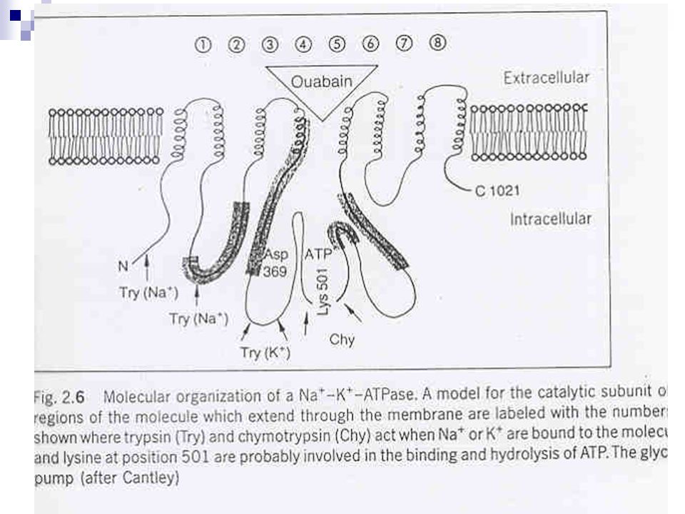
35
Maintaining the Resting Potential Na+ ions are actively transported (this uses energy) to maintain the resting potential. The sodium-potassium pump (a membrane protein) exchanges three Na + ions for two K + ions. inside outside Na + K+ Na +
 exchanges three Na + ions for two K + ions. inside outside Na + K+ Na +.")
37
Integration of information PSPs are small. An individual EPSP will not produce enough depolarization to trigger an action potential. IPSPs will counteract the effect of EPSPs at the same neuron. Summation means the effect of many coincident IPSPs and EPSPs at one neuron. If there is sufficient depolarization at the axon hillock, an action potential will be triggered. axon hillock

38
Neuronal firing: the action potential The action potential is a rapid depolarization of the membrane. It starts at the axon hillock and passes quickly along the axon. The membrane is quickly repolarized to allow subsequent firing.

39
Before Depolarization

40
Action potentials: Rapid depolarization When partial depolarization reaches the activation threshold, voltage-gated sodium ion channels open. Sodium ions rush in. The membrane potential changes from -70mV to +40mV. Na + - + + -

41
Depolarization

43
Action potentials: Repolarization Sodium ion channels close and become refractory. Depolarization triggers opening of voltage-gated potassium ion channels. K+ ions rush out of the cell, repolarizing and then hyperpolarizing the membrane. K+K+ K+K+ K+K+ Na + + -

44
Repolarization

45
Action potentials: Resuming the Resting Potential Potassium channels close. Repolarization resets sodium ion channels. Ions diffuse away from the area. Sodium-potassium transporter maintains polarization. The membrane is now ready to “fire” again. K+K+ K+K+ K+K+ K+K+ Na + K+ Na+ K+

46
The Action Potential The action potential is “all-or-none”. It is always the same size. Either it is not triggered at all - e.g. too little depolarization, or the membrane is “refractory”; Or it is triggered completely.

48
Conduction of the action potential. Passive conduction will ensure that adjacent membrane depolarizes, so the action potential “travels” down the axon. But transmission by continuous action potentials is relatively slow and energy-consuming (Na + /K + pump). A faster, more efficient mechanism has evolved: saltatory conduction. Myelination provides saltatory conduction.
. A faster, more efficient mechanism has evolved: saltatory conduction. Myelination provides saltatory conduction..")
49
Action Potential

50
Myelination Most mammalian axons are myelinated. The myelin sheath is provided by oligodendrocytes and Schwann cells. Myelin is insulating, preventing passage of ions over the membrane.

51
Saltatory Conduction Myelinated regions of axon are electrically insulated. Electrical charge moves along the axon rather than across the membrane. Action potentials occur only at unmyelinated regions: nodes of Ranvier. Node of RanvierMyelin sheath

52
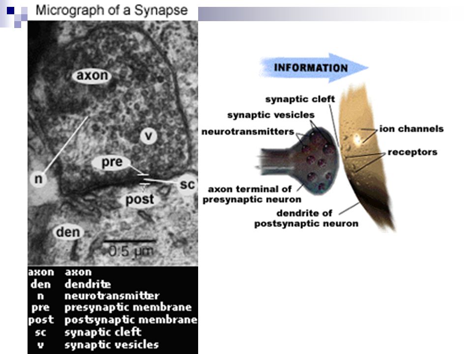
postsynaptic neuron science-education.nih.gov

53
Synaptic transmission Information is transmitted from the presynaptic neuron to the postsynaptic cell. Chemical neurotransmitters cross the synapse, from the terminal to the dendrite or soma. The synapse is very narrow, so transmission is fast.

54
terminal dendritic spine synaptic cleft presynaptic membrane postsynaptic membrane extracellular fluid Structure of the synapse An action potential causes neurotransmitter release from the presynaptic membrane. Neurotransmitters diffuse across the synaptic cleft. They bind to receptors within the postsynaptic membrane, altering the membrane potential.

55
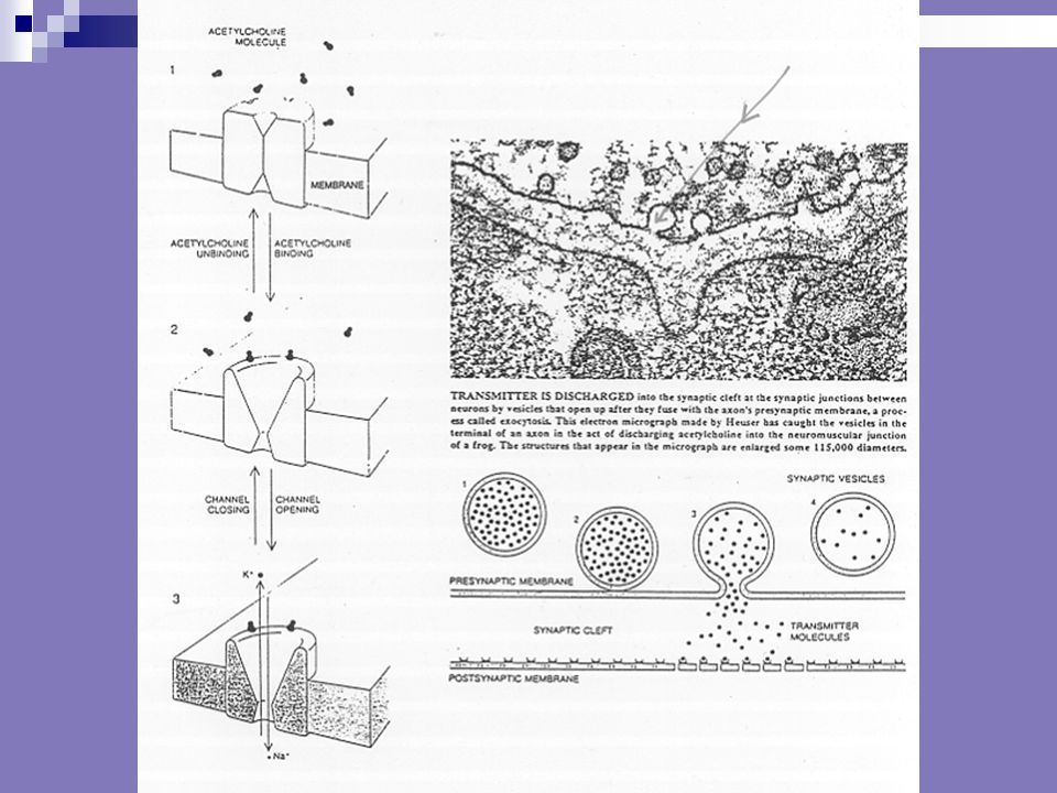
Neurotransmitter release Synaptic vesicles, containing neurotransmitter, congregate at the presynaptic membrane. The action potential causes voltage-gated calcium (Ca 2+ ) channels to open; Ca 2+ ions flood in. vesicles Ca 2+
 channels to open; Ca 2+ ions flood in. vesicles Ca 2+.")
56
Neurotransmitter release Ca 2+ causes vesicle membrane to fuse with presynaptic membrane. Vesicle contents empty into cleft: exocytosis. Neurotransmitter diffuses across synaptic cleft. Ca 2+

59
Ionotropic receptors (ligand gated) Synaptic activity at ionotropic receptors is fast and brief (milliseconds). Acetylcholine (Ach) works in this way at nicotinic receptors. Neurotransmitter binding changes the receptor’s shape to open an ion channel directly. ACh
 works in this way at nicotinic receptors. Neurotransmitter binding changes the receptor’s shape to open an ion channel directly. ACh.")
60
Ionotropic Receptors

62
Metabotropic Receptors (G-Protein)
")
63
Postsynaptic potentials Depending on the type of ion channel which opens, the postsynaptic cell membrane becomes either depolarized or hyperpolarized. Ions will tend to follow the concentration gradient from high to low concentration, and the electrostatic gradient towards the opposite charge.

64
Excitatory postsynaptic potentials (EPSPs) Opening of ion channels which leads to depolarization makes an action potential more likely, hence “excitatory PSPs”: EPSPs. Inside of post-synaptic cell becomes less negative. Na + channels (NB remember the action potential) Ca 2+. (Also activates structural intracellular changes -> learning.) inside outside Na + Ca 2+ + -
 Ca 2+. (Also activates structural intracellular changes -> learning.) inside outside Na + Ca")
65
Inhibitory postsynaptic potentials (IPSPs) Opening of ion channels which leads to hyperpolarization makes an action potential less likely, hence “inhibitory PSPs”: IPSPs. Inside of post-synaptic cell becomes more negative. K + (NB remember termination of the action potential) Cl - (if already depolarized) K+K+ Cl - + - inside outside
 Cl - (if already depolarized) K+K+ Cl inside outside.")
66
Postsynaptic Ion motion

67
Requirements at the synapse For the synapse to work properly, six basic events need to happen: Production of the Neurotransmitters Synaptic vesicles (SV) Storage of Neurotransmitters SV Release of Neurotransmitters Binding of Neurotransmitters Lock and key Generation of a New Action Potential Removal of Neurotransmitters from the Synapse reuptake
 Storage of Neurotransmitters SV Release of Neurotransmitters Binding of Neurotransmitters Lock and key Generation of a New Action Potential Removal of Neurotransmitters from the Synapse reuptake")
68
Three Nobel Prize Winners on Synaptic Transmission Arvid Carlsson discovered dopamine is a neurotransmitter. Carlsson also found lack of dopamine in the brain of Parkinson patients. Paul Greengard studied in detail how neurotransmitters carry out their work in the neurons. Dopamine activated a certain protein (DARPP-32), which could change the function of many other proteins. Eric Kandel proved that learning and memory processes involve a change of form and function of the synapse, increasing its efficiency. This research was on a certain kind of snail, the Sea Slug (Aplysia). With its relatively low number of 20,000 neurons, this snail is suitable for neuron research.
, which could change the function of many other proteins. Eric Kandel proved that learning and memory processes involve a change of form and function of the synapse, increasing its efficiency. This research was on a certain kind of snail, the Sea Slug (Aplysia). With its relatively low number of 20,000 neurons, this snail is suitable for neuron research..")
69
Fast Brain ~ Slow Neurons Mental Connections are Active Neural Connections There is No Erasing in the Brain

70
How does it all work?

Similar presentations


 Nervous system functions Structure of a neuron Sensory, motor, inter- neurons Membrane potential Sodium.>")


