Download presentation
Presentation is loading. Please wait.

1
http://www.brain-map.org

2
A Big Thanks Prof. Jason Bohland Quantitative Neuroscience Laboratory Boston University Dr. Luis Ibanez Open Source Proponent, ITK Kitware Inc.

4
Supplemental Material

5
Genome-wide atlas of gene expression throughout the mouse brain (N=1,2 or a few mice/gene) 56 day-old (young adult) C57BL/6J mice High-throughput experiments using in situ hybridization Pipeline - sectioning, ISH, digital microscopy, image analysis, atlas registration Allen Mouse Brain Atlas
 56 day-old (young adult) C57BL/6J mice High-throughput experiments using in situ hybridization Pipeline - sectioning, ISH, digital microscopy, image analysis, atlas registration Allen Mouse Brain Atlas")
6
The Process Construction and representation of the Anatomic Gene Expression Atlas (AGEA).
.")
7
Nissl-Stained Atlas – Ground Truth (a) Level 53 coronal plate (bregma 0.145 mm) from the The Allen Reference Atlas (ARA) delineating 2D anatomic boundaries of a Nissl-stained mouse brain section.
 Level 53 coronal plate (bregma mm) from the The Allen Reference Atlas (ARA) delineating 2D anatomic boundaries of a Nissl-stained mouse brain section.")
9
Bregma – Neurological Context http://en.wikipedia.org/wiki/Bregma bregma located at the intersection of the coronal and sagittal sutures. Level 53 coronal plate (bregma 0.145 mm)
.")
11
Image

12
Nissl http://en.wikipedia.org/wiki/File:NisslHippo2.jpg Nissl-stained histological section through the rodent hippocampus showing various classes of cells (neurons and glia). Motor nerve cell from ventral horn of medulla spinalis of rabbit. The angular ande spindle- shaped Nissl bodies are well shown Nissl stains the cell body esp. endoplasmic reticulum. Basic dyes (e.g. aniline, thionine, or cresyl violet) to stain negatively charged RNA blue, Nissl substance (rough endoplasmic reticulum) appears dark blue from ribosomal RNA DNA stains a similar color
 to stain negatively charged RNA blue, Nissl substance (rough endoplasmic reticulum) appears dark blue from ribosomal RNA DNA stains a similar color.")
13
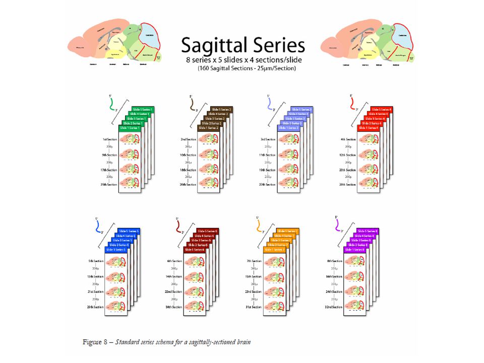
Atlas Assembly (b) 3D assembly of high-level ARA structures formed by 3D reconstruction of the Nissl sections. The 3D ARA space is partitioned into 200-mm^3 voxels forming the smallest spatial unit for analysis. New annotated anatomical reference atlas (Hong-Wei Dong, 2007) 528 coronal Nissl sections: unfixed, frozen mouse brain (25μm thick) 132 sections, with 100μm spacing, annotated over1000 brain All image data are mapped to common coordinate space Waxholm - http://en.wikipedia.org/wiki/Waxholm_space
 528 coronal Nissl sections: unfixed, frozen mouse brain (25μm thick) 132 sections, with 100μm spacing, annotated over1000 brain All image data are mapped to common coordinate space Waxholm -")
15
Creating Geometry from Images Placenta H+E SlidesAlignment Segmentation Visualization/Surface Extraction Aperio

16
Digital Placenta

17
Virtual Cellular Reconstructions Before using cellular segmentationUsing cellular segmentations

18
Plane-by-Plane Reconstruction Mammary duct segmentationVisualization: N-point function feature space

19
What We Did …

20
Sub-Sampling by Half Origin (Ox,Oy) New Origin (O’x,O’y) New Spacing S’y New Spacing S’x
 New Origin (O’x,O’y) New Spacing S’y New Spacing S’x")
21
Resampling in ITK Transform Interpolator Origin Spacing Region Start Region Size Resample Filter

22
Image Registration

23
Formulation Assume correspondences are known Find such f() and g() such that the images are best matched I 2 (x,y)=g(I 1 (f(x,y)) f() – spatial transformation g() – intensity transformation
 and g() such that the images are best matched I 2 (x,y)=g(I 1 (f(x,y)) f() – spatial transformation g() – intensity transformation")
24
24 General Formulation The general formulation for registration with regularization is: where is the Error term is the regularization parameter is the penalty term

25
Registration Fixed Image Moving Image Metric Transform Interpolator Optimizer

26
Image Metrics Mean Squares Normalized Correlation Mean Reciprocal Square Difference Mutual Information - Viola-Wells - Mattes - Histogram based - Histogram normalized

27
Plotting the Metric Mean Squared Differences Transform Parametric Space

28
Plotting the Metric Mean Squared Differences Transform Parametric Space

29
Plotting the Metric Mean Squared Differences – A PROBLEM Transform Parametric Space

30
Registration Fixed Image Moving Image Metric Transform Interpolator Optimizer

31
Transforms Translation Scaling Rotation Rigid3D Rigid2D Affine BSplines Splines: TPS, EBS, VS

32
Rigid Transformation Rotation(R) Translation(t) Similarity(scale)
 Translation(t) Similarity(scale)")
33
Registration Fixed Image Moving Image Metric Transform Interpolator Optimizer

34
Interpolators Nearest Neighbor Linear BSpline

35
Optimizers Gradient Descent Regular Step Gradient Descent Conjugate Gradient Levenberg-Marquardt One plus One Evolutionary Algorithm

36
Gradient Descent Optimizer f( x, y ) S = L ∙ G( x, y ) f( x, y ) ∆ G( x, y ) =
 S = L ∙ G( x, y ) f( x, y ) ∆ G( x, y ) =")
37
Gradient Descent Optimizer f( x, y ) S = L ∙ G( x, y ) f( x, y ) ∆ G( x, y ) = L too large
 S = L ∙ G( x, y ) f( x, y ) ∆ G( x, y ) = L too large")
38
Gradient Descent Optimizer f( x, y ) S = L ∙ G( x, y ) f( x, y ) ∆ G( x, y ) = L too small
 S = L ∙ G( x, y ) f( x, y ) ∆ G( x, y ) = L too small")
39
Registration in ITK Image Registration Framework Multi Resolution Registration Framework PDE Based Registration FEM Based Registration Components

40
Construction of ARA and ISH

41
Allen Reference Atlas

42
3D Nissl volume comes from rigid reconstruction Each section reoriented to match adjacent images as closely as possible A 1.5T low resolution 3D average MRI volume used to ensure reconstruction is realistic Reoriented Nissl section down-sampled, converted to grayscale Isotropic 25μm grayscale volume.

43
Anatomy 208 large structures and structural groupings extracted Projected & smoothed onto 3D atlas volume to for structural annotation Additional decomposition of cortex into an intersection of 202 regions and areas

Similar presentations


 and g() such that the images are best.>")


 785-803 Reporter: Xingwang Zhang June 19, 2005.>")


 –division or separation of the image into segments (connected regions) of similar properties.>")


 Other applications: Intra-subject:>")

 Slides are from RPI Registration Class.>")


