Download presentation
Presentation is loading. Please wait.

1
Salivary Stones and Lab Prep (for sialendoscopy)
Missouri Sialendoscopy Course Columbia Missouri April 16, 2015 Henry Hoffman MD MS 3,000+ page handout with videos/photos: search for “Iowa Protocols” search: “Sialendoscopy Course LSU 2015”

2
Missouri Sialendoscopy Course
H Hoffman Disclosures -- Consultant to COOK Medical -- research in laryngology, surgical management of salivary disorders and other ENT applications The University of Iowa Research Foundation (UIRF)and Dr. Hoffman filed a provisional patent application April “Transilluminating Obturator” UIK The University of Iowa Research Foundation and Dr. Hoffman filed a patent application March (US Patent) and March (International Patent: CT/US2010/027995) “Arytenoid Repositioning Device” Portions of possible future profits from devices associated with these patents will be disbursed through the UIRF with Henry Hoffman listed as one of the recipients Missouri Sialendoscopy Course Columbia, Missouri April 16, 2015 Henry Hoffman MD MS
and Dr. Hoffman filed a provisional patent application April Transilluminating Obturator UIK The University of Iowa Research Foundation and Dr. Hoffman filed a patent application March (US Patent) and March (International Patent: CT/US2010/027995) Arytenoid Repositioning Device Portions of possible future profits from devices associated with these patents will be disbursed through the UIRF with Henry Hoffman listed as one of the recipients. Missouri Sialendoscopy Course. Columbia, Missouri. April 16, Henry Hoffman MD MS.")
3
Endoscopic Salivary Stone Fragmentation with Pneumatic Lithotripsy
Henry Hoffman, MD, Jack Kolenda MD, Rohan R Walvekar MD R Walvekar MD Conclusion: Modification to the evolving technology of intracorporeal pneumatic management of nephrolithiasis was successfully applied in an ex vivo model to manage sialolithiasis. Take Home Message: We should copy, modify, and apply that which has been successfully used by Urologists.

5
“Today it is hard to believe, but in early- and mid-Victorian Britain
it was possible to walk into a chemist's shop and buy without prescription laudanum, cocaine, and even arsenic. Opium preparations were also sold freely in towns on market halls and in the countryside by travelling hawkers. Victorian Drug Use”

9
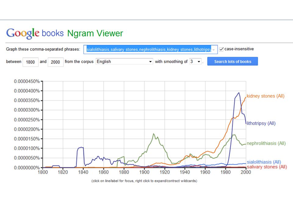
Incidence/Prevalence of Stones
Nephrolithiasis Sialolithiasis 11.7% prevalence by age 70 (a) 10% lifetime risk in U.S. ( and increasing) (b) Est. annual cost of 5.3 billion in US (c) Lifetime risk 13%(male) 7%(female) (d) 1 per 10-20,000 population per year (1) 1 per 15-30,000 population per year (2) 1 per 300,000 population per year (3) 1.2% Prevalence in postmortem study (0.45% symptomatic prevalence) (4) lifetime risk of 1-2% 0.1 to 1.0% of adult population (a) Brenner and Rector’s the Kidney Ch 39 (b) Stamatelou KK, Francis ME, Jones CA et al: Time trends in reported prevalence of kidney stones in the United States: 1976–1994. Kidney Int 2003; 63:1817 (c) Saigal CS et al Urologic Disease in America Project: Direct and indirect costs of nephrolithiasis … Kidney Int 2005;68:pp d. Preminger GM, Tiselius HG, Assimos DG, et al. Guideline for the management of ureteral calculi. J Urol :187: Marchal F, Dulguerov P. Sialolithiasis management: the state of the art.Arch Otolaryngol Head Neck Surg 2003;129: 951–956. Escudier MP, McGurkM:Symptomatic sialoadenitis and sialolithiasis in the English population: an estimate of the cost of hospital treatment. Br Dent J 1999;186: Rauch from Rice DH> chronic inflammatory disorders of the salivary glands. Otolaryngol Clin North Am. 1999;32: McGurk M, Escudier MP, Brown JE. Modern management of salivary calculi. Br. J. Surg. 2005;92:
 10% lifetime risk in U.S. ( and increasing) (b) Est. annual cost of 5.3 billion in US (c) Lifetime risk 13%(male) 7%(female) (d) 1 per 10-20,000 population per year (1) 1 per 15-30,000 population per year (2) 1 per 300,000 population per year (3) 1.2% Prevalence in postmortem study. (0.45% symptomatic prevalence) (4) lifetime risk of 1-2% 0.1 to 1.0% of adult population. (a) Brenner and Rector’s the Kidney Ch 39. (b) Stamatelou KK, Francis ME, Jones CA et al: Time. trends in reported prevalence of kidney stones in. the United States: 1976–1994. Kidney Int 2003; 63:1817. (c) Saigal CS et al Urologic Disease in America Project: Direct and indirect costs of nephrolithiasis … Kidney Int 2005;68:pp d. Preminger GM, Tiselius HG, Assimos DG, et al. Guideline for the management of ureteral calculi. J Urol :187: Marchal F, Dulguerov P. Sialolithiasis management: the state of the art.Arch Otolaryngol Head Neck Surg 2003;129: 951–956. Escudier MP, McGurkM:Symptomatic sialoadenitis and sialolithiasis in the English population: an estimate of the cost of hospital treatment. Br Dent J 1999;186: Rauch from Rice DH> chronic inflammatory disorders of the salivary glands. Otolaryngol Clin North Am. 1999;32: McGurk M, Escudier MP, Brown JE. Modern management of salivary calculi. Br. J. Surg. 2005;92:")
10
Kidney Stone Composition
Calcium Oxalate and Calcium Phosphate Uric Acid Calcium oxalate Uric acid Cystine Struvite Number of stones 34 5 33 1 3 19 1000 Prien 30 21 13 27 — 15 155 Mellick 44 8 2 28 460 Lagergren 46 25 243 Nordin

11
Salivary Stone Mineral Composition
Calcium phosphate Hydroxyl apatite (HA) only Hydroxyl apatite & Whitlockite (WI) Brushite (BR) Number of stones 100% (23) All from SMG ‘intra-glandular were all HA only’ 52% (12) 12 (HA only) 43% (10) 10 (HA+WI) 4%(1) 1 (HA + BR) 23 Teymoortash (2003) 100% (19) 68% ()13 6 19 Anneroth (1975) When a stone is available, clinicians should obtain a stone analysis at least once. ( Clinical Principle) Stone composition of uric acid, cystine or struvite implicates specific metabolic or genetic abnormalities, and knowledge of stone composition may help direct preventive measures. Journal of Urology 192, Issue Pearle M et al “Medical Management of Kidney Stones: AUA Guideline” Teymoortash A, Buck P, Jepsen H, Wernereymoranra: Sialolith crystals localized intraglandularly and in the Wharton’s duct of the human submandibular gland: an X-ray diffraction analysis Archives of Oral Biology (2003)48, Anneroth G, Eneroth CM, Isacsson G: Crystalline structure of salviary calculi. A microradiographic and microdiffractometric study J Oral Pathol 1975 Nov;4(5):266-72
 only. Hydroxyl. apatite. & Whitlockite. (WI) Brushite. (BR) Number of stones. 100% (23) All from SMG. ‘intra-glandular were all HA only’ 52% (12) 12 (HA only) 43% (10) 10 (HA+WI) 4%(1) 1 (HA + BR) 23. Teymoortash (2003) 100% (19) 68% () Anneroth (1975) When a stone is available, clinicians should obtain a stone analysis at least once. ( Clinical Principle) Stone composition of uric acid, cystine or struvite implicates specific metabolic or genetic abnormalities, and knowledge of stone composition may help direct preventive measures. Journal of Urology 192, Issue Pearle M et al Medical Management of Kidney Stones: AUA Guideline Teymoortash A, Buck P, Jepsen H, Wernereymoranra: Sialolith crystals localized intraglandularly. and in the Wharton’s duct of the human submandibular gland: an X-ray diffraction analysis. Archives of Oral Biology (2003)48, Anneroth G, Eneroth CM, Isacsson G: Crystalline structure of salviary calculi. A microradiographic. and microdiffractometric study. J Oral Pathol 1975 Nov;4(5):")
12
Definitions Calculus: a hard, stone-like concretion that forms in the body usually made from mineral salts ex: dental calculus, kidney stones, salivary stones Lithogenesis: formation of a calculus Mineral salts: apatite … whitlockite and brushite Lithotripsy is a procedure that uses shock waves to break up stones in the kidney, bladder, or ureter (tube that carries urine from your kidneys to your bladder). After the procedure, the tiny pieces of stones pass out of the body in your urine. Pneumatics is a section of technology that deals with the study and application of pressurized gas to produce mechanical motion. Pneumatic systems used extensively in industry are commonly powered by compressed air or compressed inert gases Picture removed Micro CT scan of sialolith organic nucleus surround by laminated layers of organic and inorganic substances British Dental Journal 217, E23 (2014) Published on-line
. After the procedure, the tiny pieces of stones pass out of the body in your urine. Pneumatics is a section of technology that deals with the study and application of. pressurized gas to produce mechanical motion. Pneumatic systems used extensively. in industry are commonly powered by compressed air or compressed inert gases. Picture. removed. Micro CT scan of sialolith. organic nucleus surround by. laminated layers of organic. and inorganic substances. British Dental Journal 217, E23 (2014) Published on-line.")
13
Extracorporeal shock wave lithotripsy: high energy shock waves are passed through the body and to fragment kidney stones – ideally to a small size permitting them to pass from the body along with the urine. Can be administered in tub of water or with the patient lying on a soft cushion through which the waves pass. ~ 1-2 thousand shock waves are used for a treatment that takes minutes. Photo by DiverDave - Own work. Licensed under CC BY-SA 3.0 via Wikimedia Commons –

14
Submandibular (N=60) and Parotid (N=34) small to medium size
Shock waves disconnect the stone from the ductal wall and reduce stone volume and provide revascularization angiogenesis Picture removed Picture removed After External Lithotripsy: 32% total elimination of stone 29% sialendoscopic assistance 39% combined with external U/S and endoscopy assist in locating the stone The endoscope irrigates and inflates gland (saline lidocaine) External lithotripsy applide low-energy levels up to 130 atm 1,000 to 1,5000 shock waves per session 3 sessions administered at one month intervals Low energy – therefore not painful, no anesthesia Photo depicts sialendoscope intraorally and a miniature external lithotripter –Nahlieli O, Shacham R, and Zaguir A: Combined External Lithotripsy and Endoscopic Techniques for Advanced Sialolithiasis Cases J Oral Maxillofac Surg 68: , 2010
 External lithotripsy applide. low-energy levels up to 130 atm. 1,000 to 1,5000 shock waves per session. 3 sessions administered at one month intervals. Low energy – therefore not painful, no anesthesia. Photo depicts sialendoscope intraorally and a miniature external lithotripter –Nahlieli O, Shacham R, and Zaguir A: Combined External Lithotripsy and Endoscopic Techniques for Advanced Sialolithiasis Cases J Oral Maxillofac Surg 68: ,")
15
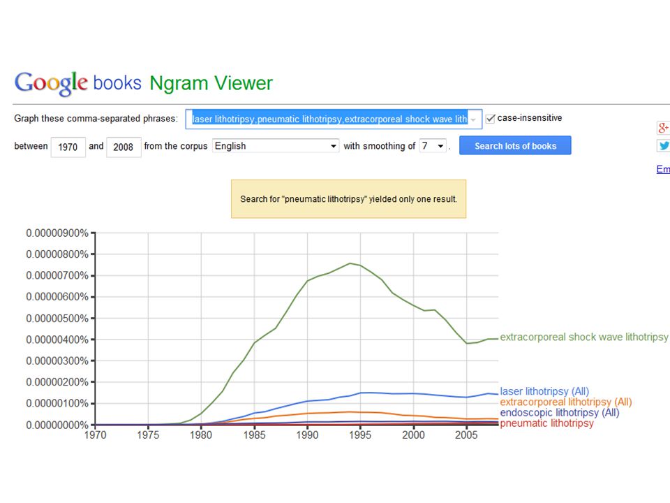
Pneumatic Lithotripsy vs Laser Lithotripsy
Pneumatic lithotripsy will transmit mechanical energy via a rigid probe on the stone Pneumatic Lithotripsy vs Laser Lithotripsy Prospective Randomized Trial Comparing Pneumatic Lithotripsy and Holmium Laser For Management of Middle and Distal Ureteral Calculi J of Endourology 2014 Li et al Three-year study of 9482 pts ending 2012 Stone free rate equal after 3 months Operative time shorter for laser Laser group = 28 minutes Pneumatic = 41 minutes Higher stricture rate (p=0.02) with laser Pneumatic versus laser ureteroscopic lithotripsy: a comparison of initial outcomes and cost Int Urol Nephrol (2014) Demir et al Retrospective study of 187 patients Treated with either holmium laser ($1,350 per probe) or pneumatic Device ($60 per probe) Laser more expensive but more effective Higher stricture and perforation rate with pneumatic device 3. Concept of ‘retropulsion’ -- less with laser Salivary Laser Issues 1. Special oral protection - +/- laser tube 2. Extra nurse (‘laser nurse’) 3. Extra set-up time 4. Eye protection (personnel, patient) 5. Risk of laser fire
 with laser. Pneumatic versus laser ureteroscopic. lithotripsy: a comparison of initial. outcomes and cost. Int Urol Nephrol (2014) Demir et al. Retrospective study of 187 patients. Treated with either holmium laser. ($1,350 per probe) or pneumatic. Device ($60 per probe) Laser more expensive but more effective. Higher stricture and perforation rate. with pneumatic device. 3. Concept of ‘retropulsion’ -- less with laser. Salivary Laser Issues. 1. Special oral protection - +/- laser tube. 2. Extra nurse (‘laser nurse’) 3. Extra set-up time. 4. Eye protection (personnel, patient) 5. Risk of laser fire.")
16
Email I received two weeks ago:
From: xxxxxxx Sent: Wednesday, March 04, :52 AM To: Hoffman, Henry Subject: RE: HH surgery for (Team 5 updated list) Dr. Hoffman, They moved your sialendoscopy to be the 2nd case in room 28. I need to know if you need holmium yag laser for this case as this room does not have the plug in to accommodate that laser. Thanks, xxx
 Dr. Hoffman, They moved your sialendoscopy to be the 2nd case in room 28. I need to know if you need. holmium yag laser for this case as this room does not have the plug in to accommodate. that laser. Thanks, xxx.")
17
Pneumatic Lithotripsy for
Sialolithiasis

18
Endoscopic Salivary Stone Fragmentation with Pneumatic Lithotripsy
Henry Hoffman, MD, Jack Kolenda MD, Rohan R Walvekar MD Feasibility of stone Fragmentation with Pneumatic Lithotripsy (StonebreakerTM) in a live porcine model Rohan R Walvekar, MD*; Jack Kolenda, MD; Henry Hoffman, MD
 in a live porcine model. Rohan R Walvekar, MD*; Jack Kolenda, MD; Henry Hoffman, MD.")
20
A flexible nitanal contact probe adapted to the Stonebreaker™ urologic CO2 gas-driven hand-held pneumatic lithotripser was deployed through a sialendoscope to disrupt 8 parotid and submandbular stones embedded in separate 3-D printed plastic models of the mouth and submandibular glands. Simulation included endoscopic removal of small stone fragments by standard basket retrieval supplement by irrigation and suction through a salivary duct introducer system.

21
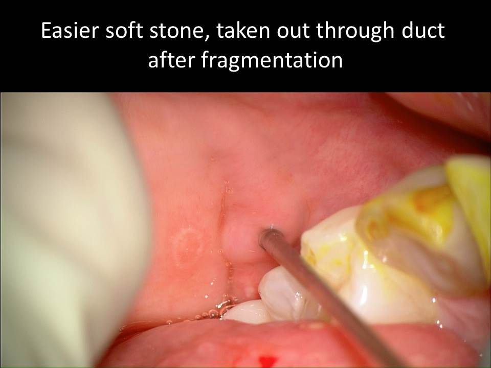
Use of CO2 cartridge powered
pneumatic lithotripsy to fragment stone with central portion removed with basket

22
Endoscopic Salivary Stone Fragmentation with Pneumatic Lithotripsy
Henry Hoffman, MD, Jack Kolenda MD, Rohan R Walvekar MD R Walvekar MD Correlations were made between stone appearance, volume, and density with the duration of the procedures and number of pneumatic pulses required to completely disrupt and remove stone fragments. Results Among the seven stones fragmented sufficiently to permit full endoscopic removal, the average procedure time (32 minutes) and the average number of pneumatic pulses (98) correlated with stone density (range 0.4 to 1.5 g/ml) and stone volume (0.05 to 0.4 ml). One stone was sufficiently resistant to fragmentation as to prevent successful removal. Conclusion: Modification to the evolving technology of intracorporeal pneumatic management of nephrolithiasis was successfully applied in an ex vivo model to manage sialolithiasis.
 and the average number of. pneumatic pulses (98) correlated with stone density (range 0.4 to 1.5 g/ml) and stone volume (0.05 to 0.4 ml). One stone was sufficiently resistant to fragmentation as to prevent successful removal. Conclusion: Modification to the evolving technology of intracorporeal. pneumatic management of nephrolithiasis was successfully. applied in an ex vivo model to manage sialolithiasis.")
35
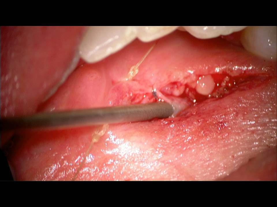
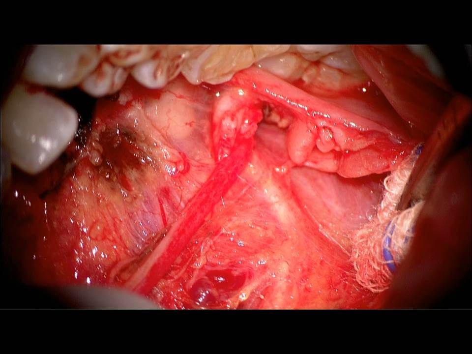
Simulation Model Cadaver Dissection
Lab Simulation Model Cadaver Dissection

36
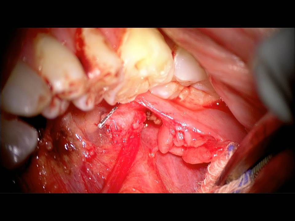
Simulation Model Cadaver Dissection
Lab Simulation Model Cadaver Dissection Submandibular duct (Wharton’s duct) cannulation dilation, sialendoscopy Identify duct through floor of mouth incision sialendoscopy through FOM duct incision Identify lingual nerve Remove sublingual gland Dissect duct to hilum * extra points: identify submandibular ganglion Ann Otol Rhinol Laryngol May;124(5): doi: / Epub 2014 Nov 25. Transoral submandibular ganglion neurectomy: an anatomical feasibility study. Spock T1, Hoffman HT2, Joshi AS3.
 cannulation. dilation, sialendoscopy. Identify duct through floor of mouth incision. sialendoscopy through FOM duct incision. Identify lingual nerve. Remove sublingual gland. Dissect duct to hilum. * extra points: identify submandibular ganglion. Ann Otol Rhinol Laryngol May;124(5): doi: / Epub 2014 Nov 25. Transoral submandibular ganglion neurectomy: an anatomical feasibility study. Spock T1, Hoffman HT2, Joshi AS3.")
Similar presentations


>")


>")


