Download presentation
Presentation is loading. Please wait.

1
Assoc. Prof. Ales BARTOS, MD, PhD
Movement disorders (Extrapyramidal and cerebellar syndromes) (seminar, teaching unit 60) Assoc. Prof. Ales BARTOS, MD, PhD Department of Neurology, Charles University in Prague
 (seminar, teaching unit 60) Assoc. Prof. Ales BARTOS, MD, PhD. Department of Neurology, Charles University in Prague.")
2
The questions for the oral state exam in neurobehavioral sciences :
Parkinson´s disease and syndromes: causes, symptoms, diagnosis, treatment (a neurology part) Disturbances of motor functions, including drug-induced syndromes Vascular dementia: clinical signs and symptoms, classification, differential diagnosis, treatment and prevention Dementia: clinical signs and symptoms, classification, differenial diagnosis
 Disturbances of motor functions, including drug-induced syndromes. Vascular dementia: clinical signs and symptoms, classification, differential diagnosis, treatment and prevention. Dementia: clinical signs and symptoms, classification, differenial diagnosis.")
3
The questions for the oral state exam in neurobehavioral sciences :
Hydrocephalus: types, causes, symptoms, diagnosis, treatment. CSF hypotension Nervous system disorders in metabolic and autoimmune diseases, CO toxicity: causes, symptoms, diagnosis, treatment, complications

4
Outline movement disorders in general
case history and examination focused on movement disorders the Parkinsonian syndrome Parkinson´s disease secondary parkinsonian syndromes and a surprise in the end: questions and quizzes …

5
2 categories - syndromes:
Movement disorders observed during the interview („one look is worth a thousand words“) abnormal involuntary movements are exaggerated with anxiety and diminished during sleep 2 categories - syndromes: 1) Akinetic-rigid – paucity of spontaneous movement, increased tone (rigidity) examples: Parkinson´s disease, vascular encephalopathy / dementia 2) Hyperkinetic-hypotonic – irregular involuntary movements, often at varying sites, diminished muscle tone at rest (chorea, athetosis, ballism, dystonias) example: Huntington´s disease
 abnormal involuntary movements are exaggerated with anxiety and diminished during sleep. 2 categories - syndromes: 1) Akinetic-rigid – paucity of spontaneous movement, increased tone (rigidity) examples: Parkinson´s disease, vascular encephalopathy / dementia. 2) Hyperkinetic-hypotonic – irregular involuntary movements, often at varying sites, diminished muscle tone at rest (chorea, athetosis, ballism, dystonias) example: Huntington´s disease.")
6
Types of abnormal movements
Tremor = involuntary, rhythmic movement across a joint: 1) rest – at rest 2) static, postural – only with a specific posture 3) intention – only with motion Chorea = sudden movement usually involving the extremities and the face Athetosis = a slow, sinuous movement (the distinction is sometimes unclear – choreoathetosis)
 rest – at rest. 2) static, postural – only with a specific posture. 3) intention – only with motion. Chorea = sudden movement usually involving the extremities and the face. Athetosis = a slow, sinuous movement. (the distinction is sometimes unclear – choreoathetosis)")
7
Types of abnormal movements
(Hemi-)ballism = irregular, sudden, large-amplitude movements of an entire limb or limbs Myoclonus = spontaneous, shocklike contractions of one or more muscles across a joint (the jerk-like movements during falling asleep in dogs, cats and man) Tics = irregular, stereotyped movements, often in the face Dystonia = a sustained abnormal or inappropriate posture
ballism = irregular, sudden, large-amplitude movements of an entire limb or limbs. Myoclonus = spontaneous, shocklike contractions of one or more muscles across a joint (the jerk-like movements during falling asleep in dogs, cats and man) Tics = irregular, stereotyped movements, often in the face. Dystonia = a sustained abnormal or inappropriate posture.")
8
History of the patient with a movement disorder
symmetry, exacerbating or alleviating factors (stress, sleep, alcohol) family history – many movement disorders drug history (!) – neuroleptics → parkinsonism, tardive dyskinesias; antiemetics ! → dystonia, dyskinesia alcohol history → cerebellar degeneration / dysfunction, Wernicke encephalopathy, alleviate essential tremor medical history – hyperthyroidism → tremor, liver disease → asterixis
 family history – many movement disorders. drug history (!) – neuroleptics → parkinsonism, tardive dyskinesias; antiemetics ! → dystonia, dyskinesia. alcohol history → cerebellar degeneration / dysfunction, Wernicke encephalopathy, alleviate essential tremor. medical history – hyperthyroidism → tremor, liver disease → asterixis.")
9
Neurologic examination
have the patient draw a spiral or connect dots as a permanent record of the motor dysfunction; later this can be used to monitor the efficacy of treatment muscle tone: hypertonia – a passive movement of the arm abnormal postures – during station and gait testing

10
The Parkinsonian SYNDROME (parkinsonism)
= movement abnormalities mainly characterized by: 1) bradykinesia, hypokinesia, 2) rigidity and 3) resting tremor, 4) postural abnormalities (stance and gait) based on pathology in basal ganglia and their connections
 bradykinesia, hypokinesia, 2) rigidity and 3) resting tremor, 4) postural abnormalities (stance and gait) based on pathology in basal ganglia and their connections.")
11
The Parkinsonian syndrome
BRADYKINESIA, HYPOKINESIA („slow movements“): slowness of walking, other movements (and mental processes), delayed responses physically and mentally difficulty in initiating movements slow and smaller handwriting reduced facial reactions („masked face“)
: slowness of walking, other movements (and mental processes), delayed responses physically and mentally. difficulty in initiating movements. slow and smaller handwriting. reduced facial reactions („masked face )")
12
The Parkinsonian syndrome
RIGIDITY (increased tone): passively move the extremities increased tone as increased resistance (cogwheel phenomenon) !! (versus spasticity)
: passively move the extremities. increased tone as increased resistance (cogwheel phenomenon) !! (versus spasticity)")
13
The Parkinsonian syndrome
TREMOR: at rest most commonly in hands and fingers (a „pill rolling“ motion) often begins unilaterally and distally spreads proximally and to the other side over months or years usually asymmetric
 often begins unilaterally and distally. spreads proximally and to the other side over months or years. usually asymmetric.")
14
The Parkinsonian syndrome
POSTURAL CHANGES during station and gait : slight flexion of back the first steps are slow and small difficult turning imbalance falls

15
The Parkinsonian syndrome
OTHER FEATURES: micrographia dysarthria cognitive slowing mood lability dementia

16
Causes of parkinsonism
the problem to distinguish in a patient with parkinsonism: 1) Idiopathic Parkinson´s disease (80 %) 2) Secondary parkinsonian syndromes (20 %) vascular parkinsonism rarer causes: drug-induced (dopamin receptor blockers: neuroleptics, antiemetics) Wilson´s disease normal pressure hydrocephalus carbon monoxide poisoning trauma post-encephalitic parkinsonism (in the past) parkinson plus syndromes (corticobasal syndrome, progressive supranucelar palsy, Lewy body disease)
 Idiopathic Parkinson´s disease (80 %) 2) Secondary parkinsonian syndromes (20 %) vascular parkinsonism. rarer causes: drug-induced (dopamin receptor blockers: neuroleptics, antiemetics) Wilson´s disease. normal pressure hydrocephalus. carbon monoxide poisoning. trauma. post-encephalitic parkinsonism (in the past) parkinson plus syndromes (corticobasal syndrome, progressive supranucelar palsy, Lewy body disease)")
17
Parkinson´s DISEASE (vs. syndrome)
chronic, slowly progressive, degenerative and usually sporadic disease a) loss of dopaminergic neurons pars compacta substantiae nigrae – lack of dopamin b) Lewy bodies – deposits of alfa-synuclein (synucleinopathy) in old age over 50 years, prevalence rises with age the lecture on Parkinson´s disease:
 loss of dopaminergic neurons pars compacta substantiae nigrae – lack of dopamin. b) Lewy bodies – deposits of alfa-synuclein (synucleinopathy) in old age over 50 years, prevalence rises with age. the lecture on Parkinson´s disease: v=xRbdzgKFl10.")
18
Parkinson´s disease in pictures

19
Parkinson´s disease – clinical features
the parkinsonian syndrome – „everything is hypo- (reduced)“: upper limbs – hypokinesia and rigidity, micrographia, reduced synkinesias during gait gait disorders – steps: too small, too slow, freezing, difficult turning face – hypomimia speech – hypofonia, dysarthria
 : upper limbs – hypokinesia and rigidity, micrographia, reduced synkinesias during gait. gait disorders – steps: too small, too slow, freezing, difficult turning. face – hypomimia. speech – hypofonia, dysarthria.")
20
Parkinson´s disease – other clinical features
rest tremor – very typical, not in all patients depression cognitive disturbances (attention, executive functions), dementia in late phases
, dementia in late phases.")
21
Parkinson´s disease – diagnosis
the diagnosis is based on clinical evaluation in 3 steps: 1) the presence of hypokinetic-rigid syndrome and other features typical for Parkinson´s disease 2) red flags – suspect an alternative diagnosis – features for secondary parkinsonism: repeated strokes and step-wise onset, neuroleptics, supranuclear gaze palsy, cerebellar syndromes, early severe dementia, Babinski sign, brain tumor or hydrocephalus on CT scanning, absence of dopa responsiveness 3) the response to dopaminergic treatment
 the presence of hypokinetic-rigid syndrome and other features typical for Parkinson´s disease. 2) red flags – suspect an alternative diagnosis – features for secondary parkinsonism: repeated strokes and step-wise onset, neuroleptics, supranuclear gaze palsy, cerebellar syndromes, early severe dementia, Babinski sign, brain tumor or hydrocephalus on CT scanning, absence of dopa responsiveness. 3) the response to dopaminergic treatment.")
22
Parkinson´s disease – treatment

23
Parkinson´s disease – treatment
a replacement of decreased dopamine concentration: levodopa – is the most effective treatment, most patients have a remarkable response to levodopa, commonly used if patients have Parkinson´s disease and / or dopaminergic agonists (bromocriptine, pregolide, ropinirole, cabergoline, pramipexole) – usually a weaker effect and more peripheral adverse effects (blood pressure fluctuations, arrhythmias, nausea, vomiting) „start low, go slow“ deep brain stimulation (neuropacemaker)
 – usually a weaker effect and more peripheral adverse effects (blood pressure fluctuations, arrhythmias, nausea, vomiting) „start low, go slow deep brain stimulation (neuropacemaker)")
24
Break

25
Secondary parkinsonian syndromes
frontal type of gait (lower body parkinsonism) – symmetrical involvement of lower limbs, more than relatively spared upper limbs: vascular parkinsonian syndrome – multiple, repeated strokes in basal ganglias (postsynaptic damage, sometimes effective levodopa) – step-wise progression, Babinski +, axial phenomena, pseudobulbar syndrome, cognitive deficit, dementia normal pressure hydrocephalus – triad: 1) gait disorder, 2) cognitive deficit, 3) urine incontinence treatable condition !
 – symmetrical involvement of lower limbs, more than relatively spared upper limbs: vascular parkinsonian syndrome – multiple, repeated strokes in basal ganglias (postsynaptic damage, sometimes effective levodopa) – step-wise progression, Babinski +, axial phenomena, pseudobulbar syndrome, cognitive deficit, dementia. normal pressure hydrocephalus – triad: 1) gait disorder, 2) cognitive deficit, 3) urine incontinence. treatable condition !")
26
Vascular parkinsonian syndrome
bilateral lesions!

27
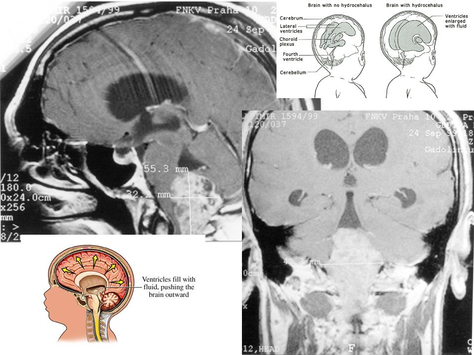
Normal pressure hydrocephalus

28
Drug-induced secondary parkinsonism (contraindicated in Parkinson´s disease)
dopamine receptor blockers: typical neuroleptics (fenothiazines – chlorpromazine, butyrophenons – haloperidole) antiemetics (neuroleptics) – metoclopramid calcium channel blockers – flunarizine, cinnarizine treatment of delirium in Parkinson´s patients with ATYPICAL antipsychotic drugs - tiapride
 antiemetics (neuroleptics) – metoclopramid. calcium channel blockers – flunarizine, cinnarizine. treatment of delirium in Parkinson´s patients with ATYPICAL antipsychotic drugs - tiapride.")
29
Wilson´s disease (=hepatolenticular degeneration)
a genetic autosomal recessive disorder of copper metabolism „parkinsonism in the young“ progressive, „wing-beating“ tremor, rigidity, dysarthria, mental changes, hepatic dysfunction, corneal Kayser-Fleischer ring less commonly tremor treatment: penicillamine, zinc sulfate

30
Wilson´s disease (=hepatolenticular degeneration)
")
31
Wilson disease in a 14-year-old girl with dysarthria
Wilson disease in a 14-year-old girl with dysarthria.A, Initial T2-weighted axial MR image shows increased signal intensity in both caudate nuclei (arrowheads) and putamen (arrows).B, Follow-up T2-weighted axial MR image obtained after 3 years shows complet... Wilson disease in a 14-year-old girl with dysarthria.A, Initial T2-weighted axial MR image shows increased signal intensity in both caudate nuclei (arrowheads) and putamen (arrows).B, Follow-up T2-weighted axial MR image obtained after 3 years shows complete resolution of the lesions. Patient’s neurologic symptoms were also improved. Kim T et al. AJNR Am J Neuroradiol 2006;27: ©2006 by American Society of Neuroradiology
 and putamen (arrows).B, Follow-up T2-weighted axial MR image obtained after 3 years shows complet... Wilson disease in a 14-year-old girl with dysarthria.A, Initial T2-weighted axial MR image shows increased signal intensity in both caudate nuclei (arrowheads) and putamen (arrows).B, Follow-up T2-weighted axial MR image obtained after 3 years shows complete resolution of the lesions. Patient’s neurologic symptoms were also improved. Kim T et al. AJNR Am J Neuroradiol 2006;27: ©2006 by American Society of Neuroradiology.")
32
Treatment of secondary parkinsonian syndromes
vascular parkinsonism – controlling vascular risk factors, rehabilitation rarer causes: normal pressure hydrocephalus – ventriculo-peritoneal shunt drugs (neuroleptics, antiemetics) – anticholinergic agents, atypical antipsychotics Wilson´s disease – penicillamine, zinc post-traumatic, carbon monoxide poisoning - rehabilitation
 – anticholinergic agents, atypical antipsychotics. Wilson´s disease – penicillamine, zinc. post-traumatic, carbon monoxide poisoning - rehabilitation.")
33
Treatment differences in two parkinsonian syndromes
idiopathic Parkinson´s disease presynaptic disease low dopamine concentrations dopaminergic responsiveness vascular parkinsonism postsynaptic (presynaptic ?) disease normal dopamine concetrations no effect of dopaminergic drugs
 disease. normal dopamine concetrations. no effect of dopaminergic drugs.")
34
Movement disorders in broader sense
Chorea = sudden movement usually involving the extremities and the face Ataxia = an impairment of coordination in the absence of significant weakness 1) limb 2) truncal = cerebellar disorders
 limb. 2) truncal. = cerebellar disorders.")
35
Chorea irregular, asymmetric, sudden, brief, shooting involuntary movements in distal segments of the extremities grimacing and lip-smacking

36
Huntington´s disease autosomal dominant inheritance
expanded CAG repeat on the chromosome 4 longer CAG repeat – earlier age of onset age: years presymptomatic diagnosis by genetic testing very poor prognosis: death after years from the diagnosis no etiologic treatment, symptomatic management

37
Hemiballism „ballistic“ movements of high amplitude involving multiple segments of a limb cause: stroke in the subthalamic nucleus neuroleptics, but usually resolves spontaneously in a few weeks

38
Cerebellar disorders syndromes:
neocerebellar (limb ataxia, intention tremor, dysdiadochokinesia) palleocerebellar (truncal ataxia, unstable stance in the Romberg test, unstable, broad-based gait, dyssynergy) mixed (global) an important neuroanatomical association of the cerebellum with the rest of the brain
 palleocerebellar (truncal ataxia, unstable stance in the Romberg test, unstable, broad-based gait, dyssynergy) mixed (global) an important neuroanatomical association of the cerebellum with the rest of the brain.")
40
Causes of cerebellar disorders
VINDICATE: Vascular (stroke) Inflammatory (encephalitis) Neoplastic (tumors – meningeoma, gliomas –oligodendroglioma, astrocytoma) Degenerative (spinocerebellar ataxias, paraneoplastic syndromes) Intoxication (alcohol, Wernicke´s encephalopathy, antiepileptics, lithium) Congenital – Arnold-Chiari, agenesis Autoimmune (multiple sclerosis, paraneoplastic cerebellar degeneration in ovarian cancer) Traumatic (contusion, sub- / epi- dural hematomas) Endocrine (metabolic – see intoxication)
 Inflammatory (encephalitis) Neoplastic (tumors – meningeoma, gliomas –oligodendroglioma, astrocytoma) Degenerative (spinocerebellar ataxias, paraneoplastic syndromes) Intoxication (alcohol, Wernicke´s encephalopathy, antiepileptics, lithium) Congenital – Arnold-Chiari, agenesis. Autoimmune (multiple sclerosis, paraneoplastic cerebellar degeneration in ovarian cancer) Traumatic (contusion, sub- / epi- dural hematomas) Endocrine (metabolic – see intoxication)")
41
Wernicke´s encephalopathy
conditons due to thiamin deficiency even in nonalcoholics, e.g. in patients vomiting, poor nutrition oculomotor disturbances (abducent palsy, nystagmus, gaze palsy) ataxia (more truncal than limb, unable to stand or walk) antero- and retrograde amnesis + confabulations brain MR: lesions in periaqueductal region and adjacent to the third ventricle (demyelination, small foci of hemorrhage) treatment: thiamin glucose solutions after thiamin has been given, otherwise it may induce acute worsening of Wernicke´s encephalopathy early treatment – regression, often residual deficits
 ataxia (more truncal than limb, unable to stand or walk) antero- and retrograde amnesis + confabulations. brain MR: lesions in periaqueductal region and adjacent to the third ventricle (demyelination, small foci of hemorrhage) treatment: thiamin. glucose solutions after thiamin has been given, otherwise it may induce acute worsening of Wernicke´s encephalopathy. early treatment – regression, often residual deficits.")
42
Wernicke´s encephalopathy

43
Figure. Rapid clearing of MRI signals in Wernicke encephalopathy.
Figure. Rapid clearing of MRI signals in Wernicke encephalopathy. T2-weighted MR images at the time of admission show abnormal hyperintense signal (arrows) in the periaqueductal gray region (A, axial view) and dorsomedial thalami (B, coronal view), which cleared after 5 days of thiamine (100 mg IV) treatment (C, D). Watson W et al. Neurology 2003;61:527 © 2013 American Academy of Neurology
 in the periaqueductal gray region (A, axial view) and dorsomedial thalami (B, coronal view), which cleared after 5 days of thiamine (100 mg IV) treatment (C, D). Watson W et al. Neurology 2003;61:527. © 2013 American Academy of Neurology.")
44
Let us verify your knowledge….

45
Quizzes distinguish different types of tremor
identify the type of abnormal movements

46
Quiz question No 1 The most common cause of the parkinsonian syndrome is: a) repeated and multiple strokes b) drug abuse c) neuroleptics d) Parkinson´s disease e) carbon monoxide poisoning
 drug abuse. c) neuroleptics. d) Parkinson´s disease. e) carbon monoxide poisoning.")
47
Quiz question No 2 Dopaminergic transmission is impaired in Parkinson´s disease due to: a) blockage of dopamin receptors b) decreased presynaptic uptake c) presynaptic degeneration and loss of dopaminergic neurons d) degeneration of nigrostriatal pathway e) postsynaptic degeneration and loss of dopaminergic neurons in striatum
 decreased presynaptic uptake. c) presynaptic degeneration and loss of dopaminergic neurons. d) degeneration of nigrostriatal pathway. e) postsynaptic degeneration and loss of dopaminergic neurons in striatum.")
48
Quiz question No 3 Parkinsonism prevailing in the upper part of the body: a) may indicate a treatable condition – normal pressure hydrocephalus b) is typical for Parkinson´s disease c) may be found in vascular encephalopathy
 is typical for Parkinson´s disease. c) may be found in vascular encephalopathy.")
49
Quiz question No 4 The onset of the parkinsonian syndrome:
a) under the age of 45 years is impossible b) in the 7th and 8th decade is usual for Parkinson´s disease c) between 50 and 60 years is typical for vascular encphalopathy d) under the age of 45 may be caused by rare disease – which one?
 under the age of 45 years is impossible. b) in the 7th and 8th decade is usual for Parkinson´s disease. c) between 50 and 60 years is typical for vascular encphalopathy. d) under the age of 45 may be caused by rare disease – which one")
50
Quiz question No 5 A response to levodopa or dopaminergic agonists (motor improvement): a) is found almost exclusively in Parkinson´s disease b) is profound in the vascular parkinsonian syndrome c) is supportive for the diagnosis of Parkinson´s disease d) is typical for all parkinsonian syndromes (Why?)
 is profound in the vascular parkinsonian syndrome. c) is supportive for the diagnosis of Parkinson´s disease. d) is typical for all parkinsonian syndromes. (Why )")
51
Quiz question No 6 Parkinsonian features prevail in lower part of the body. Which statement is true? a) dopaminergic treatment will be probably effective b) it is typical in early Parkinson´s disease c) clinical evaluation is insufficient, brain neuroimaging is not necessary d) it suggests an alternative diagnosis (Why?)
 dopaminergic treatment will be probably effective. b) it is typical in early Parkinson´s disease. c) clinical evaluation is insufficient, brain neuroimaging is not necessary. d) it suggests an alternative diagnosis. (Why )")
53
Overlapping features of various types of tremor
Parkinson´s syndrome Essential tremor Cerebellar tremor Rest tremor ++ + Postural / static tremor 0 / + Intentional tremor Increased tone Postural abnormality Incoordination

Similar presentations


 (MND) are a group of neurological disorders that selectively affect motor neurons.>")


>")

>")


