Download presentation
Presentation is loading. Please wait.

1
Jaundice – For the practitioners
Dr.R.V.S.N.Sarma., M.D., M.Sc., Consultant Physician & Chest Specialist A Lucid Understanding of Jaundice – For the practitioners

2
Jaundice – Classification
Normal Serum Bilirubin (SB) is 0.3 to 1.0 mg% Jaundice is increased levels of SB > 1.0 mg% Over production of Bilirubin (Hemolytic) From hemolysis of RBC Lysis of RBC precursors – Ineffective erythropoesis Impaired hepatic function (Hepatitic) Hepatocellular dysfunction in handling bilirubin Uptake, Metabolism and Excretion of bilirubin Obstruction to bile flow (Obstructive) Intrahepatic cholestasis Extrahepatic Obstruction (Surgical Jaundice)
 is 0.3 to 1.0 mg% Jaundice is increased levels of SB > 1.0 mg% Over production of Bilirubin (Hemolytic) From hemolysis of RBC. Lysis of RBC precursors – Ineffective erythropoesis. Impaired hepatic function (Hepatitic) Hepatocellular dysfunction in handling bilirubin. Uptake, Metabolism and Excretion of bilirubin. Obstruction to bile flow (Obstructive) Intrahepatic cholestasis. Extrahepatic Obstruction (Surgical Jaundice)")
3
Clinical Aspects of Jaundice
Clinically detectable if SB is >2.0 mg% With edema and dark skin – Jaundice is masked What is special about the sclera ? – Rich Elastin Darkening of the urine – Differential Diagnosis Skin discoloration – Yellowish, - Carotinemia – Eyes N Mucosa – hard palate (in dark skinned) Greenish hue of skin and sclera - due Biliverdin – indicates long standing jaundice Generalized Pruritus – Obstructive Jaundice – Why ?
 Greenish hue of skin and sclera - due Biliverdin – indicates long standing jaundice. Generalized Pruritus – Obstructive Jaundice – Why")
4
Clinical History – Imp clues
Duration of jaundice – Acute / Chronic Abdominal pain v/s painless jaundice Fever – Viral / bacteria /sepsis Arthralgia, rash, glands; Pruritus - obstructive Appetite – Hepatocellular / Malignancy Weight loss – Malignancy – CAH Colour of stools –chalky white –obstructive Family history – Hemolytic – Inherited dis. H/o transfusion, promiscuity, IDU Alcohol abuse, Medications – INH, EM, Largactil

5
Coloured Urine – Differ. Diagnosis
Bilirubin in urine due to Jaundice (CB) Concentrated urine in dehydration Fluid deprivation syndromes Sulfasalazine use – for Ulcerative colitis Rifampicin, Pyridium and Thiamine use Red urine – Porphyria, Hemoglobin & Myoglobinuria, Hematuria Dark black urine in Ochranosis - HGA Melanin excretion from Melanoma Red sweat in Clofazamine, Rifampicin
 Concentrated urine in dehydration. Fluid deprivation syndromes. Sulfasalazine use – for Ulcerative colitis. Rifampicin, Pyridium and Thiamine use. Red urine – Porphyria, Hemoglobin & Myoglobinuria, Hematuria. Dark black urine in Ochranosis - HGA. Melanin excretion from Melanoma. Red sweat in Clofazamine, Rifampicin.")
6
Fate of Senescent RBC RBC life span in blood stream is 90-120 days
Old RBCs are phagocytosed and/or lysed Lysis occurs extravascularly in the RE system subsequent to RBC phagocytosis Intravascular Hemolysis of young RBC This is due to hemolytic diseases of RBC

7
The Hepatobiliary & Portal System
Hepatobiliary Tree Portal Circulation

8
E V Pathway for RBC Scavanging
Liver, Spleen & Bone marrow Phagocytosis & Lysis Hemoglobin Globin Heme Bilirubin Amino acids Fe2+ Through Liver Amino acid pool Excreted

9
Bilirubin Handling

10
Bilirubin Metabolism - Summary

11
Bilirubin – And its nature
Properties Unconjugated Conjugated Normal serum fraction 90% 10% Water solubility (polarity) 0 (non polar) + (polar) Affinity to lipids (Kernicterus) +++ Renal excretion Nil + Vanden Berg Reaction Indirect Direct Temporary Albumin Binding Irreversible Delta Bilirubin ++
 0 (non polar) + (polar) Affinity to lipids (Kernicterus) +++ Renal excretion. Nil. + Vanden Berg Reaction. Indirect. Direct. Temporary Albumin Binding. Irreversible Delta Bilirubin")
12
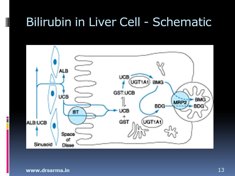
Bilirubin in the Liver Cell
1 Hepatocyte (HC) uptake of UCB Alb+UCB dissociates and UCB enters HC By passive diffusion into HC – Ligandin bound Insoluble UCB is to be made soluble in HC 2 Conjugation in ER of Hepatocyte (HC) Formation of mono and di glucuronides BMG, BDG UDP Glucuronosyl transferase is energy depend. Insoluble UCB made water soluble for excretion 3 Excretion in into biliary canaliculi Rate limiting step in metabolism CB 50% is not protein bound – no loss of albumin Remaining 50% bilirubin – Irreversibly bound
 uptake of UCB. Alb+UCB dissociates and UCB enters HC. By passive diffusion into HC – Ligandin bound. Insoluble UCB is to be made soluble in HC. 2. Conjugation in ER of Hepatocyte (HC) Formation of mono and di glucuronides BMG, BDG. UDP Glucuronosyl transferase is energy depend. Insoluble UCB made water soluble for excretion. 3. Excretion in into biliary canaliculi. Rate limiting step in metabolism. CB 50% is not protein bound – no loss of albumin. Remaining 50% bilirubin – Irreversibly bound.")
13
Bilirubin in Liver Cell - Schematic
14
Blood Bile ALT ALT ALT ALT ALT ALT ALT ALT ALT ALT ALT ALT ALT ALT ALT
AlkP AlkP AlkP AlkP AlkP AlkP AlkP AlkP AlkP AlkP AlkP AlkP AlkP AlkP AlkP Bile

15
X Blood Bile AlkP AlkP AlkP AlkP AlkP AlkP AlkP AlkP AlkP AlkP AlkP
ALT ALT ALT ALT ALT ALT ALT ALT ALT ALT ALT ALT ALT ALT ALT ALT ALT ALT ALT ALT ALT ALT ALT ALT ALT ALT ALT AlkP AlkP AlkP AlkP AlkP AlkP AlkP AlkP AlkP AlkP AlkP AlkP X Bile

16
Bilirubin in the Intestine
1. CB in bile is excreted into Duodenum CB 10% diffuses in to blood CB excreted is not reabsorbed 2. Conversion of CB into uro & stercobilinogen Urobilinogen excreted in stool Part of the UBG enters EHC 3. From gut, UBG but not CB enters EHC Kidney excretes absorbed UBG In biliary obst. UBG absent in urine

17
Bilirubin handling in Kidney
Conjugated Bilirubin Bound (20 days) Bilirubin in urine is conjugated Unconjugated Bilirubin Not filtered or secreted Nil in urine Urobilinogen in urine Normally traces ↑ in Cholestaiss
 Bilirubin in urine is conjugated. Unconjugated Bilirubin. Not filtered or secreted. Nil in urine. Urobilinogen in urine. Normally traces. ↑ in Cholestaiss.")
18
An Approach to Jaundice
Is it isolated elevation of serum bilirubin ? If so, is the↑unconjugated or conjugated fraction? Is it accompanied by other liver test abnormalities ? Is the disorder hepatocellular or cholestatic? If cholestatic, is it intra- or extrahepatic? These can be answered with a thoughtful History and physical examination Interpretation of laboratory tests and Radiological tests and procedures.

19
Bilirubin testing Van den Berg Reaction
SB + SAA Diazo compound formed Diazo is chromogenic – Colourimerty Reaction in H2O medium – Direct – CB Reaction in ethnol medium – Indirect Indirect includes CB and UCB = Total B Time is the essence of the direct test Foam test, Ictotest for urine – CB only

20
Normal values for LFT Features Healthy Normal Total Bilirubin
Less than 1.00 mg Conjugated Bilirubin Less than 0.15 mg AST (SGOT) Less than 31 i.u/L ALT (SGPT) Less than 35 i.u/L Alkaline phosphatase Less than 112 i.u /L GGT and 5’ Nucleosidase, CDT Significantly ↑ in ALD Urine Bilirubin Absent Urine Urobilinogen In trace quantity Urine Bile Salts
 Less than 31 i.u/L. ALT (SGPT) Less than 35 i.u/L. Alkaline phosphatase. Less than 112 i.u /L. GGT and 5’ Nucleosidase, CDT. Significantly ↑ in ALD. Urine Bilirubin. Absent. Urine Urobilinogen. In trace quantity. Urine Bile Salts.")
21
Lab Diagnosis of Jaundice – D.D
Features Prehepatic (Heamolytic) Intrahepatic (Hepatocellular) Posthepatic (Obstructive) Unconjugated ↑ Normal Conjugated AST or ALT ↑ ↑ Alkaline phos. and GGT Urine bilirubin Absent Present Increased Urobilinogen
 Intrahepatic. (Hepatocellular) Posthepatic. (Obstructive) Unconjugated. ↑ Normal. Conjugated. AST or ALT. ↑ ↑ Alkaline phos. and GGT. Urine bilirubin. Absent. Present. Increased. Urobilinogen.")
22
Liver Function Tests (LFT)
Normal Range Value Bilirubin Total Conjugated 0.1 to 1.0 mg < 0.2 mg Dx. Of Jaundice, Severity Alkaline phosphatase iu/L Dx of Obstructive Jaundice Aspartate transaminase (AST/SGOT) 5-31 iu/L Early Dx and follow up Alanine transaminase (ALT/SGPT) 5-35 iu/L AST/ALT > 1 in ALD Albumin g/dL Assess severity of disease Prothrombin time (PT) 12-16 s
 5-31 iu/L. Early Dx and follow up. Alanine transaminase. (ALT/SGPT) 5-35 iu/L. AST/ALT > 1 in ALD. Albumin g/dL. Assess severity of disease. Prothrombin time (PT) s.")
23
Utility of Liver Function Tests
LFT Utility of the test ALT/SGPT ALT ↓than AST in alcoholism Albumin Assess severity / chronicity Alk. phosphatase Cholestasis, hepatic infiltrations AST/SGOT Early Dx. of Liver disease, F/up Bilirubin (Total) /Conjug. Diagnose jaundice Gamma-globulin Dx. F/up Chronic hepatitis & cirrhosis GGT Dx alcohol abuse, Dilantin toxicity
 /Conjug. Diagnose jaundice. Gamma-globulin. Dx. F/up Chronic hepatitis & cirrhosis. GGT. Dx alcohol abuse, Dilantin toxicity.")
24
Non Hepatic causes of abnormal LFT
Albumin PLE, Nephrotic syndrome Malnutrition, CHF AKP Bone disease, Pregnancy, Malignancy , Adv age AST MI, Myositis, I.M.injections Bilirubin Hemolysis, Sepsis, Ineffective erythropoiesis PTT Antibiotics, Anticoagulant, Steatorrhea, Dietary

25
Algorithmic approach for Jaundice
How to clinically evaluate the patient ? What tests will help us in D.D ? What imaging modalities will be useful ? How to monitor the progress ?

26
First Step Estimate Serum Bilirubin Is it less than 1 mg % - Normal
Is it more than 1 mg % - Elevated

27
Second Step : If SB > 1.0 mg
Is it unconjugated bilirubin ? Haemolytic Jaundice Is it Conjugated Bilirubin ? (> 20%) Hepatocellular jaundice Obstructive jaundice
 Hepatocellular jaundice. Obstructive jaundice.")
28
↑ in Unconjugated Bilirubin
Hemolytic Jaundice - Uncommon 1. Hemolytic Disorders + Anemia Inherited – Sphero, SS, G6PD, PK Acquired – MAHA, PNH 2. Ineffective Erythropoesis –B12, Fe, F 3. Drugs – Rifampicin, Probenecid 4. Inherited –Crigler Najjar, Gilberts

29
Third Step : If CSB is increased
Do - AST and ALT (SGOT and SGPT) Elevated AST and ALT Hepatocellular jaundice AKP, 5N, GGT will be normal Do - Alkaline Phosphatase and GGT AKP, GGT ↑↑ in Obstructive Jaundice AST and ALT will be normal
 Elevated AST and ALT. Hepatocellular jaundice. AKP, 5N, GGT will be normal. Do - Alkaline Phosphatase and GGT. AKP, GGT ↑↑ in Obstructive Jaundice. AST and ALT will be normal.")
30
Fourth Step : Hepatocellular
Hepatocellular – Features and D.D Conjugated SB is increased AST and ALT are increased AKP, 5NS, GGT are normal Hepititis – A,B,C,D,E, CMV,EBV Toxic Hepatitis – Drugs, Alcohol Malignancy – Primary Ca Cirrhosis – ALD, NAFLD

31
What imaging we need Ultrasonography – 98% Sp, 90% Sen.
For GB stones USG better than CT For duct stones –only 40% seen in USG PTC – Extrahepatic obstr. – drainage ERCP – Distal biliary obstruction Dx.Rx. MRCP – Most useful for duct stones

32
Neonatal Jaundice Neonatal jaundice is common 50% healthy term infants
Re-emergence of kernicterus In utero bilirubin is handled by placenta and mother’s liver After birth, neonate to has cope with increase in bilirubin production and the immature liver cannot handle for a few days

33
Treatment options for neonatal jaundice

34
Basis of photo therapy ? UCB is not water soluble in its form
Blue light confrontational change in UBG Its Photo Isomers are water soluble Blue light converts the UCG into its photo isomers The soluble photo isomers pass through the Glomerular filter and get excreted Thus conjugation in liver is by passed.

35
Post hepatic Obstructive Jaundice
Painful v/s painless Obstruction can be Luminal (stone) Stricture (benign v/s cholangiocarcinoma) Extra luminal pancreatic cancer, Sec. lymph nodes Investigate & treat with Radiology (US, CT, MRCP) ERCP / PTC
 Stricture (benign v/s cholangiocarcinoma) Extra luminal pancreatic cancer, Sec. lymph nodes. Investigate & treat with. Radiology (US, CT, MRCP) ERCP / PTC.")
36
Chronic Liver Disease (CLD)
Alcoholic Liver (ALD) Chronic viral hepatitis Hepatitis B Hepatitis C Autoimmune liver disease: Autoimmune hepatitis Primary Biliary Cirrhosis (PBC) Inherited conditions Haemochromatosis Wilson’s Disease Alpha1-Antitrypsin Deficiency (AATD) Non-alcoholic steato- hepatitis (NASH) Budd-Chiari syndrome Cryptogenic
 Chronic viral hepatitis. Hepatitis B. Hepatitis C. Autoimmune liver disease: Autoimmune hepatitis. Primary Biliary Cirrhosis (PBC) Inherited conditions. Haemochromatosis. Wilson’s Disease. Alpha1-Antitrypsin Deficiency (AATD) Non-alcoholic steato- hepatitis (NASH) Budd-Chiari syndrome. Cryptogenic.")
37
Hepato toxic drugs Conventional Drugs Natural Substances
Acetaminophen, Alpha-methyldopa Vitamins, Hypervitaminosis A Amiodarone, Dantrolene, Diclofenac Niacin, Cocaine, Mushrooms Disulfiram, Fluconazole, Glipizide Aflatoxins, Herbal remedies Glyburide, Isoniazid, Ketaconazole Senecio, crotaliaria, Labetalol, Lovastatin, Nitrofurantoin Pennyroyal oil, Chapparral, Thiouracil, Troglitazone, Trazadone Germander, Senna, Herbal mix.

38
GB wall is thickened and striated. Courtesy of Udo Schmiedl, M.D.
Acute Cholecystitis GB wall is thickened and striated. Courtesy of Udo Schmiedl, M.D.

39
Causes of Cholestatic Jaundice
Intrahepatic Extrahepatic Acute liver injury, Viral hepatitis Choledocholithiasis Alcohol hepatitis, Drugs Stone obstructing CBD, CD Chronic liver injury, PBC, PSC Biliary strictures Autoimmune cholangiopathy Cholangiocarcinoma Drugs, Total parenteral nutrition Pancreatic carcinoma Systemic infection, Postoperative Pancreatitis, Periampullary Ca Benign causes, Amyloid, lymphoma PSC, Biliary atresia, duct cysts

40
Drugs causing Cholestasis
Anabolic steroids (testosterone, norethandrolone) Antithyroid agents (methimazole) Azathioprine (Immunosuppressive drug) Chlorpromazine HCI (Largactil) Clofibrate, Erythromycin estolate Oral contraceptives (containing estrogens) Oral hypoglycemics (especially chlorpropamide)
 Antithyroid agents (methimazole) Azathioprine (Immunosuppressive drug) Chlorpromazine HCI (Largactil) Clofibrate, Erythromycin estolate. Oral contraceptives (containing estrogens) Oral hypoglycemics (especially chlorpropamide)")
41
Complications of CLD Portal hypertension Varices Ascites Hypersplenism
Synthetic dysfunction Coagulopathy Encephalopathy Immunodeficiency Malnutrition Hepato-cellular carcinoma

42
Manifestations of Wilson's Disease
Hepatic Psychiatric CAH, Cirrhosis, Fulminant hepatitis Behavioral, organic dementia, Early Neurological Psychoneurosis, manic-depressive Incoordination, dysarthria, Schizophrenic psychosis Resting and intention tremors Ophthalmic Excessive salivation, dysphagia KF ring, sunflower cataract Mask-like facies, ataxia Hematologic and others Late Neurological IV hemolysis, Hypersplenism Dystonia, spasticity, Rigidity, TCS Distal RTA, Osteomalacia, OS

43
KF Ring of Periphery of Iris
Courtesy of Robert L. Carithers, Jr., M.D.

44
Magnetic Resonance Cholangio-Pancreatography (MRCP)
Two stones in the common bile duct Courtesy of Udo Schmiedl, M.D.

45
Retrograde Cholangiogram - ERCP
Bile leak from the cystic duct after cholecystectomy Courtesy of Michael Kimmey, M.D.

46
Retrograde Cholangiogram - ERCP
Primary sclerosing cholangitis (PSC) with stricture due to cholangiocarcinoma. Courtesy of Robert L. Carithers, Jr., M.D.
 with stricture due to cholangiocarcinoma. Courtesy of Robert L. Carithers, Jr., M.D.")
47
Retrograde Cholangiogram - ERCP
Irregular dilation of intrahepatic and extrahepatic ducts. Courtesy of Charles Rohrmann, M.D.

48
Primary Sclerosing Cholangitis
Narrowed abnormal intra-heptic bile ducts. Normal Extra hepatic BD

49
Alcoholic Cirrhosis of Liver
The cut surface of a autopsy liver of a patient with alcoholic cirrhosis - multiple small nodules and diffuse scarring. Courtesy of Robert L. Carithers, Jr., M.D.

50
CT Abdomen A large mass with a hepatoma.
Courtesy of Udo Schmiedl, M.D.

51
Causes of Jaundice - Frequency

52
When to refer to GE Specialist
Unexplained jaundice Suspected biliary obstruction Acute hepatitis - severe or fulminant Unexplained abnormal LFTs persisting (for 6 months or greater) Unexplained cholestatic liver disease Cirrhosis (in non-alcoholic) for consideration of liver transplant Suspected hereditary hemochromatosis Suspected Wilson's disease Suspected autoimmune hepatitis Chronic hepatitis C for consideration of antiviral therapy
 Unexplained cholestatic liver disease. Cirrhosis (in non-alcoholic) for consideration of liver transplant. Suspected hereditary hemochromatosis. Suspected Wilson s disease. Suspected autoimmune hepatitis. Chronic hepatitis C for consideration of antiviral therapy.")
53
Conclusions Jaundice and liver injury are very common
Careful history and physical examination are a must Acute hepatocellular diseases with jaundice Chronic hepatocellular jaundice (CLD) Cholestasis and obstructive jaundice LFT – SB, CB, – AST. ALT, AKP, 5’NS, GGT, Alb, PT Ultasonography, MRCP, ERCP, PTC Laparoscopy and liver biopsy Treatment as per the cause
 Cholestasis and obstructive jaundice. LFT – SB, CB, – AST. ALT, AKP, 5’NS, GGT, Alb, PT. Ultasonography, MRCP, ERCP, PTC. Laparoscopy and liver biopsy. Treatment as per the cause.")
Similar presentations


>")
 LIVER FUNCTION AND THE BILIARY TRACT LECTURE FOUR Dr. Essam H. Aljiffri.>")
 LIVER FUNCTION AND THE BILIARY TRACT LECTURE THREE Dr. Essam H. Aljiffri.>")

 LIVER FUNCTION AND THE BILIARY TRACT LECTURE FIVE Dr. Essam H. Aljiffri.>")


