Download presentation
Presentation is loading. Please wait.

2
Anatomy GOHARI .M. MD

3
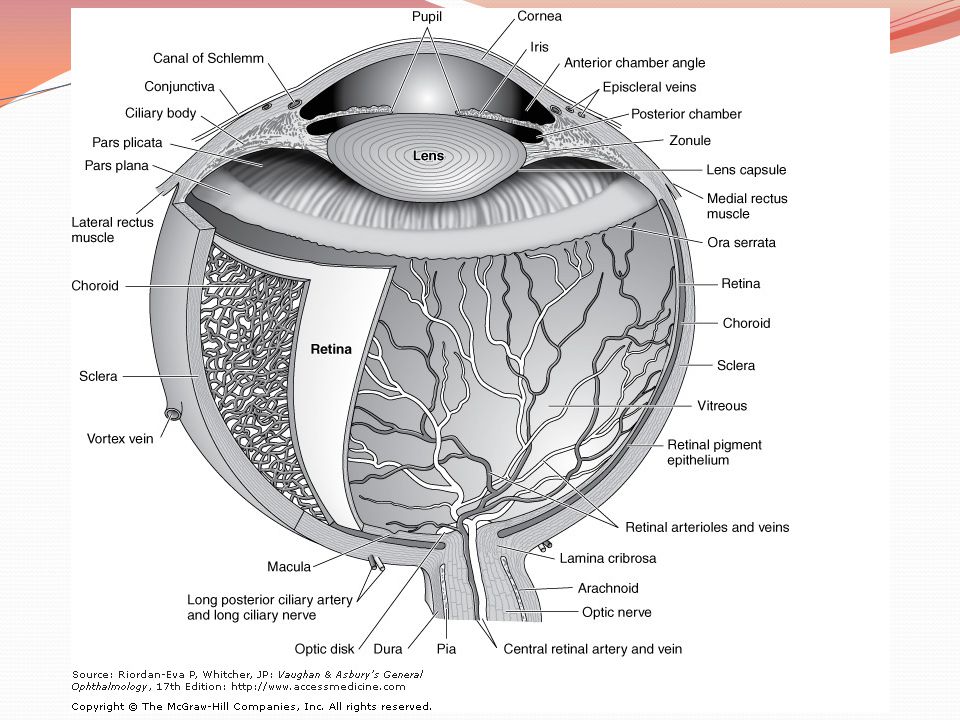
A thorough understanding of the anatomy of the eye, orbit, visual pathways, upper cranial nerves, and central pathways for the control of eye movements is a prerequisite for proper interpretation of diseases having ocular manifestations. Furthermore, such anatomic knowledge is essential to the proper planning and safe execution of ocular and orbital surgery.

4
The Ocular Adnexa Eyebrows Eyelids Lacrimal Apparatus

5
Eyelids

6
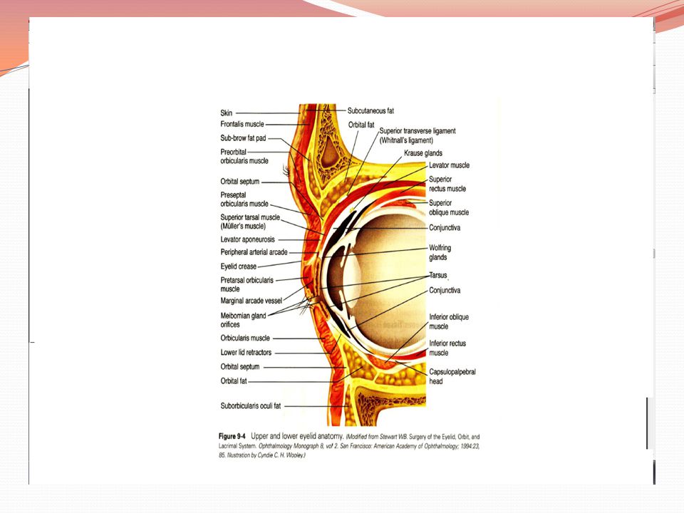
From superficial to deep, they are the skin layer, a layer of striated muscle (orbicularis oculi), areolar tissue, fibrous tissue (tarsal plates), and a layer of mucous membrane (palpebral conjunctiva)
, areolar tissue, fibrous tissue (tarsal plates), and a layer of mucous membrane (palpebral conjunctiva)")
9
Lid Margins Anterior Margin Eyelashes Glands of Zeis Glands of Moll
Posterior Margin small orifices of modified sebaceous glands (meibomian, or tarsal, glands).
.")
10
Orbital Septum the fascia behind that portion of the orbicularis muscle that lies between the orbital rim and the tarsus and serves as a barrier between the lid and the orbit.

11
Lid Retractors The lid retractors are responsible for opening the eyelids. In the upper lid: levator palpebrae superioris & Muller's (superior tarsal) muscle In the lower lid:the main retractor is the inferior rectus The smooth muscle components of the lid retractors are innervated by sympathetic nerves. The levator and inferior rectus muscles are supplied by the third cranial (oculomotor) nerve. Ptosis is thus a feature of both Horner's syndrome and third nerve palsy
 muscle. In the lower lid:the main retractor is the inferior rectus. The smooth muscle components of the lid retractors are innervated by sympathetic nerves. The levator and inferior rectus muscles are supplied by the third cranial (oculomotor) nerve. Ptosis is thus a feature of both Horner s syndrome and third nerve palsy.")
12
Sensory Nerve Supply first and second divisions of the trigeminal nerve (V).
.")
13
Blood Supply & Lymphatics
the lacrimal and ophthalmic arteries by their lateral and medial palpebral branches. Venous drainage from the lids empties into the ophthalmic vein and the veins that drain the forehead and temple Lymphatics from the lateral segment of the lids run into the preauricular and parotid nodes. Lymphatics draining the medial side of the lids empty into the submandibular lymph nodes

14
The Lacrimal Apparatus

15
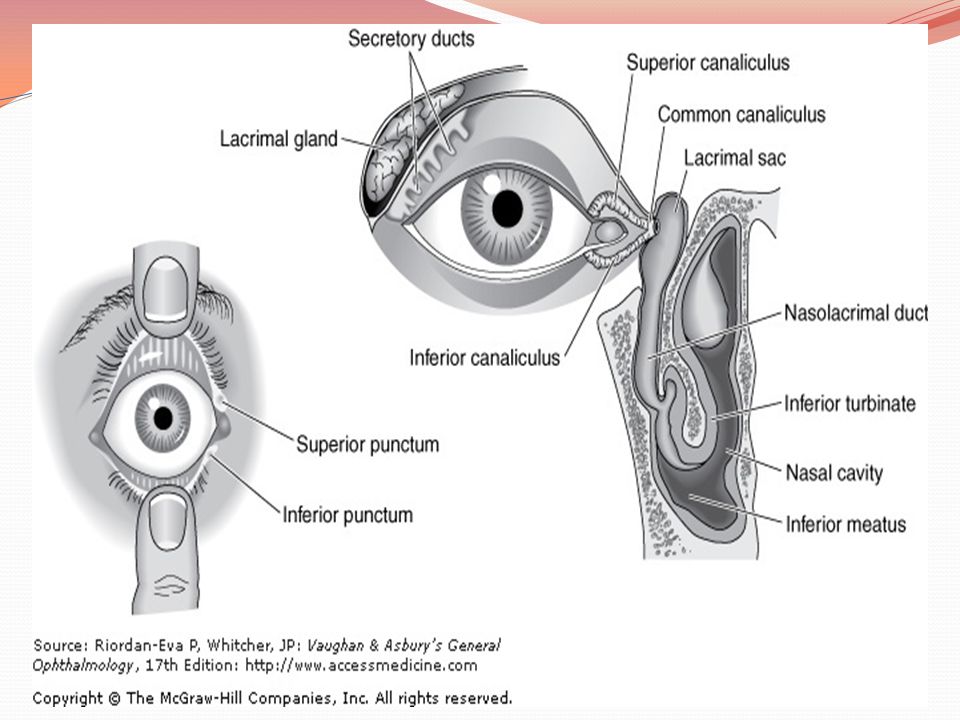
consists of the lacrimal gland, the accessory lacrimal glands, the canaliculi, the lacrimal sac.
The accessory lacrimal glands (glands of Krause and Wolfring) are located in the substantia propria of the palpebral conjunctiva
 are located in the substantia propria of the palpebral conjunctiva.")
16
The blood supply of the lacrimal gland is derived from the lacrimal artery.
The nerve supply to the lacrimal gland is by (1) the lacrimal nerve (sensory), a branch of the trigeminal first division; (2) the great superficial petrosal nerve (secretory), which comes from the superior salivary nucleus; and (3) sympathetic nerves accompanying the lacrimal artery and the lacrimal nerve
 the lacrimal nerve (sensory), a branch of the trigeminal first division; (2) the great superficial petrosal nerve (secretory), which comes from the superior salivary nucleus; and (3) sympathetic nerves accompanying the lacrimal artery and the lacrimal nerve.")
17
The angular vein and artery lie just deep to the skin, 8 mm to the nasal side of the inner canthus.
Skin incisions made in surgical procedures on the lacrimal sac should always be placed 2–3 mm to the nasal side of the inner canthus to avoid these vessels.

20
The Orbit

21
a pyramid of four walls that converge posteriorly
a pyramid of four walls that converge posteriorly. The medial walls of the right and left orbit parallel and are separated by the nose. In each orbit, the lateral and medial walls form an angle of 45 degrees, which results in a right angle between the two lateral walls. The orbit is compared to the shape of a pear, with the optic nerve representing its stem.

22
volume of the adult orbit :30 mL
eyeball occupies only about one-fifth of the space. Fat and muscle account for the bulk of the remainder.

23
The anterior limit of the orbital cavity is the orbital septum, which acts as a barrier between the eyelids and orbit

24
frontal sinus above, the maxillary sinus below, and the ethmoid and sphenoid sinuses medially.

25
The thin orbital floor is easily damaged by direct trauma to the globe, resulting in a "blowout" fracture with herniation of orbital contents into the maxillary antrum.

26
Infection within the sphenoid and ethmoid sinuses can erode the paper-thin medial wall (lamina papyracea) and involve the contents of the orbit.
 and involve the contents of the orbit.")
27
Defects in the roof (eg, neurofibromatosis) may result in visible pulsations of the globe transmitted from the brain.
 may result in visible pulsations of the globe transmitted from the brain.")
28
Orbital Walls

29
The roof : frontal bone. Posteriorly, the lesser wing of the sphenoid bone containing the optic canal completes the roof. The lacrimal gland is located in the lacrimal fossa in the anterior lateral aspect of the roof.

30
The lateral wall is separated from the roof by the superior orbital fissure, which divides the lesser from the greater wing of the sphenoid bone. The anterior portion of the lateral wall is formed by the orbital surface of the zygomatic (malar) bone. This is the strongest part of the bony orbit.
 bone. This is the strongest part of the bony orbit..")
31
Suspensory ligaments, the lateral palpebral tendon, and check ligaments have connective tissue attachments to the lateral orbital tubercle.

32
The orbital floor is separated from the lateral wall by the inferior orbital fissure. The orbital plate of the maxilla forms the large central area of the floor and is the region where blowout fractures most frequently occur. The frontal process of the maxilla medially and the zygomatic bone laterally complete the inferior orbital rim. The orbital process of the palatine bone forms a small triangular area in the posterior floor.

33
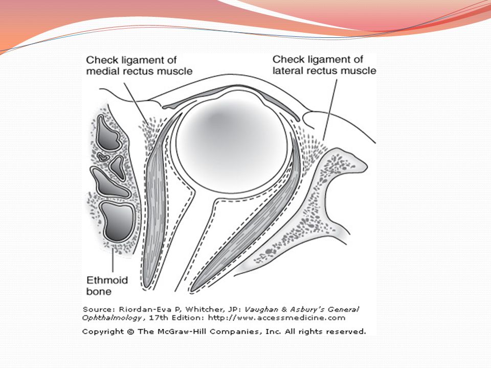
medial wall : ethmoid bone ; lacrimal bone; The body of the sphenoid ( most posterior aspect of the medial wall) the angula r process of the frontal bone forms the upper part of the posterior lacrimal crest. The lower portion of the posterior lacrimal crest is made up of the lacrimal bone. The anterior lacrimal crest is easily palpated through the lid and is composed of the frontal process of the maxilla. The lacrimal groove lies between the two crests and contains the lacrimal sac
. the angula r process of the frontal bone forms the upper part of the posterior lacrimal crest. The lower portion of the posterior lacrimal crest is made up of the lacrimal bone. The anterior lacrimal crest is easily palpated through the lid and is composed of the frontal process of the maxilla. The lacrimal groove lies between the two crests and contains the lacrimal sac.")
35
Orbital Apex

36
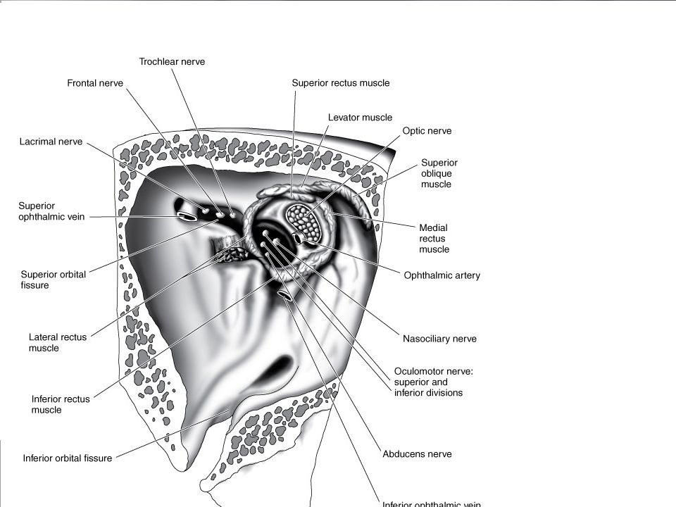
entry portal for all nerves and vessels to the eye and the site of origin of all extraocular muscles except the inferior oblique.

37
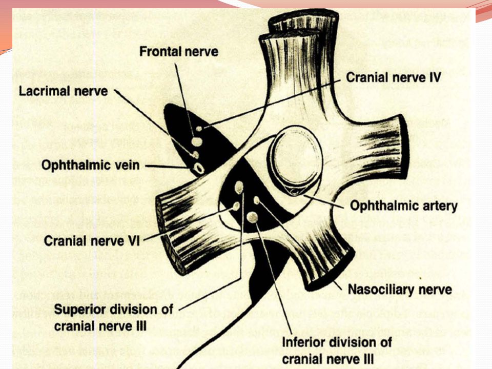
The superior ophthalmic vein and the lacrimal, frontal, and trochlear nerves pass through the lateral portion of the fissure that lies outside the annulus of Zinn. The superior and inferior divisions of the oculomotor nerve and the abducens and nasociliary nerves pass through the medial portion of the fissure within the annulus of Zinn. The optic nerve and ophthalmic artery pass through the optic canal, which also lies within the annulus of Zinn

40
Blood Supply

41
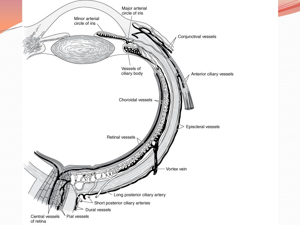
The principal arterial supply of the orbit : ophthalmic artery( the first major branch of the intracranial portion of the internal carotid artery). This branch passes beneath the optic nerve and accompanies it through the optic canal into the orbit. The first intraorbital branch is the central retinal artery, which enters the optic nerve . Other branches of the ophthalmic artery include the lacrimal artery, supplying the lacrimal gland and upper eyelid; muscular branches to the various muscles of the orbit; long and short posterior ciliary arteries; medial palpebral arteries to both eyelids; and the supraorbital and supratrochlear arteries. The short posterior ciliary arteries supply the choroid and parts of the optic ner ve. The two long posterior ciliary arteries supply the ciliary body and anastomose with each other and with the anterior ciliary arteries to form the major arterial circle of the iris. The anterior ciliary arteries are derived from the muscular branches to the rectus muscles. They supply the anterior sclera, episclera, limbus, and conjunctiva and contribute to the major arterial circle of the iris. The most anterior branches of the ophthalmic artery contribute to the formation of The most anterior branches of the ophthalmic artery contribute to the formation of the arterial arcades of the eyelids, which make an anastomosis with the external carotid circulation via the facial artery

42
The venous drainage of the orbit is primarily through the superior and inferior ophthalmic veins, into which drain the vortex veins, the anterior ciliary veins, and the central retinal vein. The ophthalmic veins communicate with the cavernous sinus via the superior orbital fissure and the pterygoid venous plexus via the inferior orbital fissure. The superior ophthalmic vein is initially formed from the supraorbital and supratrochlear veins and from a branch of the angular vein, all of which drain the skin of the periorbital region. This provides a direct communication between the skin of the face and the cavernous sinus, thus forming the basis of the potentially lethal cavernous sinus thrombosis secondary to superficial infection of the periorbital skin.

45
The Extraocular Muscles

46
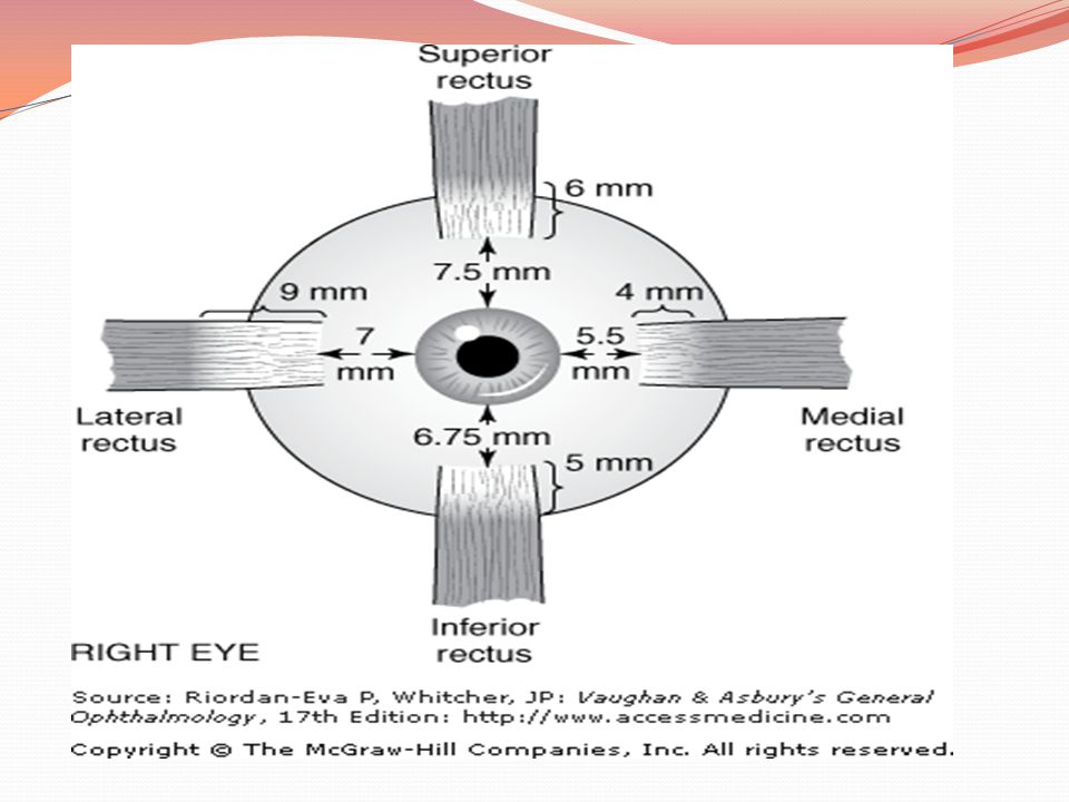
Rectus Muscles originate at a common ring tendon (annulus of Zinn) . Function distances from the limbus

47
Oblique Muscles control primarily torsional movement and, to a lesser extent, upward and downward movement of the globe

48
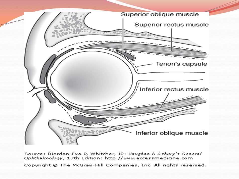
The superior oblique is the longest and thinnest of the ocular muscles.

49
The inferior oblique muscle originates from the nasal side of the orbital wall just behind the inferior orbital rim and lateral to the nasolacrimal duct. The insertion is into the posterotemporal segment of the globe and just over the macular area.

50
Nerve Supply The oculomotor nerve (III) innervates the medial, inferior, and superior rectus muscles and the inferior oblique muscle. The abducens nerve (VI) innervates the lateral rectus muscle; the trochlear nerve (IV) innervates the superior oblique muscle.
 innervates the lateral rectus muscle; the trochlear nerve (IV) innervates the superior oblique muscle.")
55
The Conjunctiva

56
thin, transparent mucous membrane that covers the posterior surface of the lids (the palpebral conjunctiva) and the anterior surface of the sclera (the bulbar conjunctiva).
 and the anterior surface of the sclera (the bulbar conjunctiva).")
57
The palpebral conjunctiva lines the posterior surface of the lids and is firmly adherent to the tarsus. Except at the limbus (where Tenon's capsule and the conjunctiva are fused for about 3 mm), the bulbar conjunctiva is loosely attached to Tenon's capsule and the underlying sclera
, the bulbar conjunctiva is loosely attached to Tenon s capsule and the underlying sclera.")
58
The accessory lacrimal glands (glands of Krause and Wolfring): in the stroma.
Most of the glands of Krause are in the upper fornix. The glands of Wolfring lie at the superior margin of the upper tarsus.

59
Blood Supply& Nerve Supply
anterior ciliary and palpebral arteries. the first (ophthalmic) division of the fifth nerve.
 division of the fifth nerve.")
60
Tenon's Capsule (Fascia Bulbi)
")
61
fibrous membrane that envelops the globe from the limbus to the optic nerve. Adjacent to the limbus, the conjunctiva, Tenon's capsule, and episclera are fused together. More posteriorly, the inner surface of Tenon's capsule lies against the sclera, and its outer aspect is in contact with orbital fat and other structures within the extraocular muscle cone. At the point where Tenon's capsule is pierced by tendons of the extraocular muscles in their passage to their attachments to the globe, it sends a tubular reflection around each of these muscles. These fascial reflections become continuous with the fascia of the muscles, the fused fasciae sending expansions to the surrounding structures and to the orbital bones. The fascial expansions are quite tough and limit the action of the extraocular muscles and are therefore known as check ligaments. They regulate the direction of action of the extraocular muscles and act as their functional mechanical origins. The lower segment of Tenon's capsule is thick and fuses with the fascia of the inferior rectus and the inferior oblique muscles to form the suspensory ligament of the eyeball (Lockwood's ligament), upon which the globe rests.
, upon which the globe rests.")
63
The Sclera & Episclera

64
The sclera is the fibrous outer protective coating of the eye, consisting almost entirely of collagen . The outer surface of the anterior sclera is covered by a thin layer of fine elastic tissue, the episclera, which contains numerous blood vessels that nourish the sclera. The brown pigment layer on the inner surface of the sclera is the lamina fusca, which forms the outer layer of the suprachoroidal space.

65
At the insertion of the rectus muscles, the sclera is about 0
At the insertion of the rectus muscles, the sclera is about 0.3 mm thick; elsewhere it is about 0.6 mm thick.

66
nerve supply to the sclera :
ciliary nerves

67
The histologic structure of the sclera is remarkably similar to that of the cornea.
The reason for the transparency of the cornea and the opacity of the sclera is the relative deturgescence of the cornea.

70
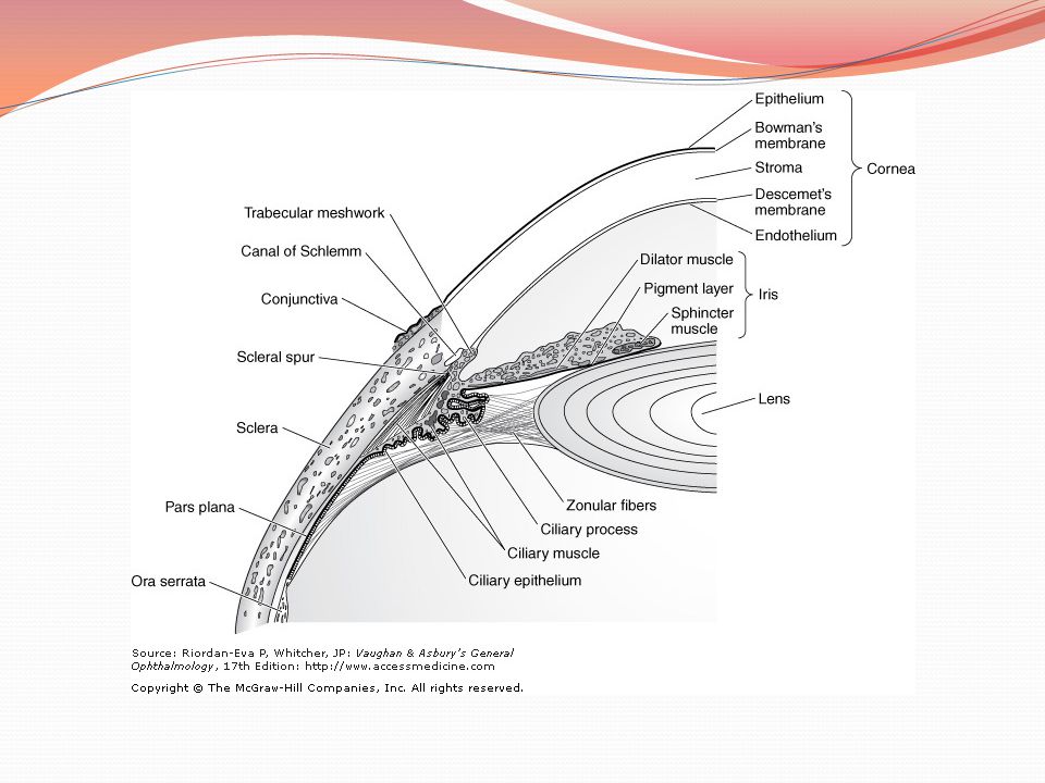
The Cornea

71
The average adult cornea is 550 m thick in the center, although there are racial variations, and about mm in diameter horizontally and 10.6 mm vertically.

72
From anterior to posterior, it has five distinct layers : the epithelium , Bowman's layer, the stroma, Descemet's membrane, and the endothelium. The corneal stroma accounts for about 90% of the corneal thickness.

74
The endothelium has only one layer of cells, but this is responsible for maintaining the essential deturgescence of the corneal stroma. The endothelium is quite susceptible to injury as well as undergoing loss of cells with age. Failure of endothelial function leads to corneal edema.

75
Sources of nutrition for the cornea :
vessels of the limbus, the aqueous, and the tears. The superficial cornea also gets most of its oxygen from the atmosphere. The sensory nerves of the cornea are supplied by the first (ophthalmic) division of the fifth (trigeminal) cranial nerve.
 division of the fifth (trigeminal) cranial nerve.")
76
The transparency of the cornea :
uniform structure, avascularity, and deturgescence.

78
The Uveal Tract

79
iris, the ciliary body, and the choroid
It contributes blood supply to the retina.

80
Iris

81
dividing the anterior chamber from the posterior chamber, each of which contains aqueous humor.

82
The blood supply : major circle of the iris .
Iris capillaries have a nonfenestrated endothelium and hence do not normally leak intravenously injected fluorescein. Sensory nerve supply: fibers in the ciliary nerves.

83
The Ciliary Body

84
It consists of a corrugated anterior zone, the pars plicata , and a flattened posterior zone, the pars plana . The ciliary processes arise from the pars plicata .

85
The capillaries are large and fenestrated and hence leak intravenously injected fluorescein.

86
The ciliary processes and their covering ciliary epithelium are responsible for the formation of aqueous.

87
The ciliary muscle is composed of a combination of longitudinal, circular, and radial fibers.
The function of the circular fibers is to contract and relax the zonular fibers, which originate in the valleys between the ciliary processes.

88
The blood vessels supplying the ciliary body are derived from the major circle of the iris.

89
The Choroid

90
the posterior segment of the uveal tract, between the retina and the sclera.
The internal portion of the choroid vessels is known as the choriocapillaris. The choroid is bounded internally by Bruch's membrane and externally by the sclera. serves to nourish the outer portion of the underlying retina

92
The Lens

93
The lens is a biconvex, avascular, colorless, and almost completely transparent structure, about 4 mm thick and 9 mm in diameter. It is suspended behind the iris by the zonule, which connects it with the ciliary body

94
lens gradually becomes larger and less elastic throughout life.

95
The lens consists of about 65% water, about 35% protein (the highest protein content of any tissue of the body). Potassium is more concentrated in the lens than in most tissues. There are no pain fibers, blood vessels, or nerves in the lens

98
The Retina

99
The retina is a thin, semitransparent, multilayered sheet of neural tissue that lines the inner aspect of the posterior two-thirds of the wall of the globe. It extends almost as far anteriorly as the ciliary body, ending at that point in a ragged edge, the ora serrata . The outer surface of the sensory retina is apposed to the retinal pigment epithelium and thus related to Bruch's membrane, the choroid, and the sclera.

100
In most areas, the retina and retinal pigment epithelium are easily separated to form the subretinal space, such as occurs in retinal detachment. But at the optic disk and the ora serrata, the retina and retinal pigment epithelium are firmly bound together, thus limiting the spread of subretinal fluid in retinal detachment. This contrasts with the potential suprachoroidal space between the choroid and sclera, which extends to the scleral spur. Choroidal detachments thus extend beyond the ora serrata, under the pars plana and pars plicata.

101
The layers of the retina, starting from its inner aspect, are as follows: (1) internal limiting membrane; (2) nerve fiber layer, containing the ganglion cell axons passing to the optic nerve; (3) ganglion cell layer; (4) inner plexiform layer, containing the connections of the ganglion cells with the amacrine and bipolar cells; (5) inner nuclear layer of bipolar, amacrine, and horizontal cell bodies; (6) outer plexiform layer, containing the connections of the bipolar and horizontal cells with the photoreceptors; (7) outer nuclear layer of photoreceptor cell nuclei; (8) external limiting membrane; (9) photoreceptor layer of rod and cone inner and outer segments; and (10) retinal pigment epithelium .
 internal limiting membrane; (2) nerve fiber layer, containing the ganglion cell axons passing to the optic nerve; (3) ganglion cell layer; (4) inner plexiform layer, containing the connections of the ganglion cells with the amacrine and bipolar cells; (5) inner nuclear layer of bipolar, amacrine, and horizontal cell bodies; (6) outer plexiform layer, containing the connections of the bipolar and horizontal cells with the photoreceptors; (7) outer nuclear layer of photoreceptor cell nuclei; (8) external limiting membrane; (9) photoreceptor layer of rod and cone inner and outer segments; and (10) retinal pigment epithelium .")
102
The 1.5-mm-diameter fovea corresponds to the retinal avascular zone of fluorescein angiography.
Histologically it is characterized by thinning of the outer nuclear layer and absence of the other parenchymal layer

103
thinnest part of area of the retina (0
thinnest part of area of the retina (0.25 mm), containing only cone photoreceptor
, containing only cone photoreceptor.")
104
blood supply from two sources: the choriocapillaris which supplies the outer third of the retina, and branches of the central retinal artery, which supply the inner two-thirds . The fovea is supplied entirely by the choriocapillaris and is susceptible to irreparable damage when the retina is detached.

105
The retinal blood vessels have a nonfenestrated endothelium, which forms the inner blood-retinal barrier. The endothelium of choroidal vessels is fenestrated. The outer blood-retinal barrier lies at the level of the retinal pigment epithelium.

106
The Vitreous

107
clear, avascular, gelatinous body that comprises two-thirds of the volume and weight of the eye.
The outer surface of the vitreous—the hyaloid membrane—is normally in contact with the following structures: the posterior lens capsule, the zonular fibers, the pars plana epithelium, the retina, and the optic nerve head. The base of the vitreous maintains a firm attachment throughout life to the pars plana epithelium and the retina immediately behind the ora serrata. The attachment to the lens capsule and the optic nerve head is firm in early life but soon disappears.

108
99% water. The remaining 1% includes two components, collagen and hyaluronic acid

109
The External Anatomic Landmarks

110
Accurate localization of the position of internal structures with reference to the external surface of the globe is important in many surgical procedures. Externally, the ora serrata is situated approximately 5.5 mm from the limbus on the medial side and 7 mm on the temporal side of the globe. This corresponds to the level of insertion of the rectus muscles.

111
Injections into the vitreous cavity through the pars plana should be given 3.5–4.0 mm from the limbus in the phakic eye and 3–3.5 mm from the limbus in the pseudophakic or aphakic eye. The pars plicata, which is the target for cyclodestructive procedures in the treatment of intractable glaucoma, occupies the 2–3 mm directly posterior to the limbus.

112
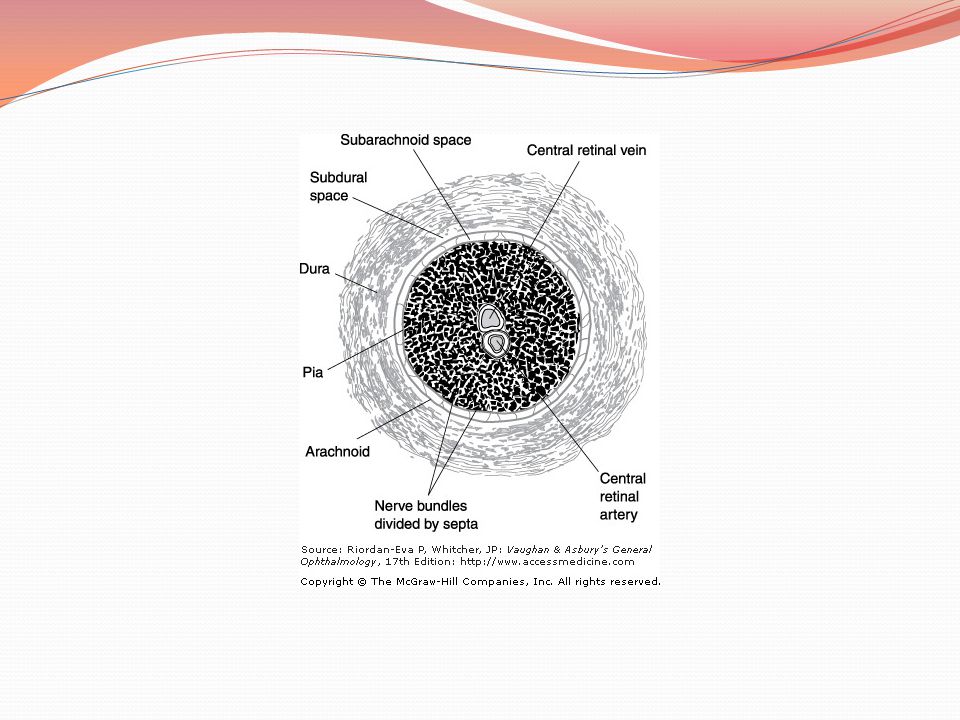
The Optic Nerve

113
consists of about 1 million axons that arise from the ganglion cells of the retina (nerve fiber layer). The orbital segment of the nerve is 25–30 mm long; it travels within the optic muscle cone, via the bony optic canal, and thus gains access to the cranial cavity. The intracanalicular portion measures 4–9 mm. After a 10-mm intracranial course, the nerve joins the opposite optic nerve to form the optic chiasm. Eighty percent of the optic nerve consists of visual fibers that synapse in the lateral geniculate body on neurons whose axons terminate in the primary visual cortex of the occipital lobes. Twenty percent of the fibers are pupillary and bypass the geniculate body en route to the pretectal area.

114
Blood Supply The surface layer of the optic disk receives blood from branches of the retinal arterioles. In the region of the lamina cribrosa, comprising the prelaminar, laminar, and retrolaminar segments of the optic nerve, the arterial supply is from the short posterior ciliary arteries. The anterior intraorbital optic nerve receives some blood from branches of the central retinal artery. The remainder of the intraorbital nerve, as well as the intracanalicular and intracranial portions, are supplied by a pial network of vessels derived from the various branches of the ophthalmic artery and other branches of the internal carotids

117
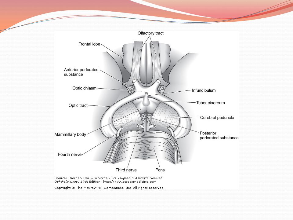
The Optic Chiasm

118
near the top of the diaphragm of the sella turcica.
The chiasm is made up of the junction of the two optic nerves and provides for crossing of the nasal fibers to the opposite optic tract and passage of temporal fibers to the ipsilateral optic tract. The macular fibers are arranged similarly to the rest of the fibers except that their decussation is farther posteriorly and superiorly. The chiasm receives many small blood vessels from the neighboring circle of Willis.

120
The Oculomotor Nerve (III)
")
121
leaves the brainstem between the cerebral peduncles and passes near the posterior communicating artery of the circle of Willis. Lateral to the pituitary gland, it is closely approximated to the optic tract, and here it pierces the dura to course in the lateral wall of the cavernous sinus. As the nerve leaves the cavernous sinus, it divides into superior and inferior divisions. The superior division enters the orbit within the annulus of Zinn at its highest point and adjacent to the trochlear nerve. The inferior division enters the annulus of Zinn low and passes below the optic nerve to supply the medial and inferior rectus muscles. A large branch from the inferior division extends forward to supply the inferior oblique. A small twig from the proximal end of the nerve to the inferior oblique carries parasympathetic fibers to the ciliary ganglion

122
The Trochlear Nerve (IV)
")
123
Although the thinnest of the cranial nerves, the trochlear nerve has the longest intracranial course, and it is also the only nerve to originate on the dorsal surface of the brain stem. The nerve pierces the dura behind the sella turcica and travels within the lateral walls of the cavernous sinus to enter the superior orbital fissure medial to the frontal nerve. From this point it travels within the periorbita of the roof over the levator muscle to the upper surface of the superior oblique muscle.

124
The Trigeminal Nerve (V)
")
125
originates from the pons
originates from the pons. The first (ophthalmic) of the three divisions passes through the lateral wall of the cavernous sinus and divides into the lacrimal, frontal, and nasociliary nerves.
 of the three divisions passes through the lateral wall of the cavernous sinus and divides into the lacrimal, frontal, and nasociliary nerves.")
126
The Abducens Nerve (VI)
")
127
originates between the pons and medulla
passes within the cavernous sinus. (All other nerves course through the lateral wall of the cavernous sinus.) After passing through the superior orbital fissure within the annulus of Zinn, the nerve continues laterally to innervate the lateral rectus muscle.
 After passing through the superior orbital fissure within the annulus of Zinn, the nerve continues laterally to innervate the lateral rectus muscle.")
128
Thanks for your attention

Similar presentations


>")


