Download presentation
Presentation is loading. Please wait.

1
HYPERSENSITIVITY REACTIONS

2
Innocous materials can cause hypersensitivity in certain individuals leading to unwanted inflammation damaged cells and tissues Non-proper reaction of the immune system to foreign substances Mainly harmless substances – after second or multiple exposure HYPERSENSITIVITY REACTIONS

3
Type I. „immediate” Type II.Type III.Type IV. „late” Antibody mediatedT cell mediated AN OVERVIEW OF HYPERSENSITIVITY REACTIONS

4
TYPES OF ANTIBODY MEDIATED HYPERSENSITIVITY REACTIONS Fc RIα)
")
5
TYPE I HYPERSENSITIVITY REACTION ALLERGY

6
SENSITISATION PROCESS

7
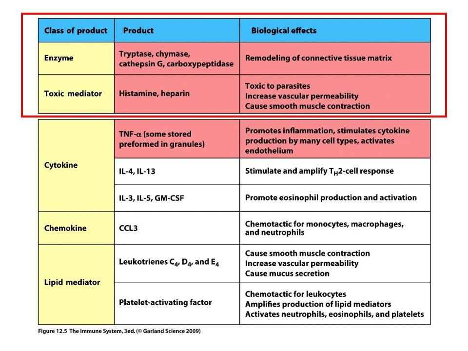
MAST CELL RESPONSE TO SURFACE FcRεI CROSSLINKING EARLY MEDIATORS Biogenic amins – histamin Enzymes – triptase, chymase, carboxypeptidase LATE MEDIATORS

9
THE EFFECT OF MAST CELL DEGRANULATION VARIES WITH THE TISSUE EXPOSED TO ALLERGEN

10
SYSTEMIC ANAPHYLAXIS IS CAUSED BY ALLERGENS THAT REACH THE BLOOD STREAM

11
TYPES OF IgE-DERIVED ALLERGIC RESPONSES SYNDROMEALLERGENSROUTE OF ENTRYRESPONSE systemic anaphylaxis drugs anti-serum intravenousedema, increased vascular permeability tracheal occlusion circulatory collapse, death acute urticaria bug bite allergy test subcutanlocal increase in blood flow and vascular permeability allergic rhinitis pollen dust mite drops inhaledirritation and edema of nasal mucosa airway inflammation asthmaanimal fur pollen dust mite drops inhaledbronchial constriction, increased mucus production food allergynut, peanuts, fish, shellfish milk, eggs oralvomiting, diarrhea pruritis (itching) urticaria (hives) anaphylaxia (rare)
 urticaria (hives) anaphylaxia (rare)")
12
Short/Common ragweed (Ambrosia artemisiifolia)
")
13
Mugwort (Artemisia vulgaris) Green leaf back White leaf back
 Green leaf back White leaf back")
14
Mugwort (Artemisia vulgaris) –? Wormwood (Artemisia absinthium) – Absinthe (thujone: max 35 mg/l)
 – Wormwood (Artemisia absinthium) – Absinthe (thujone: max 35 mg/l)")
15
PRICK TEST MAST CELL DEGRANULATION, ALLERGIC REACTION IN THE SKIN OF A SENSIBILIZED INDIVIDUAL

16
Prick test

17
ImmunoCAP Specific IgE Blood Test Anti-IgE Serum IgE Allergen Solid phase

18
Skin Prick testSpecific IgE allergensmanyalmost all speed20 min1-2 days (result) medicationno antihistaminesno problem diseasesevere eczema:no problem difficult cost€ 20 (total)€ 20 per specific IgE sensitivityhighslightly lower COMPARISON OF SKIN TEST TO SPECIFIC IgE TESTING
 medicationno antihistaminesno problem diseasesevere eczema:no problem difficult cost€ 20 (total)€ 20 per specific IgE sensitivityhighslightly lower COMPARISON OF SKIN TEST TO SPECIFIC IgE TESTING")
19
TYPE II HYPERSENSITIVITY IgG type antibodies bound to cell surface or tissue antigens cells expressing the antigen become sensitive to complement mediated lysis or to opsonized phagocytosis frustrated phagocytosis tissue damage the antibody inhibits or stimulates target cell function no tissue damage (e.g. M. gravis – receptor-blocking antibodies)
.")
20
MECHANISMS OF TYPE II HYPERSENSITIVITY REACTIONS

21
The tissue, which can not be phagocytosed, is damaged Absorbed antigen (drug) Binding Opsonization Internalization Enzyme release Opsonized surface Binding Frustrated Enzyme release phagocytosis FRUSTRATED PHAGOCYTOSIS MEDIATED BY IgG TYPE ANTIBODIES
 Binding Opsonization Internalization Enzyme release Opsonized surface Binding Frustrated Enzyme release phagocytosis FRUSTRATED PHAGOCYTOSIS MEDIATED BY IgG TYPE ANTIBODIES")
22
EXAMPLES - TYPE II HYPERSENSITIVITY Newborn haemolytic anaemia Transfusion reaction Hyperacut allograft rejection Drug-derived Haemolitic anaemia Thrombocytopenia Agranulocytosis Penicillin-based antibiotics Anti-arithmic quinidine Goodpasture syndrome (kidney, basal membrane - collagen type IV) Pemphigus vulgaris (mucosal bubbles) against desmosomal antigens, interruption of epidermal and mucosal connections, acantolysis (desintegration into single cells) Myasthaenia gravis (anti-acetyl-choline receptor antibodies) Graves-Basedow-Flajani disease (anti-TSH-receptor antibodies)
 Pemphigus vulgaris (mucosal bubbles) against desmosomal antigens, interruption of epidermal and mucosal connections, acantolysis (desintegration into single cells) Myasthaenia gravis (anti-acetyl-choline receptor antibodies) Graves-Basedow-Flajani disease (anti-TSH-receptor antibodies)")
23
DEVELOPMENT OF DRUG SENSITIVITY I.

24
DEVELOPMENT OF DRUG SENSITIVITY II.

25
TYPE III HYPERSENSITIVITY Antibodies binding to soluble antigens forming small circulating immune complexes which are deposited in various tissues Depends on: Size of immune complexes Antigen-antibody ratio Affinity of antibody Isotype of antibody

26
THE PROCESS OF TISSUE DAMAGE CAUSED BY IMMUNE COMPLEXES Immune complexes activate the complement system, neutrophils, basophils and thrombocytes Blood vessel wall permeability Frustrated phagocytosis

27
SYPMPTPOMES CAUSED BY TYPE III HYPERSENSITIVITY REACTIONS DEPEND ON THE SITE OF IMMUNECOMPLEX DEPOSITION

28
ARTHUS-REACTION Localized Type III hypersensitivity Local vasculitis develops as a result of immune complex deposition Inhaled antigens (fungi, animal feces) may induce similar reaction in the lung IgG type antibody ‘Farmers lung’ and ‘piegeon-breeder’s lung’
 may induce similar reaction in the lung IgG type antibody ‘Farmers lung’ and ‘piegeon-breeder’s lung’")
29
Facial, malar "butterfly" rash with characteristic shape across the cheeks. Discoid lupus erythematosus (DLE) involves mainly the skin, it is relatively benign compared to systemic lupus erythematosus (SLE). In either case, sunlight exposure accentuates this erythematous rash. A small number (5 to 10%) of DLE patients go on to develop SLE (usually the DLE patients with a positive ANA). Here is a more severe inflammatory skin infiltrate in the upper dermis of a patient with SLE in which the basal layer is undergoing vacuolization and dissolution, and there is purpura with RBC's in the upper dermis (which are the reason for the rash). MANIFESTATION OF TYPE III HYPERSENSITIVITY IN LUPUS ERYTHEMATOSUS
 involves mainly the skin, it is relatively benign compared to systemic lupus erythematosus (SLE). In either case, sunlight exposure accentuates this erythematous rash. A small number (5 to 10%) of DLE patients go on to develop SLE (usually the DLE patients with a positive ANA). Here is a more severe inflammatory skin infiltrate in the upper dermis of a patient with SLE in which the basal layer is undergoing vacuolization and dissolution, and there is purpura with RBC s in the upper dermis (which are the reason for the rash). MANIFESTATION OF TYPE III HYPERSENSITIVITY IN LUPUS ERYTHEMATOSUS.")
30
When immunofluorescence staining with an antibody to complement or immunoglobulin is performed, a brightly fluorescent signal staining the dermal epidermal junction is visible indicating immune complex deposition. Immunofluorescence staining pattern with antibody to IgG staining immune complexes at the dermal-epidermal junction. If such a pattern is seen only in skin involved by a rash, then the diagnosis is probably DLE, but if this pattern appears even in skin uninvolved by a rash, then the diagnosis is SLE. DEPOSITION OF IMMUNE COMPLEXES IN THE SKIN OF SLE PATIENTS

31
One of the feared complications of the systemic autoimmune diseases is renal failure. This is most likely to occur in SLE. Here is a glomerulus in which the capillary loops are markedly pink and thickened such that capillary lumens are hard to see. This is lupus nephritis. RENAL FAILURE IN IMMUNECOMPLEX DISEASES Glomerulus of a healthy individual. Relatively wide spaces between capillary loops.

32
This is the so-called "rim" pattern that is more characteristic of SLE. This is the so-called "speckled" pattern of staining which is more characteristic of the presence of autoantibodies to extractable nuclear antigens, particularly ribonucleoprotein. This pattern is not very specific, but may be seen with an entity called "mixed connective tissue disease" which is a mix between SLE, scleroderma, and polymyositis, but without serious renal or pulmonary disease. The autoimmune diseases are very hard to classify, even for the experts. This is the so-called "nucleolar pattern" of staining in which the bright fluorescence is seen within the nucleoli of the Hep2 cells. This pattern is more suggestive of progressive systemic sclerosis.ANA Anti-nuclear antibody

33
TYPE IV HYPERSENSITIVITY REACTION T CELL MEDIATED PROCESS

34
Chemokines, cytokines, cytotoxins TYPE IV HYPERSENSITIVITY REACTION

35
DELAYED-TYPE (TYPE IV) HYPERSENSITIVITY
 HYPERSENSITIVITY")
37
T H 1 from a previous immunization (memory) DELAYED-TYPE HYPERSENSITIVITY (DTH) DELAYED-TYPE HYPERSENSITIVITY (DTH) (e.g. tuberculin skin test)
.")
38
Tuberculin skin test Ag = antigen Purified protein derivate (PPD) Introduction of Ag
 Introduction of Ag")
39
CHEMICAL MEDIATORS OF DTH

40
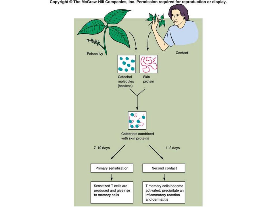
*a contact-sensitizing agent is usually a small molecule that penetrates the skin then binds to self-proteins, making them “look” foreign DTH as a result of a contact-sensitizing agent* CONTACT DERMATITIS

41
Poison ivy Anacardiaceae (family), Toxicodendron (genus) Toxicodendron radicans or Rhus toxicodendron
, Toxicodendron (genus) Toxicodendron radicans or Rhus toxicodendron")
45
Delayed-type hypersensitivity is mediated by T cells

46
CELIAC DISEASE

Similar presentations


 I) Anaphalactic II) Cytotoxic III) Immune Complex IV) Cell-mediated (Delayed) Autoimmune Diseases.>")


 leading to damage Require sensitizing dose(s) Introduction to Lab Ex. 24:>")


