Download presentation
Presentation is loading. Please wait.

1
SKELETAL RADIONUCLIDE IMAGING III Dr. Hussein Farghaly Nuclear Medicine Consultant PSMMC

2
CONTENTS Bone and BM physiology & anatomy Bone scan Radiopharmaceutical, preparation, uptake and pharmacokinetics dosimetry, protocols, normal and altered distribution Clinical indication and Skeletal pathology Bone Marrow scan

3
Soft-tissue uptake in radionuclide musculoskeletal imaging HOME WORK

5
CLINICAL USES OF SKELETAL SCINTIGRAPHY

6
Metastatic Disease The evaluation of osseous metastatic disease is the most common use of skeletal scintigraphy.

7
Metastatic Disease, cont. Patients may present with bone pain (50–80%) and elevated alkaline phosphatase (77%) but these findings are nonspecific. Bone scan may be used for staging, restaging, and monitoring therapy effectiveness. T The decision on which patients will need a bone scan depends on factors such as the type and stage of tumor, history of pain, and radiographic abnormalities. Over 90% of osseous metastasis distribute to the red marrow. In adults red marrow is found in the axial skeleton and the proximal portions of the humeri and femurs.
 and elevated alkaline phosphatase (77%) but these findings are nonspecific. Bone scan may be used for staging, restaging, and monitoring therapy effectiveness. T The decision on which patients will need a bone scan depends on factors such as the type and stage of tumor, history of pain, and radiographic abnormalities. Over 90% of osseous metastasis distribute to the red marrow. In adults red marrow is found in the axial skeleton and the proximal portions of the humeri and femurs..")
8
Metastatic Disease, cont. As the tumor enlarges, the cortex becomes involved. The body responds by attempts at repair. The Tc-99m MDP binds to these regions in areas of bone deposition. Therefore, scans image the bone response to the tumor and not the tumor itself. Even a 5% bone turnover can be detected by bone scan. Radiographs, on the other hand, require a minimum mineral loss of a 50% before a lesion is visualized. MRI is more sensitive than bone scan because signal changes in the marrow from the tumor can be visualized directly. However whole body MRI is not widely available and generally not practical at this time.

9
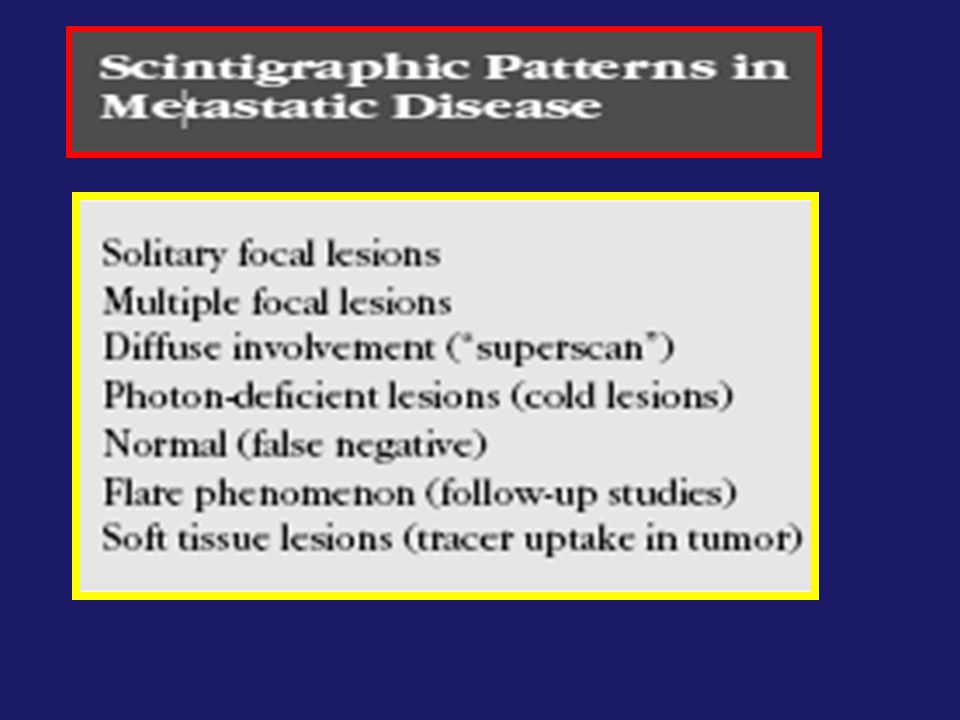
Metastatic Disease, cont.

10
Metastatic Disease in Specific Tumors Prostate Carcinoma: Until the introduction of the prostate specific antigen (PSA) blood test, bone scan was considered the most sensitive technique for detecting osseous metastasis. Serum alkaline phosphates measurement detects only half the cases detected by scintigraphy. Radiographs may be normal 30% of the time. The likelihood of an abnormal scintigram correlates with the clinical stage, Gleason score, and PSA level. Incidence of bone metastasis less than 5% early stage I disease, 10% in stage II 20% in stage III In patients with PSA levels less than 10 ng/ml, bone metastases are rarely found (<1% of the time). Skeletal scintigrams are still indicated for symptomatic patients and for evaluation of suspicious areas seen radiographically. With increasing PSA levels, the chance of detecting metastatic disease increases.
. Skeletal scintigrams are still indicated for symptomatic patients and for evaluation of suspicious areas seen radiographically. With increasing PSA levels, the chance of detecting metastatic disease increases..")
11
Breast Carcinoma: Mean survival is only 24 months among those with confirmed bone disease. Like prostate cancer, stage of disease correlates with the incidence of osseous metastases on bone scan: 0.5% in stage I, 2–3% in stage II, 8% in stage III, and 13% in stage IV. Bone scans are not generally performed in patients with stage I or II disease. Although skeletal scintigraphy has a high sensitivity for breast carcinoma, it may not detect all lesions, such as those contained in the marrow or more lytic lesions.

12
Lung Carcinoma There is no complete agreement on when to use skeletal scintigraphy. Staging is generally done with CT, surgery (including mediastinoscopy and video-assisted thoracoscopic surgery [VATS]), and increasingly with F-18 FDG PET. Skeletal scintigraphy is useful in a patient who develops pain during or after treatment and helpful in planning radiation therapy. However, it appears less useful in cases of local and mediastinal invasion or with advanced disease where therapy will be palliative.
, and increasingly with F-18 FDG PET. Skeletal scintigraphy is useful in a patient who develops pain during or after treatment and helpful in planning radiation therapy. However, it appears less useful in cases of local and mediastinal invasion or with advanced disease where therapy will be palliative..")
14
Solitary Lesions The chance that a solitary lesion is due to malignancy varies by location. Focal rib uptake is likely due to fracture, whereas uptake extending along the rib is likely tumor. Common benign causes for a solitary focus of uptake : arthritis and trauma. benign bone lesions (enchondroma, osteoma, fibrous dysplasia) osteomyelitis, monostotic Paget’s
 osteomyelitis, monostotic Paget’s.")
15
Multiple focal lesions: Is the classic pattern of metastatic disease in the skeleton Although this typical pattern provides a high degree of clinical certainty as to the diagnosis, several other etiologies can also have multiple areas of uptake These must be differentiated from osseous metastasis

16
Differential Diagnosis of Multiple Focal Lesions: The key is to recognize the different features and patterns of these other etiologies. Final diagnosis may depend on correlation with anatomical imaging. Osteoarthritic changes: Location: medial compartment of the knee, hand, and wrist (especially at the base of the first metacarpal), shoulder and bones of the feet. Bilateral: and on both sides of the joint. Patella: The patella frequently shows increased uptake due to chondromalacia and degenerative change. Spine degenerative changes: are more problematic because both metastasis and arthritic changes occur in the same location. SPECT may localize a lesion to the pedicle that is the typical location of metastasis. A bone scan lesion in the central vertebral body and disc space, could be degenerative or malignant and may require short term follow up, CT or even MRI.
, shoulder and bones of the feet. Bilateral: and on both sides of the joint. Patella: The patella frequently shows increased uptake due to chondromalacia and degenerative change. Spine degenerative changes: are more problematic because both metastasis and arthritic changes occur in the same location. SPECT may localize a lesion to the pedicle that is the typical location of metastasis. A bone scan lesion in the central vertebral body and disc space, could be degenerative or malignant and may require short term follow up, CT or even MRI..")
17
TRUMA: The findings of trauma can mimic the appearance of metastasis. Patients should be closely questioned for any history of trauma. In the ribs, a vertical alignment of focal abnormal uptake in several or successive ribs is classic for trauma. The nonrandom pattern is not expected in metastatic disease. A metastatic lesion tracks along the bone rather than remaining focal. Radiographic correlation may show the cortical disruption or callous formation. Because bone scan frequently detects fractures not seen on radiographs, correlation with CT or short-term follow-up bone scan may be needed if no fracture is seen on the radiograph. Persistently positive skeletal activity from old trauma poses another interpretive problem. Differential Diagnosis of Multiple Focal Lesions cont.: Typical appearance of rib fractures. A, Posterior views of the chest reveal focal uptake in a vertical alignment in the right lower ribs and a recent left nephrectomy with resection of some lower left ribs. B, A follow-up study 18 months later shows resolution of the right rib uptake as the fractures healed.

18
A number of other etiologies can cause multifocal abnormalities: -Infarctions in sickle cell anemia can cause multiple areas of increased and decreased uptake. - Cushing’s disease and osteomalacia, for example, frequently cause disproportionate rib lesions as compared with other areas. - Osteoporosis may result in dorsal kyphosis and classic fractures such as the vertebral insufficiency fractures and the H-type fracture of the sacrum. - Paget’s disease may be differentiated from metastasis by an expansion of the bone and classic locations. Differential Diagnosis of Multiple Focal Lesions cont.:

19
Flare Phenomenon Another potentially perplexing pattern is seen in some bone scans done on patients undergoing cyclical chemotherapy. When a patient has a good response to chemotherapy, the bone scan may paradoxically worsen, with a “flare” of increased activity. To add to the confusion, these patients may experience increased pain. If these lesions are followed radiographically, increased sclerosis is seen over 2–6 months because this is an osteoblastic response as the bone begins to heal. This is the same time frame that the bone scan typically shows increased uptake. The flare phenomenon reinforces the fact that tracer uptake is not in the tumor but rather in the surrounding bone.

20
A superscan is intense symmetric activity in the bones with diminished renal and soft tissue activity on a Tc 99 m diphosphonate bone scan This appearance can result from a range of aetiological factors: diffuse metastatic disease –prostatic carcinomaprostatic carcinoma –breast cancerbreast cancer –transitional cell carcinoma (TCC)transitional cell carcinoma (TCC) –multiple myeloma (some difference in opinion)multiple myeloma –lymphomalymphoma patchy uptake nonetheless : look at skull and ribs tends to somewhat spare the distal skeleton metabolic bone diseases –renal osteodystrophyrenal osteodystrophy –hyperparathyroidism 1 (often secondary hyperparathyroidism)hyperparathyroidism –osteomalaciaosteomalacia will involve distal skeleton smoother uptake myelofibrosis / myelosclerosis mastocytosis wide spread Paget's diseasemyelofibrosismastocytosisPaget's disease Superscan
transitional cell carcinoma (TCC) –multiple myeloma (some difference in opinion)multiple myeloma –lymphomalymphoma patchy uptake nonetheless : look at skull and ribs tends to somewhat spare the distal skeleton metabolic bone diseases –renal osteodystrophyrenal osteodystrophy –hyperparathyroidism 1 (often secondary hyperparathyroidism)hyperparathyroidism –osteomalaciaosteomalacia will involve distal skeleton smoother uptake myelofibrosis / myelosclerosis mastocytosis wide spread Paget s diseasemyelofibrosismastocytosisPaget s disease Superscan")
21
Metastatic superscan

22
Renal osteodystrophy. A–B,The absence of soft tissue uptake is striking with an appearance similar to the “superscan”seen in metastatic disease. The prominent rib end activity may help differentiate the two etiologies. The native kidneys had failed,and a renal transplant is noted in the right iliac fossa. C, Increased activity in the skull and sternum may be especially prominent. Note the increased axial skeletal uptake and paucity of soft tissue background activity.

23
Superimposed appearances of metastatic and metabolic superscan

24
Differentiation between metastatic and metabolic superscan HOME WORK

Similar presentations


 – A NEW APPROCH AND OUR EXPERIENCE Kamenetsky Natalya (1), Rachmilewitz Eliezer.>")


 M0 - No metastases M1 - Metastases present.>")


