Download presentation
Presentation is loading. Please wait.

1
Wrist and Hand Anatomy

2
Bone Anatomy Scapoid Lunate Triquetrium Pisiform Trapeziod Trapezium
Capitate Hamate

3
Hand and Wrist Anatomy 14 phalanges 5 metacarpals 8 carpal bones
2 sesamoid bones (thumb) 5 metacarpals 8 carpal bones Distal Radius Forms small ulnar notch to accept the ulnar head Radial styloid process Distal Ulna Ulnar styloid process arises from medial surface Ulnar head
 5 metacarpals. 8 carpal bones. Distal Radius. Forms small ulnar notch to accept the ulnar head. Radial styloid process. Distal Ulna. Ulnar styloid process arises from medial surface. Ulnar head.")
4
Articulations Distal Radioulnar Formed by ulnar head and ulnar notch
Allows 1 degree freedom of movement Pronation/supination Radius glides around the ulna

5
Radiocarpal joint Reinforced by ligamentous thickening
Formed by distal radius articulating with scaphoid, lunate and triangular fibrocartilage disk(TFCC) Ellipsoid joint (2 degrees freedom) Flexion/extension Radial/ulnar deviation
 Ellipsoid joint (2 degrees freedom) Flexion/extension. Radial/ulnar deviation.")
6
Intercarpal Joints Palmar/dorsal/interosseous ligaments between each carpal Very little gliding

7
Midcarpal Joints Proximal/distal carpal row separated by a single joint cavity with small fibrous projections connecting the rows Limited mobility in flex/ext, radial/ulnar deviation

8
Carpometacarpal Joint (CMC)
MC1/trapezium MC2/trapezoid MC3/capitate MC4and 5/hamate (forms 1 articulation)
")
9
1st CMC (thumb) Saddle joint 2 degrees of freedom(3)
Flexion/extension Abduction/adduction Accessory rotation Allows for opposition

10
2-4 CMC 5th CMC Plane/synovial joint 1 degree freedom 2 degree freedom
Flexion/extension 5th CMC 2 degree freedom Abduction/adduction

11
Metacarpophalangeal Joint (MCP)
Two degrees freedom of movement Flexion/extension Abduction/adduction Thumb can abduct at any point/fingers only when extended Collateral ligaments Varus/valgus force When fingers are in flexion they tighten and limit abduction/adduction

12
Interphalangeal Joint
One degree freedom of movement Flexion/extension Collateral ligaments

13
Ligament Support Volar Carpal Ligaments Volar Radiocarpal Ligament
Three bands Volar Ulnocarpal Ligament Scapholunate Interosseous Ligament Lunotriquetral Ligament

14
Ligament Support Dorsal Carpal Ligaments Dorsal Radiocarpal Ligament
Dorsal Intercarpal Ligament Radial Collateral Ligament Ulnar Collateral Ligament

15
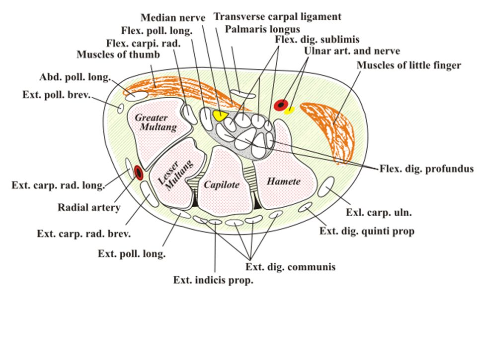
Carpal Tunnel Fibro-osseous structure Tunnel contains 10 structures
Floor is proximal carpal bones Roof is transverse carpal ligament Tunnel contains 10 structures Median n., flexor pollicis longus tendon, 4 slips of flexor digitorium superficialis, 4 flexor digitorium profundus Compression results in paresthesia 2-4 fingers and decrease grip

17
Hand

18
Wrist flexors (median n.)
Superficial Flexor carpi radialis Palmaris longus Flexor carpi ulnaris Flexor digitorium superficialis Pronator teres Deep Flexor digitorium profundus Flexor pollicis longus Pronator quadratus

19
Palmar (intrinsic) Thenar Abductor pollicis brevis
Flexor pollicis brevis Opponens Pollicis Tendon FPL Adductor pollicis

20
Hypothenar Abductor digiti minimi Opponens digiti minimi
Flexor digiti minimi brevis

21
Central Lumbricales Palmar aponeurosis Interossei Tendons FDS/FDP
Superficialis (PIP) Profundus (DIP) Lumbricales Radial side profundus tendon(extensor hood) Flex MP/ext PIP/DIP Palmar aponeurosis Interossei 4 palmar/4 dorsal
 Profundus (DIP) Lumbricales. Radial side profundus tendon(extensor hood) Flex MP/ext PIP/DIP. Palmar aponeurosis. Interossei. 4 palmar/4 dorsal.")
22
Extrinsic Hand Muscles

23
Extensor Indicis O I N F Dorsal surface lower ½ body of ulna
Interosseus membrane I Ulnar side of index finger’s EDC tendon N Radial (posterior interosseus) F MCP and IP Ext of 2nd digit
 F. MCP and IP Ext of 2nd digit.")
24
Extensor Pollicis Longus
Posterior 1/3 ulna Interosseus membrane I Posterior surface of base of thumb distal phalanx N Radial (posterior interosseus) F CMC, MCP and IP Ext of 1st digit
 F. CMC, MCP and IP Ext of 1st digit.")
25
Extensor Pollicis Brevis
Dorsal 2/3 of radius I Dorsal surface of base of proximal 1st phalanx N Radial (posterior interosseus) F CMC & MCP Ext of thumb CMC ABD of thumb
 F. CMC & MCP Ext of thumb. CMC ABD of thumb.")
26
Abductor Pollicis Longus
Posterior distal 2/3 of ulna Posterior middle 1/3 of radius Interosseus membrane I Radial side of base of 1st metacarpal N Radial (posterior interosseus) F CMC ABD & Ext of thumb
 F. CMC ABD & Ext of thumb.")
27
Flexor Pollicis Longus
Anterior middle ½ of radius Interosseus membrane I Palmar surface of base of distal 1st phalanx N Median (anterior interosseus) F IP Flexion of thumb
 F. IP Flexion of thumb.")
28
Extensor Digiti Minimi
Lateral epicondyle of humerus I Extensor expansion of 5th digit N Radial (posteior interosseus) F MCP and IP extension of 5th digit
 F. MCP and IP extension of 5th digit.")
29
Flexor Digitorum Superficialis
Medial epicondyle of humerus Coronoid process Middle ½ anterior radius I Four tendons separating into two parts that insert into sides of bases of middle 2-5 phalanxes N Median F MCP flexion digits 2-5 PIP flexion digits 2-5

30
Flexor Digitorum Profundus
Anteriomedial surface of ulna Interosseus membrane I Four tendons inserting into distal phalanxes of digits 2-5 N Media 2-3 digits Ulna 4-5 digits F DIP flexion of 2-5 digits

31
Intrinsic Hand Muscles
Thenar Eminance

32
Abductor Pollicis Brevis
Scaphoid tuberosity Trapezium ridge Transverse carpal ligament I Lateral base f proximal 1st phalanx N Median F CMC & MCP ABD of thumb

33
Flexor Pollicis Brevis
Superficial head – trapezium Deep head – trapezoid, capitate and palmar ligaments of distal carpal bones I Base of prximal 1st phalanx on radial side Extensor expansion N Superficial – median Deep – Ulnar F CMC & MCP Flexion of thumb

34
Opponens Pollicis O I N F Trapezium Transverse Carpal Ligament
Radial side of 1st metacarpal shaft N Median F Opposition

35
Intrinsic Hand Muscles
Hypothenar Eminence

36
Abductor Digiti Minimi
Pisiform I Ulnar side base of 5th proximal phalanx N Ulnar F MCP ABD of 5th digit

37
Opponen Digiti Minimi O I N F Hook of hamate
Transverse carpal ligament I Ulnar border of entire 5th metacarpal bone N Ulnar F MCP flexion & rotation of 5th digit

38
Flexor Digiti Minimi O I N F Hamate bone Transverse carpal ligament
Ulnar side of proximal 5th phalanx N Ulnar F MCP Flexion of 5th digit

39
Other Intrinsic Hand Muscles

40
Adductor Pollicis O I N F Oblique Head Transverse Head
Capitate bone Bases of 2-3 metacarpals Transverse Head Proximal 2/3 of palmar surface of 3rd metacarpal I Ulnar side of base of 1st proximal phalanx N Ulnar F CMC ADD of thumb

41
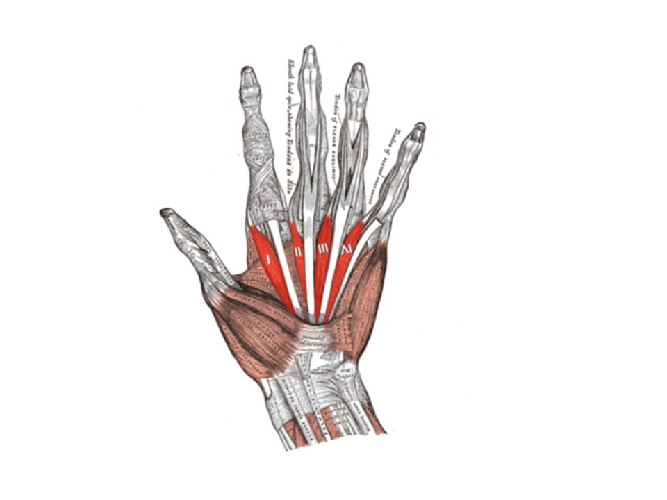
Palmar Interossei O I N F 1st – ulnar side base of 1st metacarpal bone
2nd – ulnar side of 2nd MC bone 3rd – radial side of 4th MC bone 4th – radia side of 5th MC bone I Extensor expansion of 2,4 and 5th digits N Ulnar F ADD of 1st, 2nd, 4th and 5th digits toward midline of hand

42
Dorsal Interossei O I N F
1st lateral head – ulnar side of 1st metacarpal bone 1st medial head – radial side of 2nd metacarpal bone 2nd, 3rd, 4th space between metacarpal bones I 1st – radial side 2nd proximal phalanx 2nd – radial side of 3rd 3rd – ilnar side of 3rd 4th – ulnar side of 4th N Ulnar F ABD of 2nd, 3rd, and 5th finger from midline

43
Lumbricales O I N F Tendons of FDP
Extensor expansion on dorsal aspect of each digits radial side N 1 and 2 – median 3 and 4 – ulnar F MCP flexion 2-5 digits DIP & PIP ext 2-5 digits

45
Palmaris Brevis O I N F Flexor retinaculum
Palmar surface skin on ulnar side of hand N Ulnar F Wrinkles skin of hand on ulnar side

46
Cords Give off Branches!! (in axilla)
Lateral Musculocutaneous Median Medial Ulnar Posterior Radial Axillary (thoracodorsal) (subscapular)
 (subscapular)")
47
PUT IT ALL TOGETHER…... pg 416

48
Innervation by Posterior Cord
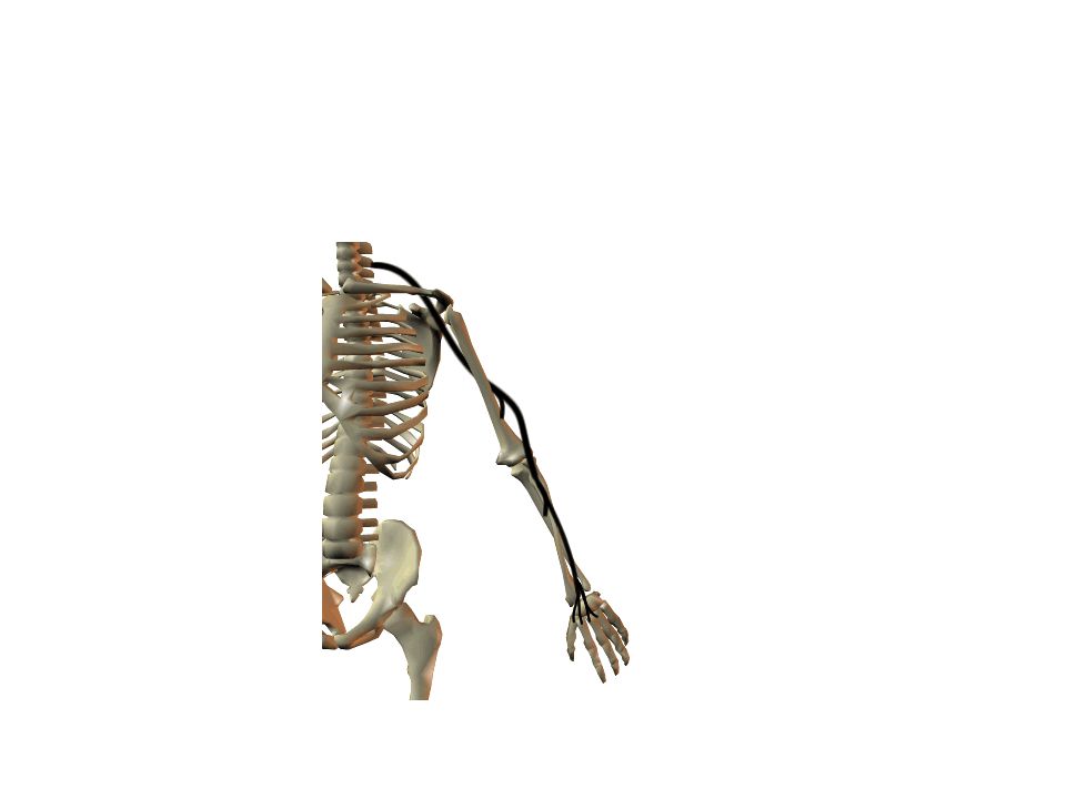
Radial Nerve (largest branch) Course: Through arm, around humerus, around lateral epicondyle, then divides Innervates: all posterior muscles of arm and forearm Triceps brachii, anconeus, supinator, brachioradialis Divides in forearm: Superficial = skin of arm and dorsolateral surface of hand Deep = extensor muscles of forearm (eg E. carpi radialis L + B) Damage to Radial Nerve = wristdrop Inability to extend the hand, st inability to fully extend forearm
 Course: Through arm, around humerus, around lateral epicondyle, then divides. Innervates: all posterior muscles of arm and forearm. Triceps brachii, anconeus, supinator, brachioradialis. Divides in forearm: Superficial = skin of arm and dorsolateral surface of hand. Deep = extensor muscles of forearm (eg E. carpi radialis L + B) Damage to Radial Nerve = wristdrop. Inability to extend the hand, st inability to fully extend forearm.")
51
Innervation by Posterior Cord (continued)
Axillary Nerve (runs w/ humeral circumflex a.) Innervates: Deltoid and Teres minor (motor inn) Capsule of shoulder, skin of shoulder (sensory inn) Subscapular Nerve {branches of C5 + C6 rami} Innervates: Subscapularis, Teres major Thoracodorsal Nerve (runs w/thoracodorsal a+v) Innervates: Latissimus dorsi
 Innervates: Deltoid and Teres minor (motor inn) Capsule of shoulder, skin of shoulder (sensory inn) Subscapular Nerve {branches of C5 + C6 rami} Innervates: Subscapularis, Teres major. Thoracodorsal Nerve (runs w/thoracodorsal a+v) Innervates: Latissimus dorsi.")
52
Innervation by Lateral Cord
Musculocutaneous Course: branches to arm, distal to elbow becomes cutaneous for lateral forearm skin Innervates Biceps brachii, brachialis, coracobrachialis (motor inn) Skin distal to elbow (sensory) Suprascapular (runs w/suprascapular a+v) {C5, C6} Innervates: Supraspinatus, Infraspinatus
 Skin distal to elbow (sensory) Suprascapular (runs w/suprascapular a+v) {C5, C6} Innervates: Supraspinatus, Infraspinatus.")
53
Innervation by both Lateral and Medial Cords
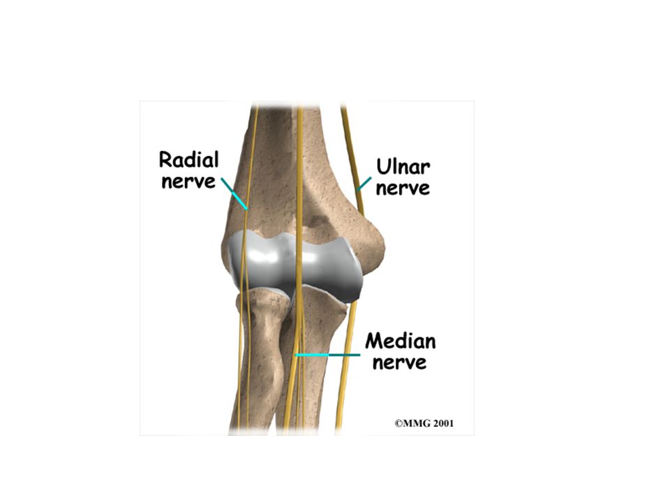
Median Course: middle of brachial plexus, does not branch in arm, distal to elbow provides many branches to most forearm flexors, passes through carpal tunnel to hand to lateral palmar intrinsics Innervates: most muscles of anterior forearm (motor inn) (eg) most flexors, some intrinsics (thumb) Innervates: skin of lateral 2/3 hand on palm side, dorsum of fingers 2+3 (sensory inn) Nerve Damage = “Ape” Hand Inability to Oppose Thumb
 (eg) most flexors, some intrinsics (thumb) Innervates: skin of lateral 2/3 hand on palm side, dorsum of fingers 2+3 (sensory inn) Nerve Damage = Ape Hand. Inability to Oppose Thumb.")
54
Innervation by Medial Cord
Ulnar Course: runs along medial side of arm, behind medial epicondyle, superficial to carpal tunnel into hand, branches to supply intrinsics and skin Innervates: FCU and part of FDP, most intrinsics (motor inn) Skin of medial 2/3 of hand A+P (sensory inn) Nerve Damage: Clawhand Inability to extend fingers at interphalangeal joints, results in permanent flexion = claw
 Skin of medial 2/3 of hand A+P (sensory inn) Nerve Damage: Clawhand. Inability to extend fingers at interphalangeal joints, results in permanent flexion = claw.")
56
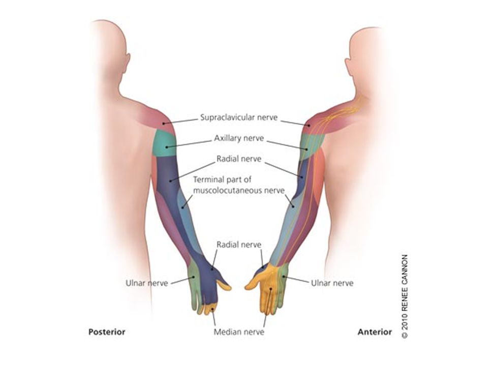
Cutaneous Innervation to the Hand

57
Ulnar Nerve Median Nerve Brachial Artery Musculocutaneous Nerve
UlnarArtery Where’s Radial Nerve? Radial Artery Median Nerve Ulnar Nerve

59
Thank you…

Similar presentations


>")


