Download presentation
Presentation is loading. Please wait.

1
The Knee (Tibiofemoral) Joint
By: Jackie, Stacey, Gabe, and scharlie

2
Bones and Surface Anatomy
By: Jackie

3
KNEE (TIBIOFEMORAL) JOINT
FEMUR: Lateral Epicondyle Lateral Condyle Medial Epicondyle Medial Condyle Intercondylar Fossa TIBIA: Tibial Tuberosity Intercondylar Eminence FIBULA: Head PATELLA

4
SURFACE ANATOMY Vastus Medialis Vastus Lateralis Patella

5
SURFACE ANATOMY Biceps Femoris Tendon Semitendinous Tendon Semimembranous Tendon

6
SURFACE ANATOMY Medial Head of Gastronemius Lateral Head of Gastronemius Soleus Popliteal Fossa

7
Ligaments, Bursae, and Cartilage
By: stacey

8
Ligament Review lig·a·ment
a short band of tough, flexible, fibrous connective tissue that connects two bones or cartilages or holds together a joint. a membranous fold that supports an organ and keeps it in position.

9
Knee Ligaments Anterior Cruciate Ligament Oblique Popliteal Ligament
Patellar Ligament Transverse Ligament Anterior Cruciate Ligament Oblique Popliteal Ligament Tibial Collateral Ligament Arcuate Popliteal Ligament Fibular Collateral Ligament Posterior Cruciate Ligament PTA OTA Fine People

10
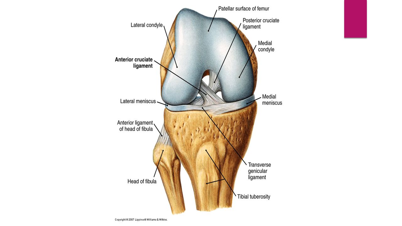
Patellar Ligament Tibial Collateral Ligament Anterior Cruciate Ligament Posterior Cruciate Ligament

11
Oblique Popliteal Arcuate Popliteal Fibular Collateral

12
Tendon Review ten·don a flexible but inelastic cord of strong fibrous collagen tissue attaching a muscle to a bone.

13
Quadriceps Femoris Tendon

14
Cartilage Review firm, whitish, flexible connective tissue found in various forms in the larynx and respiratory tract, in structures such as the external ear, and in the articulating surfaces of joints.

15
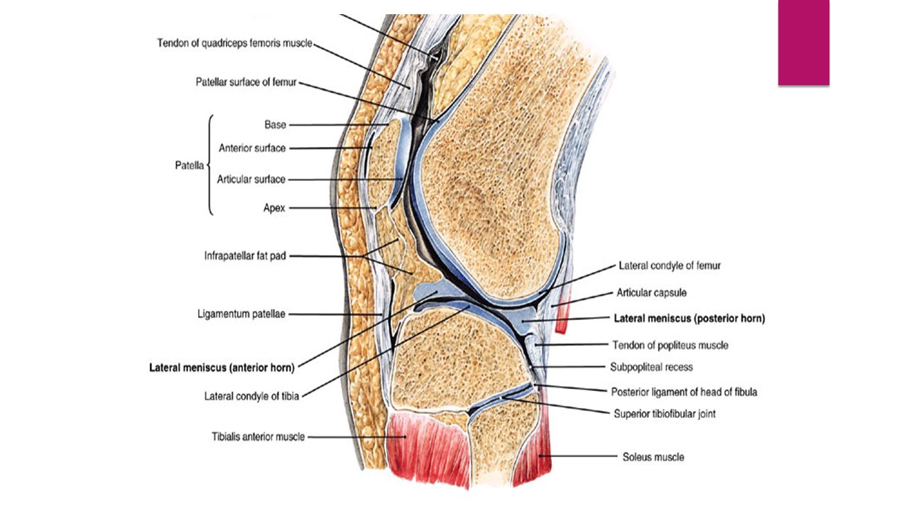
Lateral and Medial Menisci

16
Bursa Review bur·sa plural noun: bursae a fluid-filled sac or saclike cavity, especially one countering friction at a joint.

17
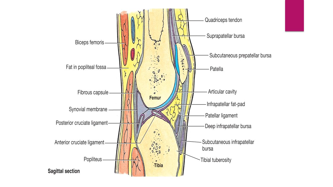
Prepatellar Bursa Deep Infrapatellar Bursa Subcutaneous Infrapatellar Bursa

18
Suprapatellar Bursa

22
Tibiofemoral Joint By: gabe
Muscles and Nerves Tibiofemoral Joint By: gabe

23
Rectus Femoris

24
Rectus Femoris Origin: ASIS
Insertion: Patella via quadriceps tendon and then the tibial tuberosity via patellar tendon (ligament) Action: Hip Flexion and knee extension Nerve: Femoral Nerve
 Action: Hip Flexion and knee extension. Nerve: Femoral Nerve.")
25
Vastus Intermedialis

26
Vastus Intermedialis Origin: Anterior 2/3 and the lateral shaft of the femur Insertion: Patella via quadriceps tendon and then tibial tuberosity via patellar ligament Action: Knee Extension Nerve: Femoral Nerve

27
Vastus Lateralis

28
Vastus Lateralis Origin: Intertrochanteric line and linea aspera of the femur Insertion: Patella via quadriceps tendon then tibial tuberosity via patellar tendon (ligament) Action: Knee Extension Nerve: Femoral Nerve
 Action: Knee Extension. Nerve: Femoral Nerve.")
29
Vastus Medialis

30
Vastus Medialis Origin: Linea aspera of the femur
Insertion: Patella via the quadriceps tendon and the tibial tuberosity via the patellar tendon (ligament) Action: Knee extension Nerve: Femoral Nerve
 Action: Knee extension. Nerve: Femoral Nerve.")
31
Biceps Femoris

32
Biceps Femoris Origin: Long Head- Ischial tuberosity
Short Head- Linea aspera of the femur Insertion: Head of the fibula and lateral condyle of the tibia Action: Long Head- Extends hip and flexes knee Short Head- Flexes knee and laterally rotates hip Nerve: Tibial Nerve

33
Semimembranosus

34
Semimembranosus Origin: Ischial tuberosity
Insertion: Posterior medial condyle of the tibia Action: Extends and medial rotates the hip; Knee flexion Nerve: Tibial Nerve

35
Semitendinosus

36
Semitendinosus Origin: Ischial tuberosity
Insertion: Proximal part of medial shaft of tibia Action: Extends and medially rotates hip; Flexes knee Nerve: Tibial nerve

37
Popliteus

38
Popliteus Origin: Lateral condyle of femur and lateral meniscus
Insertion: Posterior surface of tibia Action: Unlocks knee and weakly flexes the knee Nerve: Tibial

39
Gastrocnemius

40
Gastrocnemius Origin: Lateral head- Lateral condyle of femur
Medial Head- Superior to medial condyle of femur Insertion: Posterior calcaneus via calcaneal (Achilles) tendon Action: Plantar flexes ankle Nerve: Tibial
 tendon. Action: Plantar flexes ankle. Nerve: Tibial.")
41
Tibiofemoral Nerves Sciatic Nerve Femoral Nerve Tibial Nerve
Superficial Fibular (Peroneal) Nerve Deep Fibular (Peroneal) Nerve
 Nerve. Deep Fibular (Peroneal) Nerve.")
42
Tibiofemoral Nerves

43
Arteries and veins that pass through the knee (tibiofemoral) joint
Vascular Supply Arteries and veins that pass through the knee (tibiofemoral) joint By: Scharlie
 joint. By: Scharlie.")
44
Arteries: Inferior Gluteal Artery
Inferior Gluteal Artery (CUT) Inferior Gluteal Artery Branches from the Internal Iliac Artery. Leaves pelvis through the greater sciatic foramen and passes inferior to the piriformis. Posterior View Medial Circumflex Femoral Artery Femoral Artery
 Inferior Gluteal Artery. Branches from the Internal Iliac Artery. Leaves pelvis through the greater sciatic foramen and passes inferior to the piriformis. Posterior View. Medial Circumflex Femoral Artery. Femoral Artery.")
45
Medial Circumflex Femoral Artery
Branches Posteriorly behind Femur Lateral Circumflex Femoral Artery Branches Anteriorly in front of Femur Both may branch from profundus femoris or femoral arteries Both encircle thigh, anastomose, and supply thigh muscles and proximal end of Femur. Anterior View Inferior Gluteal Artery

46
Femoral Artery Femoral Continuation of External Iliac Artery distal to inguinal ligament. Passes through adductor hiatus and becomes Popliteal Artery Popliteal Continuation of Femoral Artery Passes behind Posterior Knee Divides into anterior and posterior tibial arteries Gives Rise to Genicular Arteries Popliteal Artery Anterior View Anterior Tibial Artery Posterior Tibial Artery

47
Anterior Tibial Artery
Popliteal Anterior Tibial Artery Branches from Popliteal Artery Passes anteriorly between Tibia and Fibula Runs down anterior/lateral aspect of Tibia Posterior Tibial Artery Continues down posterior aspect of lower leg Anterior Tibial Posterior Tibial Anterior View Posterior View Fibular (Peroneal) Anterior Tibial
 Anterior Tibial.")
48
Anterior Tibial Artery Perforating Fibular Artery
Anterior View of foot Anterior Tibial Artery Fibular (Peroneal) Artery Braches from Popliteal Artery Runs down lateral aspect of lower leg Perforating braches supply distal muscles Dorsalis Pedis Artery Continuation of Anterior Tibial Artery Begins at ankle joint between malleoli to top of foot Perforating Fibular Artery Dorsalis Pedis Artery
 Artery. Braches from Popliteal Artery. Runs down lateral aspect of lower leg. Perforating braches supply distal muscles. Dorsalis Pedis Artery. Continuation of Anterior Tibial Artery. Begins at ankle joint between malleoli to top of foot. Perforating Fibular Artery. Dorsalis Pedis Artery.")
49
Veins: Anterior Tibial Vein Posterior Tibial Vein
Superior continuation of dorsalis Pedis Vein Runs up anterior tibia until moving posterior to join posterior tibial vein Drains into Popliteal Vein Posterior Tibial Vein Drains into Popliteal Vein after joining the Anterior Tibial Vein Both supply calf and foot Anterior Tibial Vein Posterior Tibial Vein Anterior View

50
Fibular (Peroneal) Vein
More posterior/lateral in lower leg Drains into the Popliteal Vein Popliteal Vein Anterior and Posterior Tibial Veins join to form the Popliteal Vein Crosses back of knee and becomes Femoral Vein at thigh Femoral Vein Superior continuation of Popliteal Vein Becomes the External Iliac Vein in the inguinal region Femoral Vein Popliteal Vein Fibular (Peroneal)Vein Anterior View
Vein. Anterior View.")
51
Small Saphenous Vein Great Saphenous Vein
Runs along lateral aspect of foot and through the calf muscle Drains into Popliteal Vein at the knee Great Saphenous Vein Superficial and longest vein in the body Begins in common with Small Saphenous Vein in ankle Extends up medial side of calf, knee, and thigh Drains into the Femoral Vein Great Saphenous Vein Small Saphenous Vein Anterior View

52
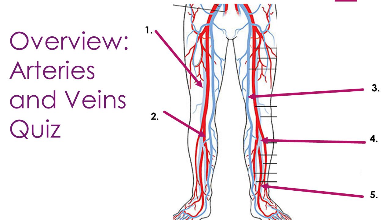
Overview: Arteries and Veins Quiz
1. 3. 2. 4. 5.
53
Clinical Concerns for the Knee (Tibiofemoral) Joint
Torn acl, plantar fasciitis, and tkr By Scharlie TKR By Gabe

54
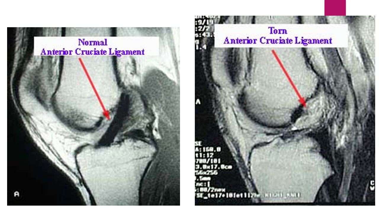
Torn ACL The injury may occur with or without contact. Women have an increased risk of ACL injury because of differences in anatomy, muscle mass, and training. Symptoms of ACL tear include hearing a loud pop as the ligament tears, pain, knee swelling, and difficulty walking

56
Torn ACL Treatment Treatment options include: Acute treatment: R.I.C.E
Nonsurgical treatment only: a physical rehab program. ACL surgery to reconstruct the ACL (will still need physical therapy) Recovery from an ACL injury varies for each person. Your treatment should continue until your knee is stable and strong rather than for a certain length of time.
 Recovery from an ACL injury varies for each person. Your treatment should continue until your knee is stable and strong rather than for a certain length of time.")
57
Plantar Fasciitis

58
Signs and Symptoms: When plantar fasciitis occurs, the pain is typically sharp and unilateral. Heel pain worsens by bearing weight on the heel after long periods of rest. Improvement of symptoms is usually seen with continued walking Risk Factors: Identified risk factors for plantar fasciitis include excessive running, standing on hard surfaces for prolonged periods of time, high arches of the feet, and flat feet. Obesity is seen in 70% of individuals with plantar fasciitis. Achilles tendon tightness and inappropriate footwear have also been identified as significant risk factors. Treatment: Non-surgical: (90% of cases)Include rest, heat, ice, calf-strengthening exercises, techniques to stretch the calf muscles, Achilles tendon, and plantar fascia, and weight reduction. Surgical: Plantar fasciotomy (release of fascia) is often considered after conservative treatment has failed to resolve the issue after six months and is viewed as a last resort.
Include rest, heat, ice, calf-strengthening exercises, techniques to stretch the calf muscles, Achilles tendon, and plantar fascia, and weight reduction. Surgical: Plantar fasciotomy (release of fascia) is often considered after conservative treatment has failed to resolve the issue after six months and is viewed as a last resort.")
59
Torn ACL Surgical Repair https://www.youtube.com/watch?v=9EgRHfoleLc
Videos: Torn ACL Surgical Repair Dr. E. Edward Khalfayan, M.D. Total Knee Replacement Surgery

60
Questions ??????????

61
Resources: Google Images:
X&ei=WIttVOG2DKWQigLJr4CYDQ&ved=0CAYQ_AUoAQ Moore KL, Agur AM, Dalley AF, Essential Clinical Anatomy. 4th ed. Baltimore,MD: Lippincott Williams & Wilkins;2011. Pearson Learning Solutions Lab Manual, Integrate Anatomy & Physiology: Pearson Learning Solutions;2011. Youtube: Google Web

Similar presentations


 Joint>")
>")


