Download presentation
Presentation is loading. Please wait.

1
Image Guided Surgery in Prostate Brachytherapy Rohit Saboo

2
Prostate Cancer A growing problem in US and world over with increasing longevity Methods of treatment Surgery Irradiation Many problems of existing methods

3
Where is it?

4
Brachytherapy procedure Localized and prolonged dose Brachytherapy overview

5
Brachytherapy - old way Pre-planning CT Outline prostate Develop plan for needle movement Guide needle at time of surgery with help from Ultra sound images “Dynamic Brachytherapy of the prostate under active image guidance”, Gang Cheng et. al, MICCAI 2001

6
Brachytherapy – old way Problems Prostate movement Prostate size/shape variance Due to anesthesia effects Time and hormonal therapy Drawbacks: Lots of error due to prostate movement More than necessary needle insertions

7
Brachytherapy – new way General outline of prostate from pre- planning CT Outline prostate in real-time during surgery Provide guidance for the needle with real time prostate outlining. Track needle errors in real time

8
Steps in automation Acquire volumetric ultra-sound images on the fly Automatically recognize the prostate/rectum and other structures (segmentation) Analyze dose distribution
 Analyze dose distribution")
9
Segmentation The process of outlining the prostate (or any organ) is called segmentation Two chief ways to deal with it Model-based Image based
 is called segmentation Two chief ways to deal with it Model-based Image based")
10
Segmentation problem

11
Ultrasound problems Noise! Speed of sound is not uniform Image distance incorrect in one axis

12
Approaches to segmentation Model based Shape model Probable shapes Probable intensity/texture variations Examples: ASM, AAM, M-reps Image based Outline drawn by expert on one image (atlas) Image intensity/feature based registration Outline carried over
 Image intensity/feature based registration Outline carried over")
13
Feature Model – Ruo et. al Set of boundary points i - Sample object X i – Tuple representing i th object Each object has m points on the boundary

14
Feature Model X i = (L i, r i1, r i2, … r im ) T
 T")
15
Shape variation Mean shape Covariance matrix

16
Feature Model Eigenvalue decomposition of covariance matrix p i Principal components (eigenvectors) Eigen-values Sort the eigenvalues, choose the largest t
 Eigen-values Sort the eigenvalues, choose the largest t")
17
Eigenvalues

18
New plausible models

19
Optimization - GA

20
Image match Fitness function out i and in i average of intensities along a profile (15 pixels long)
")
21
Image Match Simplify and speed it up v i unit normal

22
GA parameters

23
90% crossover rate 1% mutation rate population size 200 2000 generations repeated 15 times

24
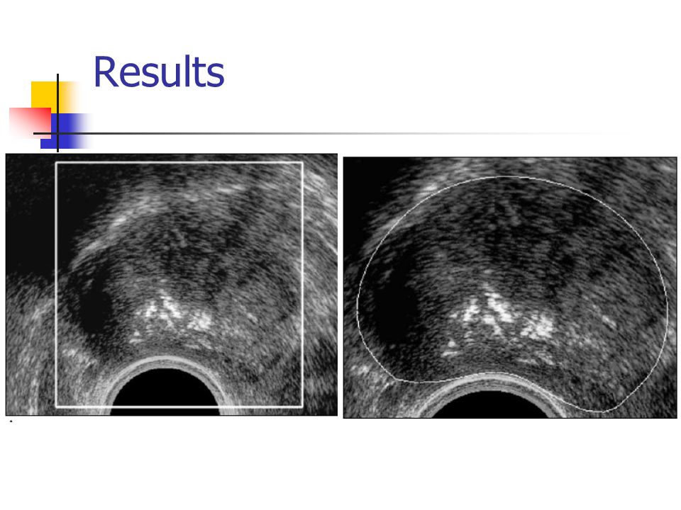
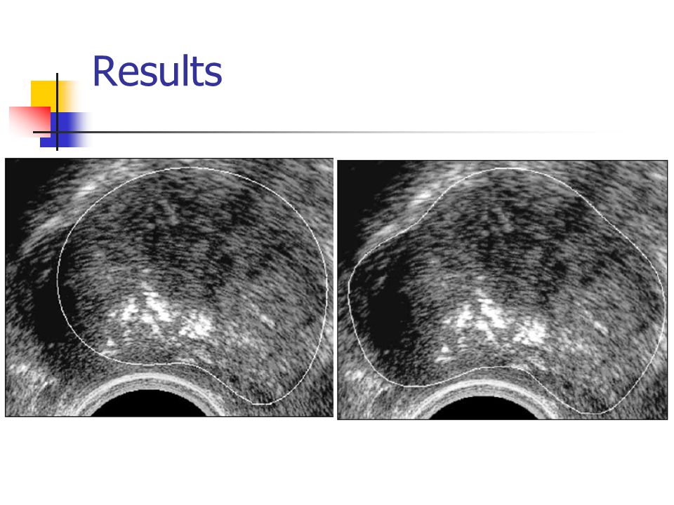
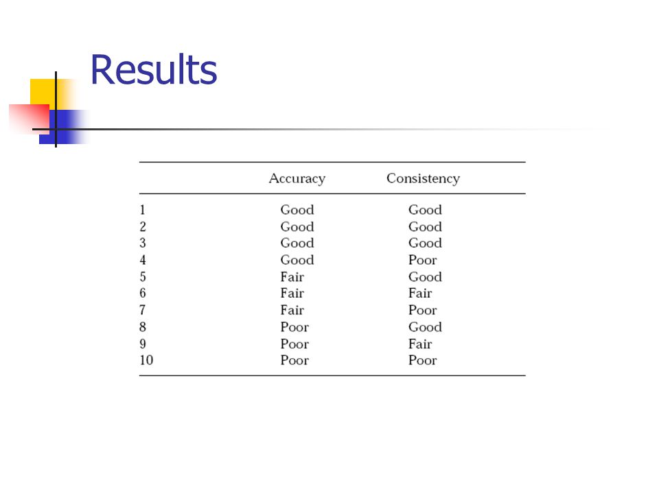
Experiment 40 images training from 27, 3 poor quality test on remaining 10 Expert segmentation by two different experts human-human disagreement vs automated-human disagreement

25
Results

29
Summary Very good analysis Point based boundary models are poor Parameter tuning No reasoning for fitness function

30
Methods Model based methods Image based methods Fully automatic, registration Deformable registration – Wei, 2004 Snakes

31
Image based Overview MRI/MRSI and US data prostate carefully outlined on MRI data US image is acquired during operation time The two images are brought into alignment during operation time. They do for biopsy, but same techniques work for brachytherapy

32
Global Alignment US data is poor Model is pre-segmented in MRI Surface to volume alignment methods are used Gradient of image is computed in US and model information is used to roughly find the correspoding boundary in US

33
Global alignment GA based optimization fitness function

34
Registration Deformation is elastic Two orthogonal directions Therefore 2-parameter model for deformation

35
Registration Obtain Curvature images Obtain mapping g for a few points Use these points to drive a TPS deformation.

36
Validation Phantom Gelatin made prostate phantom 15 fiducial markers implanted inside the prostate Soft container filled with water placed on top to simulate pubic bone.

37
Validation

38
Results on phantom

39
Patient study

40
Patient Study

41
Summary Two step registration technique Phantom Studies Only tested over one patient

42
Methods Model based methods Image based methods Fully automatic, registration Deformable registration – Wei, 2004 Snakes semi-automatic In between both

43
Snakes Give an initial approximate contour Two forces act on a snake Internal force Based on curvature External force Based on image gradients Let the model evolve using ordinary force equations till equilibrium

44
Snake based methods Approach used by Zhouping Wei, 2005

45
Snake based methods 1.19 +/- 0.14 mm on average

46
Questions?

47
References “Dynamic Brachytherapy of the prostate under active image guidance”, Gang Cheng et. al, MICCAI, 2001 “Automatic Prostate Boundary Recognition in Sonographic Images Using Feature Model and Genetic Algorithm”, Ruo Yun et. al, Journal of Ultrasound in Medicine, Vol 19, Issue 11, 2000 “Deformable Registration Between MRI/MRSI and Ultrasound Images for Targeted Robotic Prostate Biopsy”, Wei Shao et. al, Proceedings of the 2004 IEEE Conference on Cybernetics and Intelligent Systems “A Discrete Dynamic Contour Model”, Steven Lobregt et. al, IEEE Transactions on Medical Imaging, vol 14, no 1, March 1995 “Dynamic Intraoperative Prostate Brachytherapy Using 3D TRUS Guidance with Robot Assistance”, Zhouping Wei et. al, Proceedings of the 2004 IEEE Engineering in Medicine and Biology, 2005

Similar presentations

 Developed for face recognition Generalised.>")


>")


