Download presentation
Presentation is loading. Please wait.

1
Presentation and Management of Intracranial Space Occupying lesions (ICSOL)
")
2
Types of ICSOL’s Neoplasms: Primary, secondary Inflammatory:
Abscess, Tuberculoma, Syphilitic gumma, Fungal granulomas. Parasitic: Cysticercosis, Hydratid cyst, Amebic abscess, Schistosoma japonicum. Traumatic: Chronic subdural haematoma Congenital: Arachnoid and other benign cysts

3
Focal neurological deficit
Mechanisms leading to symptoms Mass effect CSF obstruction Irritation of cortex Compression Invasion Impairment of regional circulation Increased ICP Seizures Focal neurological deficit

4
Presenting Symptoms & Signs
Very important The main groups: Symptoms of raised ICP Seizures Neurological deficit (focal signs) Endocrine dysfunction Personality changes & impaired higher functions
 Endocrine dysfunction. Personality changes & impaired higher functions.")
5
I. Symptoms & Signs of Raised ICP
Headache: Worsens in Early morning Flat position Nausea and/or vomiting (projectile in the morning) Visual disturbances: Papilledema Abducent nerve palsy (False localizing) Impaired consciousness
 Visual disturbances: Papilledema. Abducent nerve palsy (False localizing) Impaired consciousness.")
6
II. Seizures: Benign lesions tend to present more with seizures
Generalized: Partial: Simple Complex

7
III. Neurological deficit (Localizing signs)
Depends on the location of the lesion Includes: Cranial nerve deficits: mainly in skull base tumors Motor weakness Sensory deficit

8
IV. Endocrine dysfunction
Mainly caused by pituitary lesions Hyperscretion: Prolactinoma Acromegaly Cushing disease Hyposecretion: Non-secreting pituitary adenoma Craniopharyngioma

9
V. Personality Changes & Impaired Higher Functions
Personality alteration Changes in mood Inability to concentrate Memory disturbance

10
Management Full medical history Complete general & neurol. Examination
Investigations Treatment

11
Neurological Examination Do not forget: PAPILLEDEMA
Normal Papilledema

12
Investigations Lab work up: CBC, U&E, PT, PTT, blood group, etc
Radiological imaging: CT-scan: Method of choice for emergency MRI: shows more anatomical details of the lesion Cerebral angiography: Mainly for vascular lesions Others: plain skull x-rays, isotope scan Neurophysiological tests: EEG, EVOP’s

13
Treatment Initial treatment (Emergency) Definitive treatment
 Definitive treatment")
14
Initial treatment Very important Often started in the ER Goals:
Control of raised ICP Control of seizures Prepare patient for definitive treatment

15
Management of raised ICP
Elevate the head to about 30° Adequate oxygenation Avoid fluid overload Dexamethasone Hyperventilation Mannitol ICP monitoring Only in patients with decreased LOC

16
Control of Seizures Interruption of a seizure or status epilepticus: Benzodiazepines Perioperative prophylaxis: Phenytoin

17
Definitive treatment Depends on the type and location of the lesion.
Most intracranial tumors need surgical excision. Radiotherapy for: Inoperable tumors Postoperative for malignancies Chemotherapy: adjuvant in malignancies.

18
Intracranial tumors Responsible for 2% of all cancer death.
Overall incidence: 8-10 cases per population per year Two peaks: Early childhood 5th-7th decades of life Benign or malignant.

19
Etiology Unknown for most of brain tumors in humans.
Chromosomal abnormalities have been noted in various benign & malignant brain tumors. Clear genetic factors play a minor role: 5% of patients have family H/o brain tumors Neurofibromatosis: autosomal dominant NF I: causative gene on the long arm of chromosome 17 NF2: causative gene on the long arm of chromosome 22 Increased incidence of brain tumors following cranial irridiation

20
Classifications Based on the cell of origin
glial(gliomas)and non-glial In relation to brain tissue: Extrinsic vs. intrinsic In relation to their site: Supratentorial vs. infratentorial (posterior fossa) Age of presentation: Pediatric vs. adult.
and non-glial. In relation to brain tissue: Extrinsic vs. intrinsic. In relation to their site: Supratentorial vs. infratentorial (posterior fossa) Age of presentation: Pediatric vs. adult.")
21
WHO Classification Based on the cell of origin
Neuroepithelial tumors: e.g. gliomas Primitive neuro-ectodermal tumors: e.g. medulloblastoma Nerve sheath tumors (neuroma): e.g. vestibular schwannoma Meningeal tumors: e.g. Meningiomas Pituitary tumors Germ cell tumors: e.g. germinoma Lymphomas Malformative tumors: e.g. craniopharyngeoma Vascular tumors Metastatic tumors Local extensions from regional tumors: e.g. Glomus jugulare
: e.g. vestibular schwannoma. Meningeal tumors: e.g. Meningiomas. Pituitary tumors. Germ cell tumors: e.g. germinoma. Lymphomas. Malformative tumors: e.g. craniopharyngeoma. Vascular tumors. Metastatic tumors. Local extensions from regional tumors: e.g. Glomus jugulare.")
22
Glioma Most common primary brain tumor (50%)
Arise from the supporting cells of the brain. Neuroectodermal in origin. Usually supratentorial. Rarely metastasize. Tumor in single lobe may be treated by lobectomy. Recurrence is common despite postoperative radiotherapy.

23
Astrocytoma Arise from the astrocyte cells
Comprise 80% of all glioma and 40% of all brain tumors Peak incidence: yrs (early middle age) Males > females All are inflitrative.
 Males > females. All are inflitrative.")
24
Histopathological Grading
Benign (Low grade) Grade 1 Grade 2 Malignant (High grade) Grade 3 Grade 4
 Grade 1. Grade 2. Malignant (High grade) Grade 3. Grade 4.")
25
Clinical presentation :
Duration of symptoms: Benign : Often long (years) Malignant : Often short (wks.) Symptoms of SOL Headache (70%) Papilledema (60%) Seizures ( %) Focal neurological deficit ( % ). Mental changes (30%)
 Malignant : Often short (wks.) Symptoms of SOL. Headache (70%) Papilledema (60%) Seizures ( %) Focal neurological deficit ( % ). Mental changes (30%)")
26
Investigations Low grade astrocytoma: High grade astrocytoma:
CT: Hypodense non-enhancing area. MRI: Hyperintense T2 + non-enhancing hypointense T1 High grade astrocytoma: CT: Mixed density area with strong, irregular contrast enhancement MRI: Hyperintense T2 + enhancing hypointense T1

28
Treatment Benign A. (grade 1 & 2):
Surgical Excision (+/- Radiotherapy) Malignant A. (grade 3 & 4): Surgical Excision + Radiotherapy (+/- Chemotherapy)
 Malignant A. (grade 3 & 4): Surgical Excision + Radiotherapy. (+/- Chemotherapy)")
29
Prognosis : Benign A. (grade 1 & 2): Malignant A. (grade 3 & 4):
Variable Cure possible after complete excision in grade I A. Malignant A. (grade 3 & 4): Generally POOR! Mean survival: about 2 years
: Generally POOR! Mean survival: about 2 years.")
30
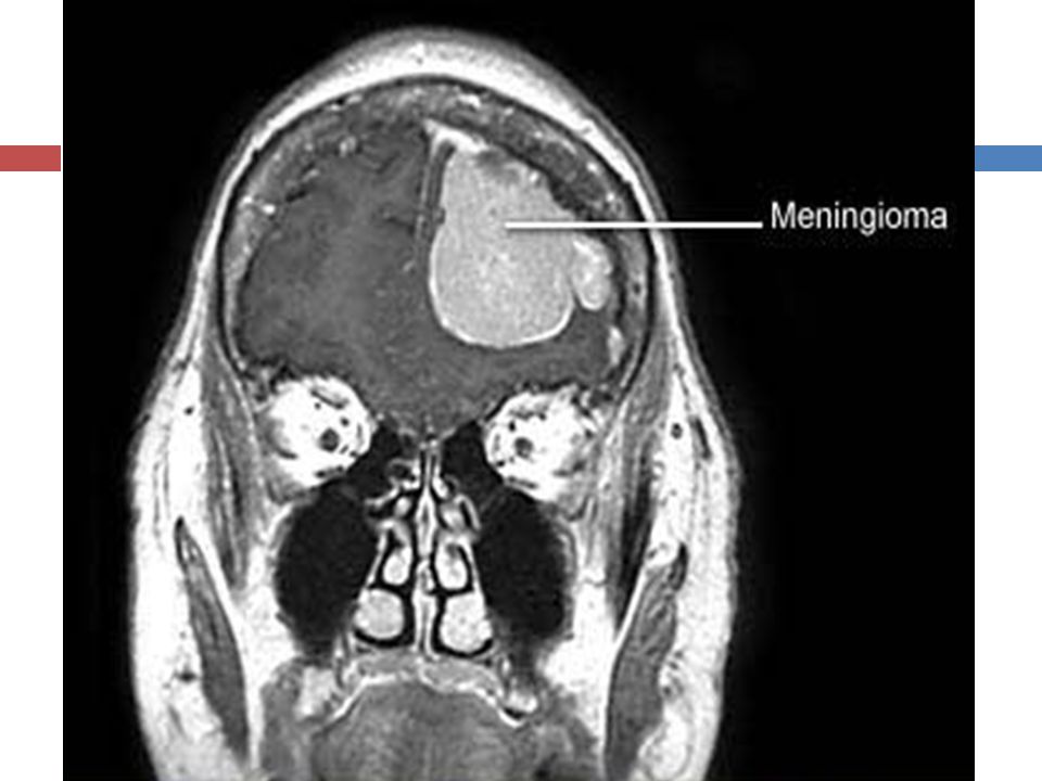
Meningioma Arises from the arachnoid villi & granulations
Comprises 15-20% of all intracranial tumors It is the most benign intracranial tumor Peak incidence: 35 – 70 yrs (Rare in children) Female preponderance (f:m ratio = 2:1)
 Female preponderance (f:m ratio = 2:1)")
31
Clinical presentation
Often long history (for many years) Symptoms of SOL: Focal neurological deficit depending on the site of meningioma Epilepsy: mainly in frontal & temporal convexity meningioma Cranial nerve deficits: common in basal M. Mental changes: in large anterior & middle fossa M.
 Symptoms of SOL: Focal neurological deficit depending on the site of meningioma. Epilepsy: mainly in frontal & temporal convexity meningioma. Cranial nerve deficits: common in basal M. Mental changes: in large anterior & middle fossa M.")
32
Investigations Plain skull X-rays:
Positive findings in up to 70% of cases Direct signs: Hyperostosis, calcification, bone erosion Indirect signs: Shifted pineal gland, sella changes CT scan MRI (Investigation of choice ) Angiography: Indicated in M. neighboring major vessels & sinuses Shows: Tumor relationship to major cerebral vessels Blood supply of tumor Whether pre-operative embolization is needed
 Angiography: Indicated in M. neighboring major vessels & sinuses. Shows: Tumor relationship to major cerebral vessels. Blood supply of tumor. Whether pre-operative embolization is needed")
34
Treatment & Prognosis High recurrence rates occur in cases of:
Treatment of choice: TOTAL EXCISION High recurrence rates occur in cases of: Incomplete excision Malignant M. Radiotherapy plays a very limited role

35
Pituitary Tumors Pituitary tumors represent between 10% and 15% of all intracranial tumors. Slightly more frequent in women Most pituitary tumors occur in young adults

36
Pituitary tumors: Classification & Presentation
Secretary : ~70-80% Prolactinoma: In women → amenorrhea-galactorrhea syndrome. In men → impotence. GH secreting: cause gigantism, acromegaly ACTH secreting: cause Cushing’s disease Non-secretary: Produce compressive symptoms: On optic chiasm → bitemporal hemianopia On secretory cells → hypopituitarism

37
Investigation: MRI: Method of choice.
CT : is contraindicated if MRI available because of radiation of optic chiasm. Ophthalmological assessment. Endocrinological assessment.

39
Treatment Prolactinomas: Medical treatment Other pituitary adenomas:
Dopamin agonists Other pituitary adenomas: Surgical excision: Transsphenoidal Transcranial Radiotherapy: as adjunct to surgery for large or recurrent tumors Hormonal substitution

40
Vestibular Schwannoma (Acoustic Neuroma)
Arise in the internal auditory meatus from the nerve sheath (Schwann) cells of the vestibular branch of the 8th cranial nerve Early symptoms: Unilateral sensori-neural deafness, tinnitus & vertigo Late symptoms Loss of corneal reflexes Facial weakness and unilateral taste loss. Dysphagia, dysarthria and hoarseness. Ataxia and nystagmus.
 cells of the vestibular branch of the 8th cranial nerve. Early symptoms: Unilateral sensori-neural deafness, tinnitus & vertigo. Late symptoms. Loss of corneal reflexes. Facial weakness and unilateral taste loss. Dysphagia, dysarthria and hoarseness. Ataxia and nystagmus.")
41
Investigations ENT assessment: Radiological imaging:
Audiometry Radiological imaging: MRI CT-scan Are helpful in determining the location and size of a tumor and also in planning its removal. Neurophysiological testing: Brainstem auditory evoked response (BAER)
")
42
Vestibular Schwannoma: MRI
Preop. Postop.

43
Treatment Standard treatment: Surgical excision
Main surgical risk in large tumors: Facial paralysis Alternative options: Observation for patients with small lesion and little symptoms Radiosurgery: Can be used to reduce the size or limit the growth of tumors up to 25 mm in diameter

44
Medulloblastoma A highly cellular, malignant tumor
Originates from the external granular layer of the fetal cerebellum (Obersteiner’s layer) Comprises ~3% of all brain tumors 2/3 of patients are children Higher involvement of males
 Comprises ~3% of all brain tumors. 2/3 of patients are children. Higher involvement of males.")
45
Clinical Presentation
Relatively short history (1-4 months) Symptoms & signs of raised ICP mainly due to obstructive hydrocephalus→ 80% Focal neurological signs → 60% Cerebellar: Truncal & gait ataxia False localizing : 6th nerve palsy Brain stem: Nystagmus
 Symptoms & signs of raised ICP mainly due to obstructive hydrocephalus→ 80% Focal neurological signs → 60% Cerebellar: Truncal & gait ataxia. False localizing : 6th nerve palsy. Brain stem: Nystagmus.")
46
Investigations CT scan MRI: Brain & spine (Diagnostic test of choice)
")
48
Treatment Surgical excision + Radiotherapy (+Chemotherapy)
Ventriculoperitoneal shunt often needed Prognosis: 5-year survival = 70%

49
Brain Metastasis It account almost 50% of brain tumors.
Lung (most common) Breast Kidney Melanoma Colon (the least)
 Breast. Kidney. Melanoma. Colon (the least)")
50
Surgical Intracranial infections
A large spectrum of microorganisms can give rise to infectious intracranial mass lesions Causative organisms include: Bacteria Parasites Fungi Most common: Brain abscess Intracranial tuberculoma

51
Brain abscess Brain abscess is a focal infection, which begins when organisms are inoculated into the brain parenchyma, usually from a site distant from the central nervous system (CNS). Male > female Peak incidence in the 3rd to 5th decades of life Up to 25% of abscesses are cryptogenic and have no clear primary source.
. Male > female. Peak incidence in the 3rd to 5th decades of life. Up to 25% of abscesses are cryptogenic and have no clear primary source.")
52
Pathogenesis There are 3 mechanisms of entry of organisms to the brain: 1- Direct extension: Infections stemming from the sinuses, teeth, middle ear, or mastoid 2- Hematogenous: Seeding of the brain occurs from distant infection sites and often results in multiple brain abscesses. 3- Following penetrating head injury or neurosurgery

53
Clinical Presentation
There are no specific symptoms for brain abscess. Mainly present with symptoms of ICSOL, including: Headache Vomiting Lethargy Neurological deficit Also can present with symptoms of toxicity include: Fever Irritability Neck stiffness Symptoms from the primary site of infection e.g. middle ear infection

54
Investigations Imaging: Lab work-up: Lumbar puncture: CT-scan MRI
Should be avoided There is a high risk of transtentorial herniation

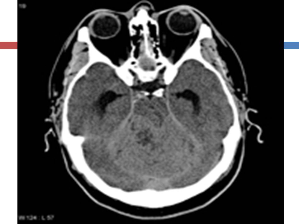
55
Right frontal brain abscess
Before contrast After contrast

56
Treatment The main treatment of brain abscess is the antimicrobial therapy. Surgery is necessary for 2 reasons: Gain samples of the infectuous material to identify the causative organism and the suitable antibiotic(s) Decrease the mass effect & ICP Surgery consists typically of: Burr hole & Aspiration of abscess
 Decrease the mass effect & ICP. Surgery consists typically of: Burr hole & Aspiration of abscess.")
57
Intracranial Tuberculoma
Tuberculosis may affect the central nervous system in which case it is called TB meningitis, TB cerebritis, and TB myelitis ,respectively. Granulomas from CSF extension or blood spread Widely distributed Usually multiple and less than 1cm diameter Central zone of caseatin

58
Clinical presentation:
The patient may be asymptomatic , have pulmonary symptoms or have Symptoms and signs of raised intracranial pressure ( the usual presenting feature).
.")
59
Incidence: In western countries: <1% of all ICSOL In Saudi Arabia: ~5% of all ICSOL In last 2 decades there was a worldwide rise in association with AIDS Often affects children & young adults

60
Intracranial Tuberculoma
Incidence: In western countries: <1% of all ICSOL In Saudi Arabia: ~5% of all ICSOL In last 2 decades there was a worldwide rise in incidence secondary to AIDS.

61
Intracranial Tuberculoma ETIOLOGY & PATHOGENSIS
Caused by human type of Mucobacterium tuberculosis, but rarely by atypical strains Secondary hematogenous spread from another lesion elsewhere in the body, usually lungs Most patients have no history of T.B. meningitis 50% of patients have a +ve history of T.B. or contact with T.B. patient Concomitant extracranial T.B. lesions detected: In 10% of patients in clinical series, and In >70% of autopsied patients

62
Intracranial Tuberculoma CLINICAL PRESENTATION
Symptoms & signs of progressive ICSOL: Raised ICP: in nearly all infratentorial and most (80%) of supratentorial tuberculomas Epileptic seizures: in 70 – 85% of patients with supratentorial tuberculomas Focal neurological deficit: ~50% of patients General symptoms (malaise, anorexia, perspiration, fever): ~50% of cases
 of supratentorial tuberculomas. Epileptic seizures: in 70 – 85% of patients with supratentorial tuberculomas. Focal neurological deficit: ~50% of patients. General symptoms (malaise, anorexia, perspiration, fever): ~50% of cases.")
63
Intracranial Tuberculoma INVESTIGATIONS
Lab work-up: Increased WBC: 60% of cases ESR: elevated or normal Mantoux test: Often positive CXR: +ve findings for T.B. in ~50% of cases Methods of choice: CT- scan MRI

64
Multiple intracranial tuberculomas

65
Treatment & Prognosis The main treatment is a combined anti-TB drug therapy for at least one year or even longer. Surgery is often required for histopathological confirmation of diagnosis Prognosis is generally good

66
Thank you ..

Similar presentations


 tumors arising from one of the many different cell types within.>")


 H. Louis Harkey Department of Neurosurgery University of Mississippi Jackson, MS.>")

