Download presentation
Presentation is loading. Please wait.

1
Cell Division Control the biochemicals that control cell division Mike Clark, M.D.

2
Why does a cell perform mitosis? 1. For organism growth – hyperplasia 2. For organism repair – to repair an organ like the liver – by producing some new functional cells 3. For replacement of cells A cell should perform mitosis – only when necessary – thus division should be controlled. If control of cell division is loss – tumors (benign or malignant) form.
 form..")
3
The eukaryotic cell cycle is regulated by a molecular control system The frequency of cell division varies with the type of cell (neurons and skeletal muscles do not divide – liver cells divide twice a year – skin cells twice a day – blood cells very fast) These cell cycle differences result from regulation at the chemical (molecular) level Copyright © 2008 Pearson Education, Inc., publishing as Pearson Benjamin Cummings
 These cell cycle differences result from regulation at the chemical (molecular) level Copyright © 2008 Pearson Education, Inc., publishing as Pearson Benjamin Cummings")
4
Evidence for Cytoplasmic Signals The cell cycle appears to be driven by specific chemical signals present in the cytoplasm – these chemicals are proteins – thus they must be formed from genes Some evidence for this hypothesis comes from experiments in which cultured mammalian cells at different phases of the cell cycle were fused to form a single cell with two nuclei

5
Fig. 12-13 Experiment 1 Experiment 2 EXPERIMENT RESULTS SG1G1 M G1G1 M M S S When a cell in the S phase was fused with a cell in G 1, the G 1 nucleus immediately entered the S phase—DNA was synthesized. When a cell in the M phase was fused with a cell in G 1, the G 1 nucleus immediately began mitosis—a spindle formed and chromatin condensed, even though the chromosome had not been duplicated.

6
The genes that code for the proteins that control cell division Proto-oncogenes code for proteins that help to regulate cell growth and differentiation. Proto-oncogenes are often involved in signal transduction and execution of mitogenic signals, usually through their protein products.proteinscell growthdifferentiationsignal transductionmitogenicprotein A tumor suppressor gene, or antioncogene, is a gene that protects a cell from one step on the path to cancer. genecell

7
Tumor-Suppressor Genes Tumor-suppressor genes help prevent uncontrolled cell growth Mutations that decrease protein products of tumor-suppressor genes may contribute to cancer onset Tumor-suppressor proteins – Repair damaged DNA – Control cell adhesion (cells should stay attached – cancer cells like to detach and move (metastasis) – Inhibit the cell cycle in the cell-signaling pathway
 – Inhibit the cell cycle in the cell-signaling pathway")
8
Mutations of the Genes A proto-oncogene is a normal gene that can become an oncogene due to mutations or increased expression. An oncogene is a gene that, when expressed at high levels, helps turn a normal cell into a tumor cell When a tumor suppressor gene is mutated to cause a loss or reduction in its function, the cell can progress to cancer, usually in combination with other genetic changes.

9
Proto-oncogenes can be converted to oncogenes by – Movement of DNA within the genome: if it ends up near an active promoter, transcription may increase – Amplification of a proto-oncogene: increases the number of copies of the gene – Point mutations in the proto-oncogene or its control elements: causes an increase in gene expression

10
Fig. 18-20 Normal growth- stimulating protein in excess New promoter DNA Proto-oncogene Gene amplification: Translocation or transposition: Normal growth-stimulating protein in excess Normal growth- stimulating protein in excess Hyperactive or degradation- resistant protein Point mutation: Oncogene within a control element within the gene

11
The Cell Cycle Control System The sequential events of the cell cycle are directed by a distinct cell cycle control system, which is similar to a clock The cell cycle control system is regulated by both internal and external controls (growth factors) The clock has specific checkpoints where the cell cycle stops until a go-ahead signal is received Copyright © 2008 Pearson Education, Inc., publishing as Pearson Benjamin Cummings
 The clock has specific checkpoints where the cell cycle stops until a go-ahead signal is received Copyright © 2008 Pearson Education, Inc., publishing as Pearson Benjamin Cummings")
12
Fig. 12-14 S G1G1 M checkpoint G2G2 M Control system G 1 checkpoint G 2 checkpoint

13
For many cells, the G 1 checkpoint seems to be the most important one (termed the restriction point) If a cell receives a go-ahead signal at the G 1 checkpoint, it will usually complete the S, G 2, and M phases and divide If the cell does not receive the go-ahead signal, it will exit the cycle, switching into a non- dividing state called the G 0 phase
 If a cell receives a go-ahead signal at the G 1 checkpoint, it will usually complete the S, G 2, and M phases and divide If the cell does not receive the go-ahead signal, it will exit the cycle, switching into a non- dividing state called the G 0 phase")
14
Fig. 12-15 G1G1 G0G0 G 1 checkpoint (a)Cell receives a go-ahead signal G1G1 (b) Cell does not receive a go-ahead signal
Cell receives a go-ahead signal G1G1 (b) Cell does not receive a go-ahead signal.")
15
Cyclins and Cyclin-Dependent Kinases Two types of regulatory proteins are involved in cell cycle control: cyclins and cyclin-dependent kinases (Cdks) What generically is a kinase enzyme? A kinase enzyme catalytically facilitates the transfer of an energized phosphate from ATP to an un- energized protein (energizes a endergonic reaction)
.")
16
Cyclin Dependent Kinases The cyclin dependent kinase enzymes maintain fairly constant concentrations in the cell during the cell division cycle The cyclin dependent kinase enzyme is generally inactive – due to not being complexed to a cyclin Since this kinase enzyme requires a cyclin to activate it – it is termed cyclin dependent kinase There are different kinds of cyclin dependent kinases and different kinds of cyclins Animal cells seem to have 3 prominent CDKs and several different cyclins

18
The Cyclins The concentrations of cyclins fluctuate (rise and lower) during the cell division cycle – this is why they are termed cyclins. The fluctuations in cyclins concentrations are due to critical timed formations and critical timed enzymatic degradations during the cell division cycle. As a the various cyclin concentrations rise – they complex with their particular cyclin dependent kinase The joining of the cyclins with their particular cyclin dependent kinase activates the kinase enzyme so that it can split ATP thus energizing the necessary structural proteins required in the operation of the cell division cycle

19
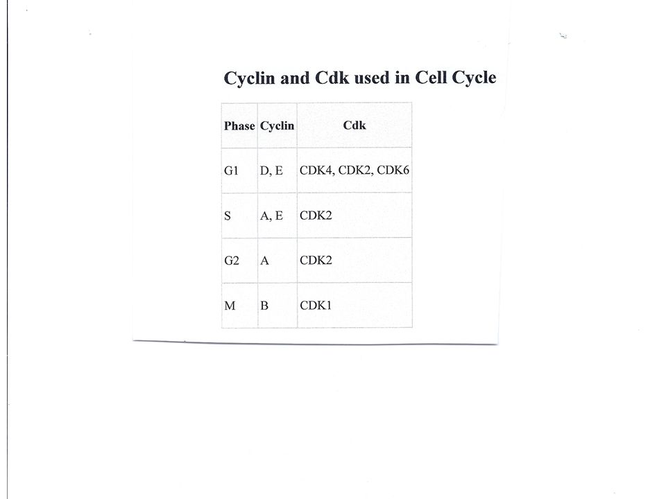
The Kinase Complexes

20
G1 Complex This is probably the most important complex in that it controls most of cell division by causing the pass through of the cell’s restriction point In order to pass the G1 checkpoint (restriction point) – the G1 cyclin must complex with its particular protein kinase This kinase energizes the proteins necessary for DNA synthesis – thus taking the cell into S-phase This is the kinase inhibited by tumor suppressor genes if a cell is damaged and should not further divide
 – the G1 cyclin must complex with its particular protein kinase This kinase energizes the proteins necessary for DNA synthesis – thus taking the cell into S-phase This is the kinase inhibited by tumor suppressor genes if a cell is damaged and should not further divide")
21
MPF A G2 cyclin can complex with its proper cyclin dependent kinase to form MPF termed by some maturation –promoting factor and by others M- phase factor MPF is a cyclin-Cdk complex that triggers a cell’s passage past the G 2 checkpoint into the M phase This kinase complex causes the proteins necessary for the start of mitosis (prophase) to become energized. It energizes the nuclear lamins which are intermediate filaments that hold the nuclear membrane in position. It energizes histone proteins which coil DNA from loop domain to chromatid/chromosome. It energizes tubulin proteins which form the microtubules of the mitotic apparatus

22
Prophase Prophase is the stage in which the cell – a. dissolves its nuclear membrane b. coils its genetic material from the loop domain fold into the chromatid /chromosome fold and c. assembles its mitotic apparatus Nuclear lamins become energized and start shaking thus dissolving the nuclear membrane Histones are proteins that the DNA coils around- thus by energizing them – they spin and coil the DNA tighter Tubulins comprise microtubules. Microtubules assemble and disassemble into tubulins when energized. Microtubules comprise the mitotic apparatus.

23
MPF MPF is a cyclin-Cdk complex that not only triggers (turns on) a cell’s passage past the G 2 checkpoint into the M phase – but it also appears to turn off some actions. During anaphase, the MPF helps switch itself off by initiating a process that leads to the destruction of its own cyclin. The MPF without its cyclin does not function. It is not until the G2 cyclin is created again during the S and G2 phases of the cell divisional cycle that it can complex with Cyclin Dependent Kinase to form G2 – thus pushing the cell into the M-phase

24
Fig. 12-17 M G1G1 S G2G2 M G1G1 SG2G2 M G1G1 MPF activity Cyclin concentration Time (a) Fluctuation of MPF activity and cyclin concentration during the cell cycle Degraded cyclin Cdk G1G1 S G2G2 M G2G2 checkpoint Cyclin is degraded Cyclin MPF (b) Molecular mechanisms that help regulate the cell cycle Cyclin accumulation G2 cyclin level rising during S and G2 and dropping during anaphase
 Fluctuation of MPF activity and cyclin concentration during the cell cycle Degraded cyclin Cdk G1G1 S G2G2 M G2G2 checkpoint Cyclin is degraded Cyclin MPF (b) Molecular mechanisms that help regulate the cell cycle Cyclin accumulation G2 cyclin level rising during S and G2 and dropping during anaphase.")
25
Fig. 12-17a Time (a) Fluctuation of MPF activity and cyclin concentration during the cell cycle Cyclin concentration MPF activity M M M SS G1G1 G1G1 G1G1 G2G2 G2G2
 Fluctuation of MPF activity and cyclin concentration during the cell cycle Cyclin concentration MPF activity M M M SS G1G1 G1G1 G1G1 G2G2 G2G2.")
26
Fig. 12-17b Cyclin is degraded Cdk MPF Cdk M S G1G1 G 2 checkpoint Degraded cyclin Cyclin (b) Molecular mechanisms that help regulate the cell cycle G2G2 Cyclin accumulation
 Molecular mechanisms that help regulate the cell cycle G2G2 Cyclin accumulation.")
27
Stop and Go Signs: Internal and External Signals at the Checkpoints An example of an internal signal is that kinetochores not attached to spindle microtubules send a molecular signal that delays anaphase Some external signals are growth factors, proteins released by certain cells that stimulate other cells to divide For example, platelet-derived growth factor (PDGF) stimulates the division of human fibroblast cells in culture
 stimulates the division of human fibroblast cells in culture")
28
External Signals for Cell Division Some researchers believe that signal to initiate cell division is almost totally controlled by outside signals Most of these signals are in the form of growth factors

29
Growth Factors A growth factor is a naturally occurring substance capable of stimulating cellular growth, proliferation and cellular differentiation. Usually it is a protein or a steroid hormone. Growth factors are important for regulating a variety of cellular processes. Growth factors typically act as signaling molecules between cells. Examples are cytokines and hormones that bind to specific receptors on the surface of their target cells. They often promote cell differentiation and maturation, which varies between growth factors. For example, bone morphogenic proteins stimulate bone cell differentiation, while fibroblast growth factors and vascular endothelial growth factors stimulate blood vessel differentiation (angiogenesis).
..")
30
A cell can only receive a chemical signal from another cell – if the receiving cell has specific receptors for that certain chemical. Once the chemical signal is attached to the receptor on the receiving cell – it turns of an internal cascade of chemical reactions inside the receiving cell The external chemical sent by the sending cell is attempting to communicate with the receiving cell’s nucleus – thus it is using a internal cascade of chemicals to communicate with the cell nucleus

31
Internal Cell Chemical Cascade

32
Partial List of Growth Factors Bone morphogenetic proteins (BMPs) Epidermal growth factor (EGF) Erythropoietin (EPO) Fibroblast growth factor (FGF) Granulocyte-colony stimulating factor (G-CSF) Granulocyte-macrophage colony stimulating factor (GM-CSF) Growth differentiation factor-9 (GDF9) Hepatocyte growth factor (HGF) Hepatoma derived growth factor (HDGF) Insulin-like growth factor (IGF) Myostatin (GDF-8) Nerve growth factor (NGF) and other neurotrophins Platelet-derived growth factor (PDGF) Thrombopoietin (TPO) Transforming growth factor alpha(TGF-α) Transforming growth factor beta (TGF-β) Vascular endothelial growth factor (VEGF) Stimulates cell cycle from G0 phase to G1 phase
 Epidermal growth factor (EGF) Erythropoietin (EPO) Fibroblast growth factor (FGF) Granulocyte-colony stimulating factor (G-CSF) Granulocyte-macrophage colony stimulating factor (GM-CSF) Growth differentiation factor-9 (GDF9) Hepatocyte growth factor (HGF) Hepatoma derived growth factor (HDGF) Insulin-like growth factor (IGF) Myostatin (GDF-8) Nerve growth factor (NGF) and other neurotrophins Platelet-derived growth factor (PDGF) Thrombopoietin (TPO) Transforming growth factor alpha(TGF-α) Transforming growth factor beta (TGF-β) Vascular endothelial growth factor (VEGF) Stimulates cell cycle from G0 phase to G1 phase")
33
Fig. 18-21a Receptor Growth factor G protein GTP Ras GTP Ras Protein kinases (phosphorylation cascade) Transcription factor (activator) DNA Hyperactive Ras protein (product of oncogene) issues signals on its own MUTATION NUCLEUS Gene expression Protein that stimulates the cell cycle (a) Cell cycle–stimulating pathway 1 1 3 4 5 2
 Transcription factor (activator) DNA Hyperactive Ras protein (product of oncogene) issues signals on its own MUTATION NUCLEUS Gene expression Protein that stimulates the cell cycle (a) Cell cycle–stimulating pathway")
34
Fig. 12-18 Petri plate Scalpels Cultured fibroblasts Without PDGF cells fail to divide With PDGF cells prolifer- ate 10 µm

35
Another example of external signals is density- dependent inhibition, in which crowded cells stop dividing Most animal cells also exhibit anchorage dependence, in which they must be attached to a substratum in order to divide Copyright © 2008 Pearson Education, Inc., publishing as Pearson Benjamin Cummings

36
Fig. 12-19 Anchorage dependence Density-dependent inhibition (a) Normal mammalian cells (b) Cancer cells 25 µm
 Normal mammalian cells (b) Cancer cells 25 µm.")
37
Cancer cells exhibit neither density-dependent inhibition nor anchorage dependence Copyright © 2008 Pearson Education, Inc., publishing as Pearson Benjamin Cummings

38
Loss of Cell Cycle Controls in Cancer Cells Cancer cells do not respond normally to the body’s control mechanisms Cancer cells may not need growth factors to grow and divide: – They may make their own growth factor – They may convey a growth factor’s signal without the presence of the growth factor – They may have an abnormal cell cycle control system Copyright © 2008 Pearson Education, Inc., publishing as Pearson Benjamin Cummings

39
A normal cell is converted to a cancerous cell by a process called transformation Cancer cells form tumors, masses of abnormal cells within otherwise normal tissue If abnormal cells remain at the original site, the lump is called a benign tumor Malignant tumors invade surrounding tissues and can metastasize, exporting cancer cells to other parts of the body, where they may form secondary tumors Copyright © 2008 Pearson Education, Inc., publishing as Pearson Benjamin Cummings

40
Fig. 18-21 Receptor Growth factor G protein GTP Ras GTP Ras Protein kinases (phosphorylation cascade) Transcription factor (activator) DNA Hyperactive Ras protein (product of oncogene) issues signals on its own MUTATION NUCLEUS Gene expression Protein that stimulates the cell cycle (a) Cell cycle–stimulating pathway MUTATION Protein kinases DNA DNA damage in genome Defective or missing transcription factor, such as p53, cannot activate transcription Protein that inhibits the cell cycle Active form of p53 UV light (b) Cell cycle–inhibiting pathway (c) Effects of mutations EFFECTS OF MUTATIONS Cell cycle not inhibited Protein absent Increased cell division Protein overexpressed Cell cycle overstimulated 1 2 3 4 5 2 1 3
 Transcription factor (activator) DNA Hyperactive Ras protein (product of oncogene) issues signals on its own MUTATION NUCLEUS Gene expression Protein that stimulates the cell cycle (a) Cell cycle–stimulating pathway MUTATION Protein kinases DNA DNA damage in genome Defective or missing transcription factor, such as p53, cannot activate transcription Protein that inhibits the cell cycle Active form of p53 UV light (b) Cell cycle–inhibiting pathway (c) Effects of mutations EFFECTS OF MUTATIONS Cell cycle not inhibited Protein absent Increased cell division Protein overexpressed Cell cycle overstimulated")
Similar presentations


 Coordination of cell division A multicellular organism needs to coordinate cell division across different tissues.>")

>")


 PHASE Cytokinesis Mitosis S G1G1.>")


