Download presentation
Presentation is loading. Please wait.

1
Stacey Sever, BSN RN CEN Clinical Nurse Educator Emergency Department Providence Alaska Medical Center With thanks to James Booth, MD, Kevin Ellis, RN, Andres Viles, RN And the University of Alabama at Birmingham

2
Indications/Contraindications Basic Anatomy of the Neck Preparation for Placement Technique for Placement What to Document

3
Obtaining IV access can often be a major problem for patients and healthcare providers. Delays in IV access lead to delays in patient care and expose patient to undue pain and anxiety, both of which are bad for patients and healthcare providers. This impacts patient satisfaction scores and consequently impacts the amount of revenue the hospital earns. The external jugular is considered a peripheral vein and can be accessed by trained RN’s

4
Shock - Hypothermic -Sepsis Difficult venous access - IV drug abuser -Renal failure -Elderly -Pediatric -Hypothermia Cardiac arrest Trauma Drowning

5
External jugular vein cannulation is indicated when emergent access is needed or for individual situations when other veins cannot be accessed. External jugular cannulation can be attempted initially in life threatening events where no obvious peripheral site is noted.

6
Patient cannot tolerate being flat. Patient is actively vomiting. Patient is agitated, moving head. Patient has a neck mass. Patient has a VP shunt on side of intended insertion

7
Cervical Spine Trauma Soft tissue neck trauma Circumferential burns to the neck Inability to identify anatomical landmarks for cannulation Evidence of infection at or near the intended insertion site

8
Anatomical location of the EJ What structures to avoid

11
The external jugular vein is superficial. Avoid inserting the IV catheter too deep in order to avoid accessing the carotid artery

12
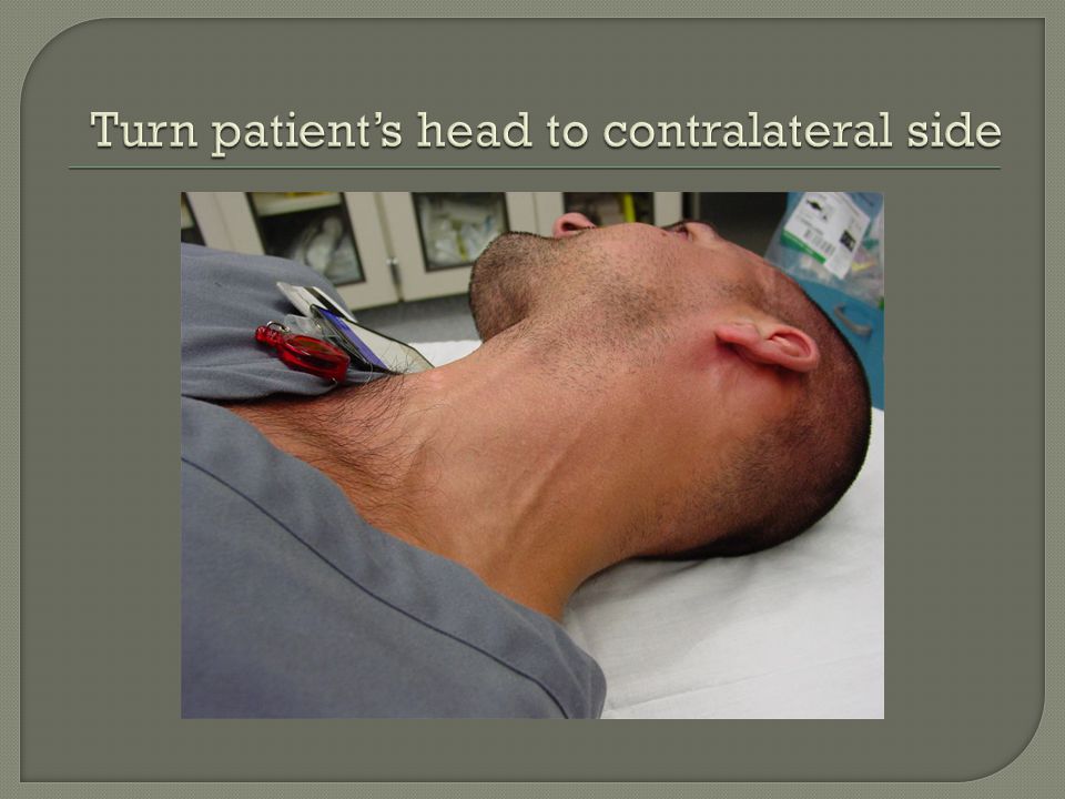
Verify MD order Assemble equipment Explain procedure to patient/family/caregiver Wash hands or use hand sanitizer Put on non-sterile gloves Place patient supine and head down if possible. This helps to distend the jugular veins and reduces the possibility of an air embolism Turn the patient’s head away from the side of the neck you intend to use

13
IV start kit IV catheter of appropriate size (14g-20g for adults) IV extension tubing (J loop) Normal saline flush
 IV extension tubing (J loop) Normal saline flush")
16
Identify the external jugular vein. Cleanse venipuncture site using chlorhexidine gluconate sponge with vigorous side-to-side prep and allow to dry. Lightly place a finger of the non-dominate hand just above the clavicle to produce a tourniqueting effect. Use the thumb of that same hand to pull traction above the puncture site. Puncture the vein midway between the angle of the jaw and the clavicle and cannulate the vein in a shallow and superficial manner.

17
Confirm placement of catheter/needle by witnessing flashback. Remove the IV catheter needle according to manufacturer’s directions, activate safety device, and discard in appropriate receptacle. Attach IV tubing or saline lock device primed with IV solution to hub. Apply transparent dressing and tape to catheter to secure, avoiding circumferential dressing or taping.

19
Hematoma Infection/phlebitis Air Embolism Infiltration /extravasation Accidental puncture of internal jugular or carotid artery Catheter shear with risk of embolus formation

20
Monitor site for signs of complications: -redness -warmth -infiltration Use IV infusion pumps only. Do not place fluids on a pressure bag. Do not use for vasoactive medications or radiographic contrast.

21
Appropriate documentation should be placed in EPIC including site location, size of catheter, and procedure toleration

22
Ask MD before attempting EJ placement. Clean the Skin! Scrub, Scrub, Scrub No blind sticks! If you can’t see it, don’t stick it. Remain shallow and superficial when inserting. Assure that you have blood return. An ED MD must observe and check-off for validation.

Similar presentations


 VTDRG: Chapter 8 (pg: 349-351)>")


 449-9330>")


