Download presentation
Presentation is loading. Please wait.

2
Body iron content – 3-4g ◦ Hb, iron containing proteins, bound to Tf, storage (ferritin, haemosiderin). Iron homeostasis is regulated strictly at level of intestinal absorption.

3
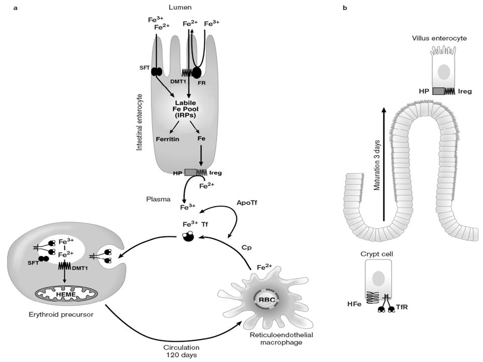
Haem diet – very readily absorbed via haem carrier protein 1 (apical bruish border membrane of duodenal enterocytes) i.e. higher bioavailability. Remainder of dietary iron poorly absorbed (10%). ◦ Ascorbic acid enhances absorption of non-animal sources of iron; tannates inhibit absorption. Fe2+ better absorbed cf. Fe3+.
. ◦ Ascorbic acid enhances absorption of non-animal sources of iron; tannates inhibit absorption. Fe2+ better absorbed cf. Fe3+..")
4
Fe3+ freed from food binding sites in stomach, binds to mucin, travels to duodenum and small bowel. ◦ Haem iron - carrier protein (endocytosis). ◦ Fe3+ - attachment to an integrin. ◦ Fe2+ - intestinal transporter DMT1.
. ◦ Fe3+ - attachment to an integrin. ◦ Fe2+ - intestinal transporter DMT1..")
5
Iron then enters cytosol, binds to cytosolic low molecular weight iron carriers and proteins e.g. Mobilferrin (shuttles iron with help of ATP) to basolateral membrane Export from basolateral membrane via duodenal iron exporter.
 to basolateral membrane Export from basolateral membrane via duodenal iron exporter..")
6
Upon release into circulation, re-oxidised to Fe3+, loaded onto transferrin. ◦ Site of influence of HFE gene product, +/- caeruloplasmin (known ferroxidase).
..")
7
Iron absorption regulated by many stimuli – ◦ Iron stores. ◦ Degree of erythropoiesis (increased with increased erythropoiesis, reticulocytosis). ◦ Ineffective erythropoiesis. ◦ Mobilferrin – mechanism of loss in iron replete state.
. ◦ Ineffective erythropoiesis. ◦ Mobilferrin – mechanism of loss in iron replete state..")
9
Transferrin and TfR. Ferritin. Iron responsive element-binding protein (IRE-BP) aka iron regulatory protein/factor (IRP/IRF). HFE. Divalent metal transporter (DMT1, Nramp2, DCT1,Slc11a) – duodenal iron transporter. Ferroportin and hephaestin, iron export proteins. Hepcidin.
 aka iron regulatory protein/factor (IRP/IRF). HFE. Divalent metal transporter (DMT1, Nramp2, DCT1,Slc11a) – duodenal iron transporter. Ferroportin and hephaestin, iron export proteins. Hepcidin..")
10
Encoded on long arm of chromosome 3. Half life 8 days. Hepatic synthesis. Complete lack incompatible with life (hypotransferrinaemia).
..")
11
Also on long arm of chromosome 3. homodimeric transmembrane protein. ◦ Found in most cells. Most dense on erythroid precursors, hepatocytes, placental cells. ◦ Restricted expression: both TfR1 and TfR2 present at high levels in hepatocytes, epithelial cells of small intestine including duodenal crypt cells.

12
Each TfR binds 2 diferric Tf molecules. Uptake by clustering on clathrin coated pits, then endocytosed. Iron off-loaded in acidified vacuoles, apotransferrin-TfR complex recycled to cell surface, apo-Tf then released back into circulation.

13
Cellular storage protein for iron. L and H chains (chromosome 19, 11). Synthesis controlled at 2 levels – ◦ DNA transcription via its promotor. ◦ mRNA translation via interactions with iron regulatory proteins. Acute phase reactant.

14
Ferritin in erythroid precursors may be of special importance in haem synthesis especially at beginning of Hb accumulation, when Tf-TfR pathway still in sufficient. When ferritin accumulates, it aggregates, proteolyzed by lysosomal enzymes,, then converted to iron-rich, poorly characterised haemosiderin, which releases iron slowly. M-ferritin – present in mitochondria. Expression correlated with tissues that have high mitochondrial number, rather than those involved in iron storage.

15
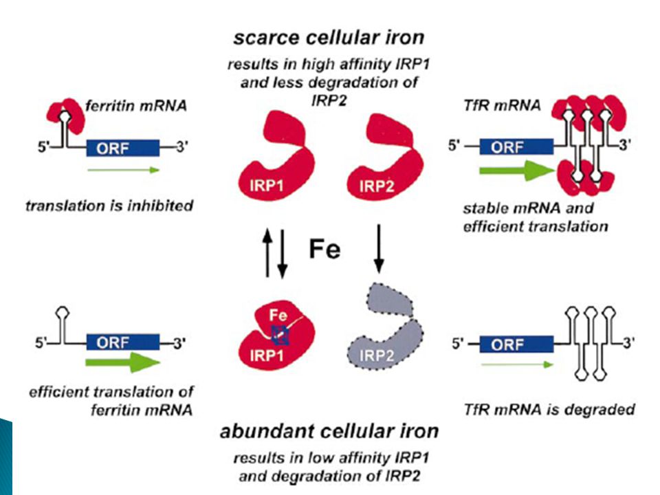
Sensing iron-regulatory proteins modulate synthesis of TfR, ferritin, DMT1. ◦ IRP1 and IRP2 – cytosolic RNA binding proteins. Bind to iron-responsive elements located in 5’ or 3’ untranslated regions of specific mRNAs encoding ferritin, TfR, DMT1 and (in erythroid cells) eALAS.
 eALAS..")
16
Binding of IRPs to IREs at 5’ end of transcrips of e.g. Ferritin, eALAS – decreases rate of synthesis; binding to 3’ end of transcripts e.g. TfR or DMT1, mRNA half life prolonged, increased synthesis. IRE-IRP complex senses state of iron balance – conformational change.

17
End result – in iron overload, increased ferritin (for adequate storage), decreased TfR (minimise further iron entry into cell), and vice versa in iron deficiency.
, decreased TfR (minimise further iron entry into cell), and vice versa in iron deficiency.")
21
Expression in GIT limited to cells in deep crypts in proximity to site of iron absorption. HFE protein associated with TfR, acts to modulate uptake of Tf-bound iron into crypt cells. Along with hepcidin, acts as iron sensor. Hereditary haemochromatosis with HFE gene mutation - inability to bind beta 2- microglobulin, impaired cellular trafficking, reduced incorporation into the cell membrane, reduced association with TfR1.

22
Divalent metal transporter protein – iron transporter (also Pb, Zn, Cu). Widely expressed, esp. in proximal duodenum. Isoform containing iron responsive element (Nramp2 isoform I) specifically upregulated in iron deficiency, greatest expression at brush border of apical pole of enterocytes in apical 2/3 of villi.
 specifically upregulated in iron deficiency, greatest expression at brush border of apical pole of enterocytes in apical 2/3 of villi..")
23
Increased body iron stores – enhanced uptake of iron from circulation into crypt cells. Increasing intracellular iron into crypt cells, differentiating enterocytes migrating up to villus tip downregulate iron transporter DMT1, reducing absorption of dietary iron from gut. Inverse relationship between ferritin levels in serum, and DMT1 levels in duodenal cells.

24
Transporting iron from basolateral membrane of enterocytes to circulation; from macrophage (from effete RBCs) into circulation for formation of new Hb. ◦ Ferroportin. ◦ Hephaestin.

25
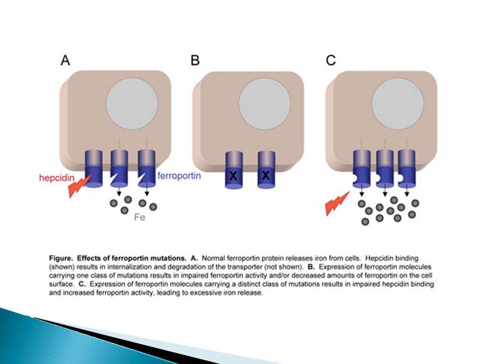
Ferroportin-1 in basal portion of placental syncytiotrophoblasts, basolateral surface of duodenal enterocytes, macrophages, hepatocytes. Upregulated by amount of available iron, downregulated through interaction with hepcidin.

27
Mutation in mice with sex-linked anaemia – enterocytes are iron loaded, but efflux through basolateral membrane inhibited. Homology to caeruloplasmin. Link between iron deficiency and copper deficiency – administration of copper facilitates egress of iron from tissue(s) into circulation.
 into circulation..")
28
SFT-mediated transport has properties defined for Tf-independent iron uptake, transporting iron across lipid bilayer. Process dependent on Cu. Has ferrireductase activity. Cytosolic localisation in recycling endosomes, stimulates Tf bound iron assimilation.

29
25 aa peptide hormone. Chromosome 19. Synthesized by hepatocyes. Intrinsic antimicrobial activity.

30
Binds ferroportin, complex internalised and degraded. Resultant decrease in efflux of iron from cells to plasma

31
Iron – stimulated with increased iron levels Inflammation, infection (and endotoxin) Hypoxia - downregulated Erythropoiesis – downregulated in anaemia, oxidative stress, ineffective erythropoiesis.
 Hypoxia - downregulated Erythropoiesis – downregulated in anaemia, oxidative stress, ineffective erythropoiesis.")
32
BMP - members of TGF-b superfamily which regulate cell proliferation, differentiation, apoptosis. Targets BMP receptors type I and II, resulting in phosphorylation of cytoplasmic R-Smads. R-Smads associate with Smad4, translocate to nucleus, activates transcription of target genes (in this case hepcidin).
..")
33
BMP2, 4, 5, 6, 7, 9 increase hepcidin expression in hepatic cells. ◦ Individual members of BMP family interact with different combinations of type I and II receptors. BMP’s effect on cellular response also modulated by BMP coreceptors. ◦ Hemojuvelin (HJV) - iron-specific, stimulates BMP2/4 pathway.
 - iron-specific, stimulates BMP2/4 pathway..")
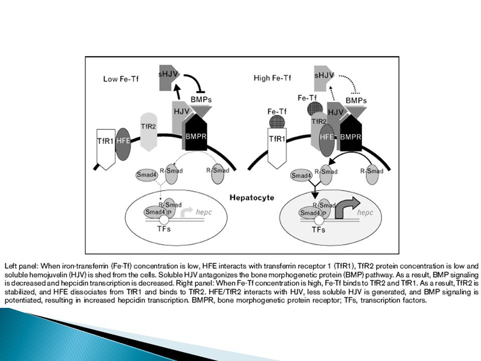
34
Member of family of repulsive guidance molecules (RGMs) - coreceptors of BMP receptors. Chromosome 1. Disruptive mutations cause juvenile haemochromatosis. 2 forms - ◦ GPI linked membrane form - stimulates BMP signalling and hepcidin expression. ◦ Soluble HJV (sHJV) - antagonist of BMP signalling.
 - antagonist of BMP signalling..")
35
Production stimulated by increased plasma iron and tissue stores. Negative feedback - hepcidin decreases release of iron into plasma (from macrophages and enterocytes). Fe-Tf increases hepcidin mRNA production (dose dependent relationship).
. Fe-Tf increases hepcidin mRNA production (dose dependent relationship)..")
36
HFE interacts with TfR1, but dissociates when Fe-Tf binds to TfR1. ◦ Amount of free HFE proportional to Tf-Fe. TfR2 – Tf-Fe stabilises TfR2 protein in dose dependent fashion. ◦ Fe-Tf binding increases fraction of TfR2 localizing to recycling endosomes, decreases fraction of TfR2 localizing to late endosomes where it is targeted for degradation. TfR2 competes with TfR1 for binding to HFE. ◦ HFE-TfR2 may regulate hepcidin expression by promoting HJV/BMP signalling, impacting upon hepcidin expression.

38
Hepcidin decreased in iron-deficiency anaemia, hereditary anaemias with ineffective erythropoiesis, and mouse models of anaemia from bleeding and haemolysis. Response not seen when erythropoiesis suppressed. ◦ Allows greater availability of iron for erythropoiesis. ◦ Degree of anaemia by itself doesn’t seem as important. Nature of erythropoietic regulator of hepcidin is unknown – proteins secreted by developing erythrocytes?

39
Mechanism particularly important in iron- loading anaemias. ◦ Urinary hepcidin very low in untransfused patients with thalassaemia intermedia, despite high serum and tissue iron levels. ◦ Very high erythropoietic activity overrides hepcidin regulation by iron. ◦ Severe hepcidin suppression leads to increased iron absorption and development of lethal iron overload.

40
Member of TGF-b superfamily, mediates hepcidin suppression in thalassaemia. Secreted during erythroblast maturation. Suppresses hepcidin mRNA production in primary human hepatocytes. Uncertain whether GDF15 plays role in pathogenesis other than that of ineffective erythropoiesis. ◦ Levels much lower in sera of sickle cell anaemia, MDS.

41
Physiological relevance uncertain. Hypoxia-inducing factor (HIF) is the main mediator of oxygen-regulated gene expression. ◦ VHL deficiency results in VHL protein deficiency, hence stabilisation of HIF. Resultant decrease in hepcidin levels.
 is the main mediator of oxygen-regulated gene expression. ◦ VHL deficiency results in VHL protein deficiency, hence stabilisation of HIF. Resultant decrease in hepcidin levels..")
42
IL-6 a prominent inducer of hepcidin, through STAT-3 dependent transcriptional mechanism. ◦ Other cytokines may also induce hepcidin independent of IL-6. Macrophage also express hepcidin in response to micobial stimulation. ◦ Hepcidin may function in autocrine manner to degrade macrophage ferroportin, causing local retention of iron in macrophages. Inflammatory stimuli acting through TNFa suppresses HJV mRNA, thus perhaps preventing iron-regulatory pathway from suppressing hepcidin during hypoferraemia of inflammation.

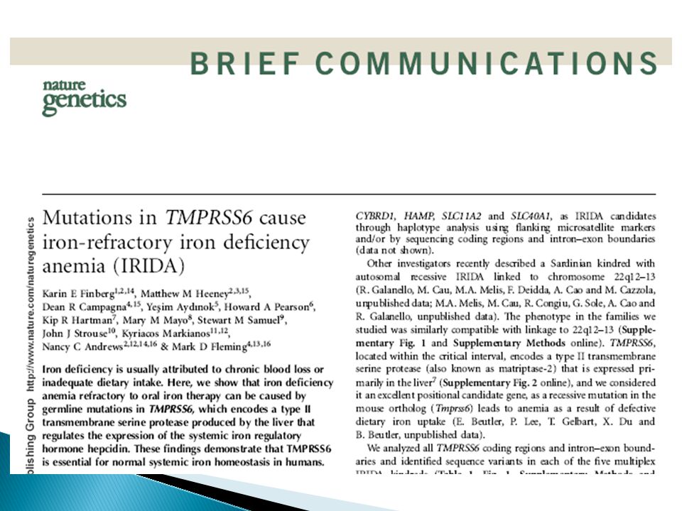
44
Iron overloadIron deficiency Hypotransferrinaemia - recessive HFE gene mutation TfR2 gene mutation – recessive Ferroportin mutation – autosomal dominant Hepcidin mutations Hemojuvelin mutations H ferritin mutation - dominant TMPRSS6 mutation - IRIDA

46
IDA unresponsive to oral iron supplementation, partially responsive to parenteral iron administration. Likely autosomal recessive. ?22q12-13 – encodes type II transmembrane serine protease (matriptase-2), primarily expressed in liver. Defect in iron uptake.
, primarily expressed in liver. Defect in iron uptake..")
48
Elevated urinary hepcidin cf. normal iron deficiency. ◦ ?reason for failure to absorb iron despite iron deficiency. Still unclear how mutations lead to ainappropriately elevated hepcidin. ◦ Negative regulator of hepcidin transcription in mouse models.

Similar presentations


 exists as bound form –IGF binding proteins (IGFBPs) IGFBPs –6 proteins and several related proteins –Serum IGFBP.>")

 Lecture 5. Signal amplification occurs in many signaling pathways Receptors are low abundance proteins The binding of few signaling.>")
 Receptors/Smad pathway BMP7 TGF β1, TGF β2, TGF β3 Dpp Inhibins Activins TGF β receptors.>")


